SISTEM KELISTRIKAN JANTUNG
Summary
TLDRThis video explains the heart's electrical conduction system, which controls the rhythm of heartbeats. It begins with a basic overview of the heart's function in pumping blood, followed by a detailed breakdown of its internal layers and how electrical impulses are generated. The SA node, acting as the heart's pacemaker, initiates the electrical signal that travels through the atria and AV node, then moves through the Bundle of His and Purkinje fibers, triggering ventricular contraction. The video highlights the significance of these electrical pathways in maintaining proper heart function and blood circulation.
Takeaways
- 😀 The heart functions as a pump, circulating blood to the entire body, including the lungs.
- 😀 The heart's contraction and blood flow require coordinated electrical activity.
- 😀 The heart has three main layers: endocardium (inner), myocardium (muscle), and pericardium (outer protective layer).
- 😀 The sinoatrial (SA) node, located near the superior vena cava, acts as the natural pacemaker of the heart.
- 😀 The SA node generates electrical impulses automatically, without influence from the nervous system or hormones.
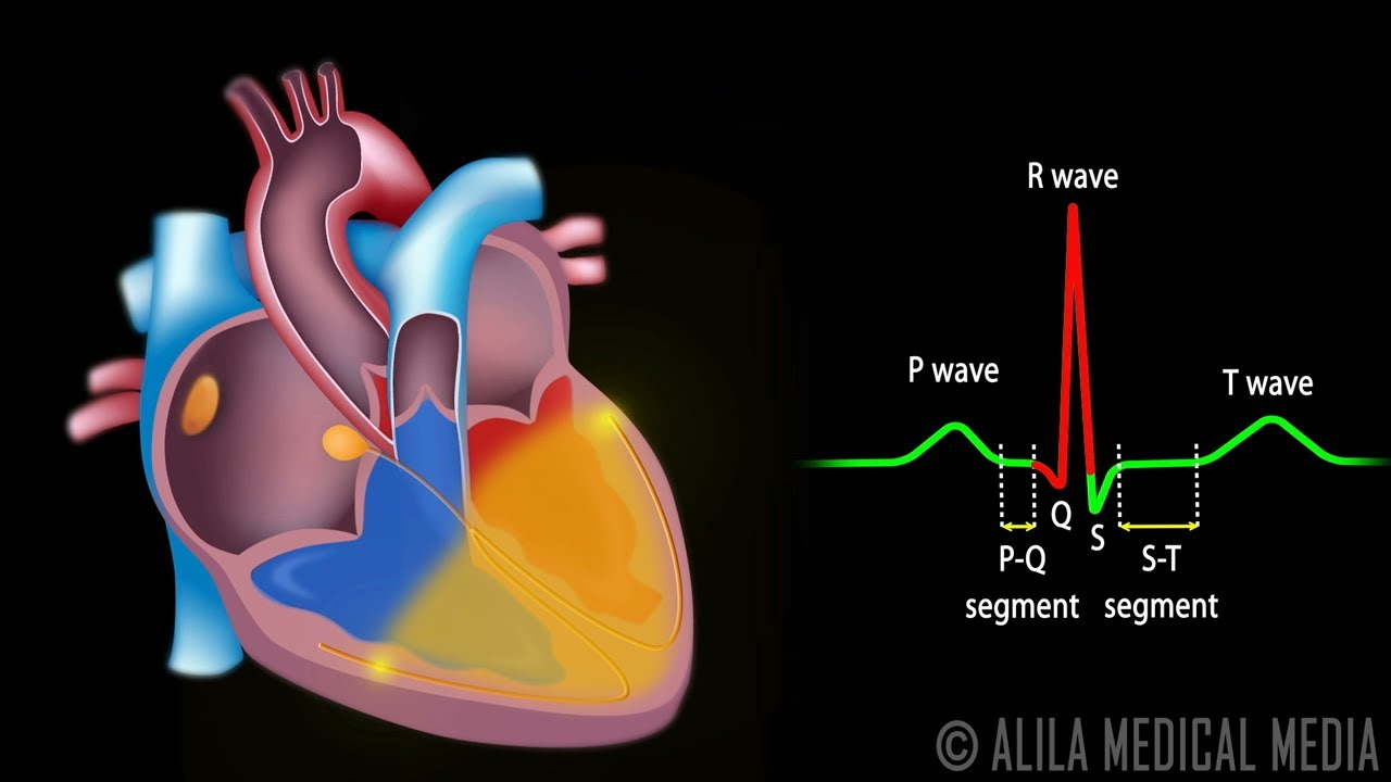
- 😀 Electrical impulses from the SA node spread through three pathways in the atria: anterior, middle, and posterior, causing atrial contraction.
- 😀 The atrioventricular (AV) node delays the electrical signal to allow the ventricles to fill with blood before contraction.
- 😀 From the AV node, impulses travel through the His bundle, which branches into the right and left bundle branches.
- 😀 The bundle branches further divide into Purkinje fibers, which conduct impulses to the ventricular muscle cells, triggering contraction.
- 😀 The synchronized electrical conduction ensures efficient pumping of blood from the atria to the ventricles and then out through the aorta and pulmonary arteries.
- 😀 Fibrous rings around the heart valves act as electrical insulators, directing the electrical impulses along proper pathways.
Q & A
What is the primary function of the heart according to the video?
-The heart's primary function is to pump blood throughout the body and lungs, ensuring oxygen and nutrients are delivered to tissues and organs.
What are the three main layers of the heart mentioned in the transcript?
-The three main layers of the heart are the endocardium (inner layer), myocardium (muscular middle layer responsible for contraction), and pericardium (outer protective layer).
What is the SA node, and why is it important?
-The SA node, or sinoatrial node, is located in the right atrium near the superior vena cava. It acts as the heart's natural pacemaker, generating electrical impulses automatically without influence from the nervous system or hormones.
How does the SA node influence other parts of the heart?
-The SA node sends electrical signals to other areas of the heart, including the atria and AV node, coordinating the timing of contractions to ensure efficient blood flow.
Why is there a delay at the AV node before ventricular contraction?
-The AV node delays the electrical impulse for about 40 milliseconds to allow the ventricles to fill completely with blood from the atria before contracting.
What are the main pathways through which the SA node conducts impulses to the atria?
-The SA node conducts impulses to the atria through three main pathways: the anterior pathway, the middle pathway, and the posterior pathway.
What is the role of the Bundle of His in heart conduction?
-The Bundle of His transmits electrical impulses from the AV node to the ventricles, branching into right and left pathways that further distribute signals to the Purkinje fibers.
What are Purkinje fibers, and what function do they serve?
-Purkinje fibers are specialized conductive fibers in the ventricles that rapidly transmit electrical impulses to ventricular muscle cells, triggering coordinated ventricular contraction.
Why can certain areas of the heart, like fibrous rings around valves, not conduct electricity?
-Fibrous rings are made of collagen-rich fibrous tissue, which is a poor conductor of electricity. This prevents electrical impulses from passing directly through valve areas, directing conduction along proper pathways.
What sequence of electrical conduction ensures coordinated heart contractions?
-The sequence is: SA node depolarization → atrial contraction → AV node delay → Bundle of His → ventricular branches → Purkinje fibers → ventricular contraction. This sequence ensures atria and ventricles contract in a coordinated manner.
How does intrinsic depolarization in the SA node differ from impulses influenced by the nervous system?
-Intrinsic depolarization occurs automatically in the SA node without external input, while nervous system signals can modify the rate and strength of heartbeats but are not required for baseline rhythm.
Why is understanding the electrical conduction system of the heart important for medical studies?
-Understanding cardiac conduction helps in diagnosing and treating arrhythmias, designing pacemakers, and comprehending how coordinated contractions maintain effective blood circulation throughout the body.
Outlines

This section is available to paid users only. Please upgrade to access this part.
Upgrade NowMindmap

This section is available to paid users only. Please upgrade to access this part.
Upgrade NowKeywords

This section is available to paid users only. Please upgrade to access this part.
Upgrade NowHighlights

This section is available to paid users only. Please upgrade to access this part.
Upgrade NowTranscripts

This section is available to paid users only. Please upgrade to access this part.
Upgrade NowBrowse More Related Video

Cardiac Arrhythmia
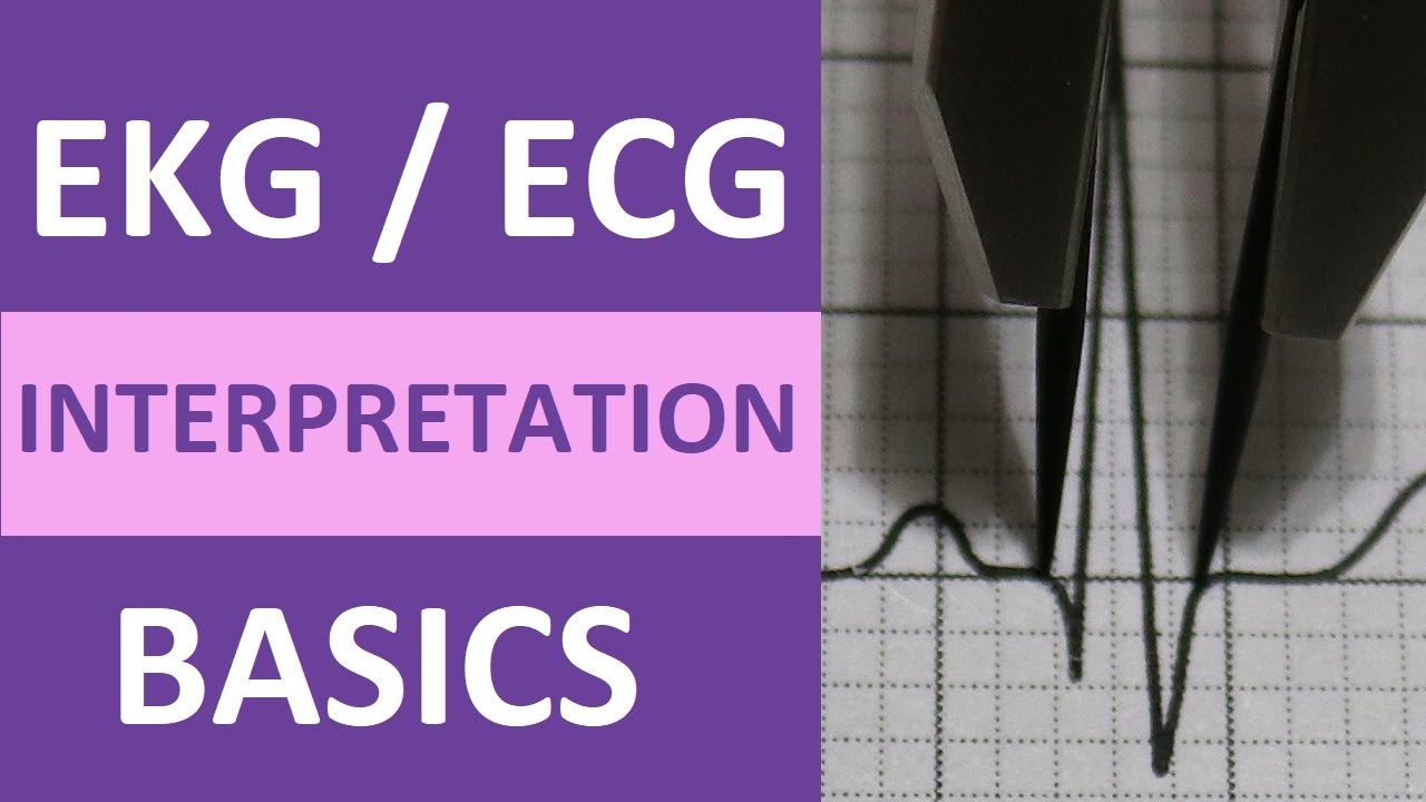
EKG/ECG Interpretation Basics Nursing NCLEX | QRS Complex, P Wave, T Wave, PR Interval
Cardiac Conduction System and Understanding ECG, Animation.

The Heart and Circulatory System - How They Work

Cardiology - Relationship of conduction system, ventricular contraction and ECG

HCL Learning | Structure of the Human Heart
5.0 / 5 (0 votes)