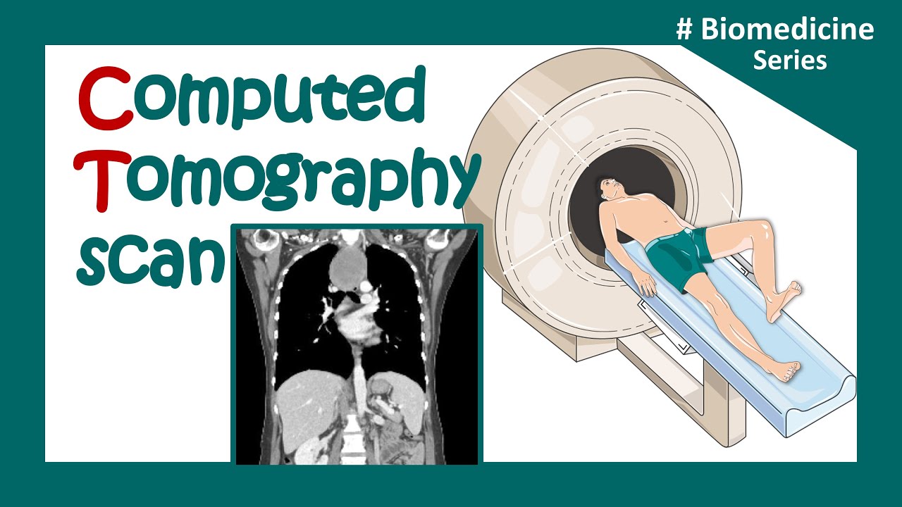
TC do Abdome: Fases do contraste venoso
Summary
TLDRThis video provides an in-depth overview of the various phases of venous contrast in computed tomography (CT), explaining the significance of each phase in diagnosing different conditions. It highlights the pre-contrast phase for detecting calcifications and fatty liver infiltration, the arterial and portal phases for vascular and tumor assessments, the pancreatic phase for identifying pancreatic tumors, and the secretory phase for detecting urothelial tumors. The speaker emphasizes the importance of selecting the appropriate phase based on clinical indications to optimize the examination, reduce radiation, and improve diagnostic accuracy in abdominal imaging.
Takeaways
- 😀 The pre-contrast phase is crucial for detecting calcifications, fatty liver infiltration, and tumors containing fat.
- 😀 Venous contrast injection is done at 3-5 ml per second, with a typical volume of 1-2 ml of contrast per kg of body weight.
- 😀 The arterial phase helps visualize hypervascular tumors and vascular abnormalities, such as aneurysms and thromboses.
- 😀 The portal phase is key for detecting hypovascular liver lesions, including hypovascular metastases.
- 😀 The pancreatic phase occurs 40 seconds after contrast injection and is important for detecting pancreatic tumors and necrosis.
- 😀 The nephrographic phase highlights the kidneys and is vital for detecting renal lesions, cysts, or infections.
- 😀 The secretory phase is used to identify filling defects in the renal collecting system, useful for detecting urothelial tumors.
- 😀 Proper selection of contrast phases based on clinical suspicion helps optimize CT exams, reducing radiation exposure and costs.
- 😀 The early arterial phase, within 15-20 seconds, is useful for detecting vascular anomalies but before contrast distribution in organs.
- 😀 Clinical data and context guide the choice of the most relevant contrast phase for each patient's diagnosis.
- 😀 In some cases, like hepatocarcinoma, hypervascular tumors need arterial phase imaging for optimal visualization of the tumor's blood supply.
Q & A
What is the purpose of the pre-contrast phase in CT imaging?
-The pre-contrast phase is crucial for detecting calcifications, fatty liver infiltrations, and abnormalities like urinary stones. It is also used for identifying fat within tumors and examining specific conditions like pneumoperitoneum, appendicitis, and diverticulitis.
Why is good venous access necessary during intravenous contrast injection?
-Good venous access is important because the contrast is injected at a rate of 3 to 5 ml per second, and the volume typically ranges from 100 to 150 ml. Proper access ensures that the contrast is administered smoothly and effectively for accurate imaging.
What is the role of the arterial phase in CT imaging?
-The arterial phase is essential for detecting vascular abnormalities such as aneurysms, thromboses, and arteriovenous fistulas. It is also crucial for identifying hypervascular tumors, including renal cell carcinoma, neuroendocrine tumors, and hepatocarcinoma.
How does the portal phase differ from the arterial phase in CT imaging?
-In the portal phase, the contrast is distributed through the portal system, enhancing the liver parenchyma. This phase is critical for detecting hypovascular lesions, such as hypovascular liver metastases, which become more visible due to the contrast difference between the normal liver tissue and the lesions.
What is the significance of the pancreatic phase in CT imaging?
-The pancreatic phase, occurring about 40 seconds after contrast injection, is important for detecting pancreatic tumors, such as pancreatic adenocarcinoma, which appears as hypodense areas. This phase highlights the pancreas maximally, enabling clear differentiation between normal tissue and tumor.
When is the nephrographic phase most useful in CT imaging?
-The nephrographic phase is valuable for detecting renal tumors, cysts, and inflammatory or infectious processes in the kidneys. It highlights the entire renal parenchyma, including both the cortex and medulla.
What is the purpose of the secretory phase in CT imaging?
-The secretory phase, which occurs 5 to 10 minutes after contrast injection, is used to assess the urinary system. It helps identify filling defects, such as urothelial tumors, and abnormalities like congenital or acquired lesions in the renal system, ureters, and bladder.
How does the use of contrast phases help optimize CT exams?
-By selecting the correct contrast phase based on the clinical indication, the exam can be optimized for accurate diagnosis. This approach reduces unnecessary radiation exposure to the patient and lowers the cost of the procedure, focusing on the most relevant phases for the pathology being investigated.
Why is the arterial phase particularly important for diagnosing hepatocarcinoma?
-Hepatocarcinoma is a hypervascular tumor that receives its blood supply from the hepatic artery, not the portal vein. The arterial phase is critical for visualizing these tumors as it highlights the hepatic artery and identifies areas of contrast enhancement in the tumor, making it more visible against the surrounding tissue.
What are some examples of hypervascular and hypovascular tumors and how are they detected in CT imaging?
-Hypervascular tumors, such as renal cell carcinoma, neuroendocrine tumors, and certain liver tumors, are best detected in the arterial phase, as they enhance more than the surrounding tissue. Hypovascular tumors, like most liver metastases, are best detected in the portal phase, where the liver parenchyma enhances significantly, highlighting the less enhanced lesions.
Outlines

This section is available to paid users only. Please upgrade to access this part.
Upgrade NowMindmap

This section is available to paid users only. Please upgrade to access this part.
Upgrade NowKeywords

This section is available to paid users only. Please upgrade to access this part.
Upgrade NowHighlights

This section is available to paid users only. Please upgrade to access this part.
Upgrade NowTranscripts

This section is available to paid users only. Please upgrade to access this part.
Upgrade NowBrowse More Related Video

Biomedical instrumentation- CT scan (Computed Tomography)

A Practical Introduction to CT

UQx Bioimg101x 3.2.4 CT Reconstruction & Back Projection
CT scan | computerized tomography (CT) scan |What is a CT scan used for? | Clinical application
What is Computed Tomography (CT) and how does it work?

CT (Computed Tomography) Scans - A Level Physics
5.0 / 5 (0 votes)