3D Tour of the Perineum
Summary
TLDRThis video delves into the intricate anatomy of the pelvis, focusing on the perineum and its components. The perineum is a diamond-shaped space between the thighs, with the pelvic floor muscles forming its ceiling. The script explores the urogenital and anal triangles, deep and superficial perineal pouches, and their roles in the body's functions. Key structures, such as the perineal membrane, external urethral sphincter, and anal sphincter muscles, are highlighted. The perineum's role in sexual and excretory health is emphasized through a detailed breakdown of its anatomical features and their functions.
Takeaways
- 😀 The perineum is a complex anatomical space located at the bottom of the pelvis, between the thighs.
- 😀 The perineum is shaped like a diamond and includes the anus, genitals, coccyx, pubic symphysis, and ischial tuberosities as its corners.
- 😀 The pelvic floor muscles form the ceiling of the perineum and are shaped like a bowl, partly convex.
- 😀 The boundaries of the pelvic outlet include the sto pubic rim and sacrohumoral ligaments.
- 😀 The urogenital triangle is the anterior half of the perineum, and the anal triangle is the posterior half.
- 😀 The anal triangle is tilted about 30 degrees from the horizontal, while the urogenital triangle is aligned with the horizontal plane in a standing position.
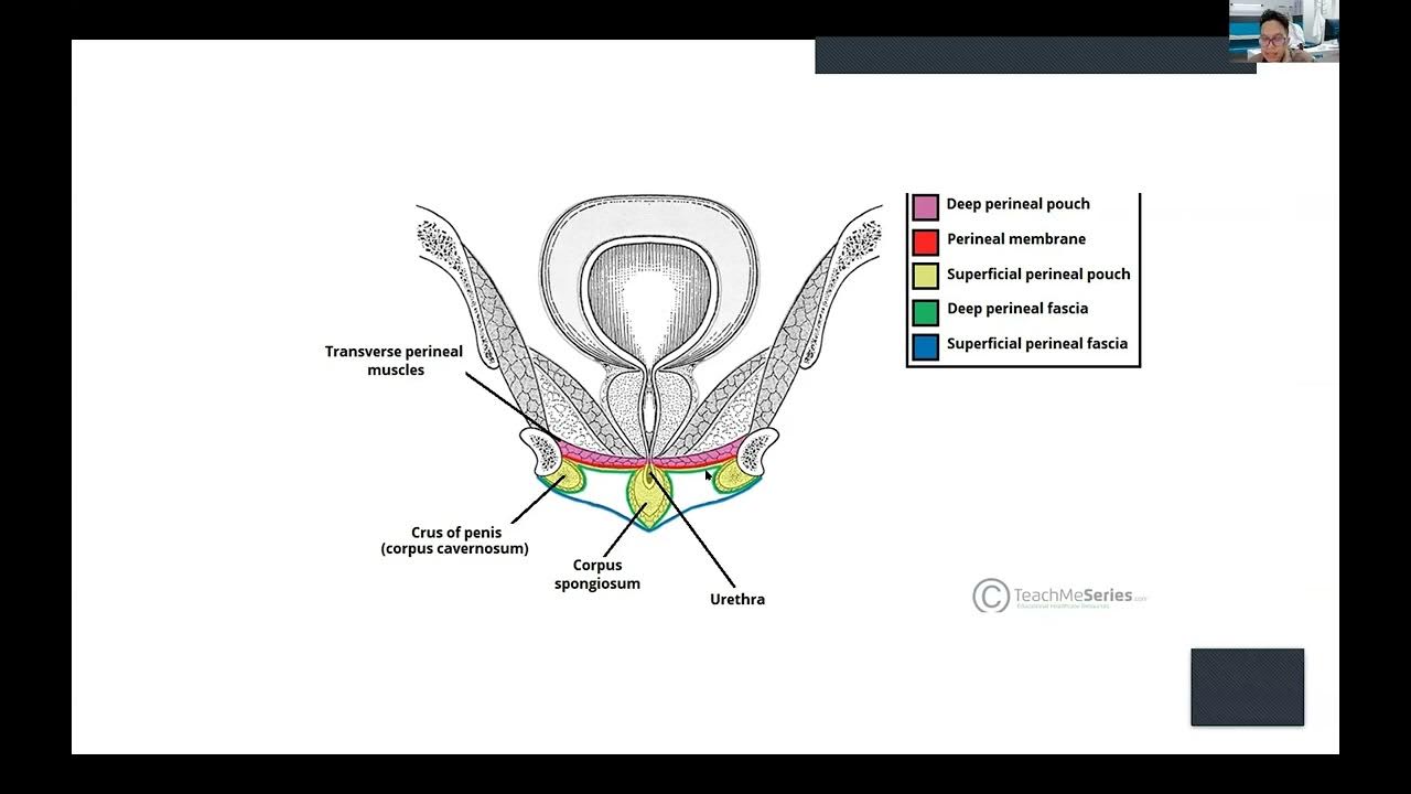
- 😀 The perineal membrane forms a fibrous sheet in the urogenital triangle and houses the deep perineal pouch.
- 😀 The deep perineal pouch is a shallow space containing muscles like the external urethral sphincter and is associated with the female reproductive system.
- 😀 The superficial perineal pouch lies between the perineal membrane and Colles' fascia, containing muscles and erectile tissues, including those of the penis and clitoris.
- 😀 The perineal body is the fibromuscular meeting point where many perineal muscles converge, and it lies posterior to the anal sphincter muscles.
Q & A
What is the perineum and where is it located?
-The perineum is the space between the thighs, encompassing the anus and genitals. It lies at the bottom of the pelvis.
What are the key anatomical landmarks that define the perineum?
-The corners of the perineum's diamond shape are defined by the coccyx, pubic symphysis, and the ischial tuberosities.
What forms the ceiling of the perineum?
-The ceiling of the perineum is formed by the pelvic floor muscles, which are shaped like a bowl.
What is the pelvic outlet and what defines its boundaries?
-The pelvic outlet is defined by the pubic symphysis, sacrotuberous ligament, and the coccyx. These form the boundaries of the pelvic outlet.
What are the two halves of the perineum, and how are they classified?
-The perineum is divided into two halves: the urogenital triangle anteriorly and the anal triangle posteriorly.
How does the urogenital triangle compare to the anal triangle in terms of position?
-In a standing position, the urogenital triangle roughly aligns with the horizontal plane, while the anal triangle is tilted upward at about 30 degrees.
What is the perineal membrane and what is its role?
-The perineal membrane is a fibrous sheet in the urogenital triangle that forms a boundary, separating the superficial and deep perineal pouches.
What structures are located in the deep perineal pouch?
-The deep perineal pouch contains muscles such as the external urethral sphincter, as well as spaces for the vagina and urethra, which pass through the lavatory ani muscles.
What muscles and tissues are found in the superficial perineal pouch?
-The superficial perineal pouch contains the bulbospongiosus, ischiocavernosus, and superficial transverse perineal muscles, along with erectile tissue of the penis and clitoris, and the continuation of the urethra and vagina.
What is the significance of the perineal body?
-The perineal body is a fibromuscular meeting point for many of the structures in the perineum, located at the center of the posterior border of the perineal membrane.
Outlines

This section is available to paid users only. Please upgrade to access this part.
Upgrade NowMindmap

This section is available to paid users only. Please upgrade to access this part.
Upgrade NowKeywords

This section is available to paid users only. Please upgrade to access this part.
Upgrade NowHighlights

This section is available to paid users only. Please upgrade to access this part.
Upgrade NowTranscripts

This section is available to paid users only. Please upgrade to access this part.
Upgrade NowBrowse More Related Video
intro la eksternal genitalia wanita_Tegar Fitriyana Sukaya Karso, dr.

Anatomi Panggul beserta ukurannya

Spermatogenesis | Blood-Testes Barrier | Hormonal Control | Structure of Sperm || Reproductive Physi

Hematologic System: Blood Components and Hemostasis - Medical-Surgical- Cardiovascular |@LevelUpRN

🥇 Anatomía del OJO 3/3 - Medios de Refracción, Cámaras del Ojo, Humor Acuoso, Cuerpo Vítreo

Internal Iliac Artery Scheme (Course, Branches, Mnemonic) | Anatomy
5.0 / 5 (0 votes)