Establishing a RESTING POTENTIAL in a neurone- Do you know what the resting potential is?
Summary
TLDRIn this informative video, Miss Esther explores the concept of resting potential in neurons, focusing on the structure of myelinated motor neurons and their function. She explains how the resting potential of -70 millivolts is maintained through the action of the sodium-potassium pump and the selective permeability of the cell membrane to potassium ions. The video is an excellent primer for understanding the electrical properties of neurons before delving into action potentials.
Takeaways
- 🧠 The video discusses the structure and function of a myelinated motor neuron, focusing on the cell body, dendrites, axon, and myelin sheath.
- 🔬 The cell body contains organelles necessary for protein and neurotransmitter synthesis, such as the nucleus, mitochondria, and ribosomes.
- 🌿 Dendrites are responsible for carrying action potentials to surrounding cells, playing a key role in neuronal communication.
- 🚀 The axon is a long conductive fiber that carries nerve impulses and is the site of multiple action potentials.
- 🛡️ The myelin sheath, made of lipid layers, acts as an insulator, allowing for faster and more efficient signal transmission along the neuron.
- 🔌 Nodes of Ranvier are gaps in the myelin sheath where action potentials can be generated, facilitating signal propagation.
- 🔋 The resting potential of a neuron, at -70 millivolts, represents the electrical charge difference between the inside and outside of the neuron when it's not conducting an impulse.
- 💡 The resting potential is maintained by the sodium-potassium pump, which actively transports ions across the membrane, creating an electrochemical gradient.
- ⚗️ The sodium-potassium pump moves three sodium ions out and two potassium ions into the axon, contributing to the resting potential.
- 🚫 The cell membrane is more permeable to potassium ions due to a higher number of potassium ion channels compared to sodium ion channels.
- 🔄 Passive diffusion of ions occurs due to the concentration gradient, with potassium ions moving out and sodium ions moving into the axon.
- 📚 The video offers additional resources for learning, including questions on miss Esther's website, and encourages viewers to subscribe for more content.
Q & A
What is the primary function of the myelin sheath in a myelinated motor neuron?
-The myelin sheath acts as an insulator, made up of lipid layers that prevent the passage of charged ions, thus facilitating the efficient transmission of electrical impulses along the neuron.
What are the gaps in the myelin sheath called, and where are action potentials generated?
-The gaps in the myelin sheath are called the nodes of Ranvier, and these are the locations where action potentials can be generated.
What is the resting potential of a neuron, and what does it measure?
-The resting potential of a neuron is the difference in electrical charge between the inside and outside of the neuron when it is not conducting an impulse. It measures the voltage, which is typically at minus 70 millivolts.
Why is the resting potential maintained at minus 70 millivolts?
-The resting potential is maintained at minus 70 millivolts due to the presence of more positive ions outside the cell compared to the inside, which is achieved by the sodium-potassium pump and the selective permeability of the cell membrane.
What role does the sodium-potassium pump play in maintaining the resting potential?
-The sodium-potassium pump actively transports three sodium ions out of the axon and two potassium ions into the axon, creating an electrochemical gradient that contributes to the resting potential of minus 70 millivolts.
How does the sodium-potassium pump create an electrochemical gradient?
-The pump creates an electrochemical gradient by actively moving more sodium ions out of the cell and more potassium ions into the cell, resulting in a higher concentration of sodium ions outside and potassium ions inside the axon.
What is the reason for the unequal distribution of sodium and potassium ions across the neuron's membrane?
-The unequal distribution is due to the cell membrane being more permeable to potassium ions, with more potassium ion channels than sodium ion channels, and the selective opening of these channels based on voltage levels.
What is the significance of the dendrites in a neuron?
-Dendrites are important for carrying action potentials to surrounding cells, playing a crucial role in the communication between neurons.
What is the function of the axon in a neuron?
-The axon is a long conductive fiber that carries the nerve impulse and is the site of multiple action potentials, essential for transmitting signals over long distances within the neuron.
Why are the nodes of Ranvier important for the transmission of action potentials?
-The nodes of Ranvier are important because they are the points where the myelin sheath is interrupted, allowing for the regeneration of the action potential as it travels along the neuron.
What is meant by 'sodium ion channels' and 'potassium ion channels' in the context of the neuron's membrane?
-Sodium and potassium ion channels are specific protein channels in the neuron's membrane that selectively allow the passage of sodium and potassium ions, respectively, playing a key role in the generation and maintenance of the neuron's resting potential and action potentials.
Outlines

This section is available to paid users only. Please upgrade to access this part.
Upgrade NowMindmap

This section is available to paid users only. Please upgrade to access this part.
Upgrade NowKeywords

This section is available to paid users only. Please upgrade to access this part.
Upgrade NowHighlights

This section is available to paid users only. Please upgrade to access this part.
Upgrade NowTranscripts

This section is available to paid users only. Please upgrade to access this part.
Upgrade NowBrowse More Related Video

Neuron materi lengkap - struktur, bentuk, fungsi, jenis - biologi sma kelas bab.sistem saraf

BTEC Applied Science: Unit 1 Biology Nerve Tissue
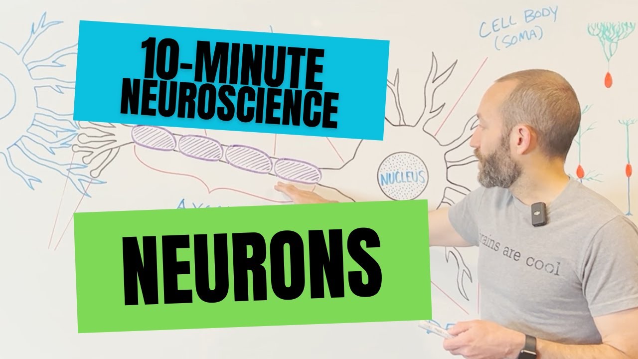
10-Minute Neuroscience: Neurons

Neurônios e Células da glia: Estruturas, funções e classificações

The Reflex Arc: Sensory, Relay and Motor Neurons - Biological Psychology [AQA ALevel]

Potenziale d'azione | NEUROSCIENZE - Lezione 4
5.0 / 5 (0 votes)