APRENDA TUDO SOBRE OS OSSOS DO QUADRIL!
Summary
TLDRThis video provides a detailed explanation of the anatomy of the hip region, focusing on key structures like the inguinal ligament and the ligaments reinforcing the hip joint (coxofemoral joint). It discusses how the inguinal ligament connects the iliac spine to the pubic tubercle, providing a pathway for important structures such as the femoral nerve, artery, and vein. The video also covers the three main ligaments of the hip joint: the pubofemoral, iliofemoral, and ischiofemoral ligaments, and their role in stabilizing the femur within the acetabulum.
Takeaways
- 😀 The inguinal ligament connects the anterior superior iliac spine to the pubic tubercle, acting as a key structure in the pelvic region.
- 😀 The inguinal ligament creates a passage for important structures: the femoral nerve, artery, and vein, in that order from lateral to medial.
- 😀 Compression of these structures due to an inguinal hernia can lead to symptoms in the lower limbs, particularly affecting circulation and nerve function.
- 😀 The inguinal ligament is a continuation of the external oblique muscle of the abdomen, providing structural support to the pelvic region.
- 😀 Three main ligaments reinforce the hip joint capsule: the pubofemoral, iliofemoral, and ischiofemoral ligaments.
- 😀 The iliofemoral ligament is the largest and most important in stabilizing the hip joint, positioned laterally.
- 😀 The pubofemoral ligament, located medially, strengthens the hip joint and prevents excessive movement.
- 😀 The ischiofemoral ligament, found posteriorly, contributes to the hip joint's stability, particularly in flexion.
- 😀 The three ligaments (pubofemoral, iliofemoral, and ischiofemoral) form a Z-shaped structure that enhances joint stability.
- 😀 The hip joint (coxofemoral joint) is reinforced by these ligaments to maintain a strong union between the acetabulum and the femur, enabling movement while preventing dislocation.
Q & A
What is the inguinal ligament and where is it located?
-The inguinal ligament is a fibrous structure that connects the anterior superior iliac spine (ASIS) of the pelvis to the pubic tubercle. It is an important landmark in the lower abdomen and plays a role in the passage of several important structures.
What structures pass through the space created by the inguinal ligament?
-The structures that pass through the space between the pelvis and the inguinal ligament include the femoral nerve, femoral artery, and femoral vein. These structures run in a lateral-to-medial sequence.
What is the clinical significance of the inguinal ligament in terms of hernias?
-Inguinal hernias can occur when there is a weakness or failure in the closure of the inguinal ligament, leading to the protrusion of abdominal contents. This can potentially compress the femoral nerve, artery, and vein, leading to symptoms in the lower limbs.
How is the inguinal ligament related to the external oblique muscle?
-The inguinal ligament is a continuation of the external oblique muscle from the abdomen. It serves as a structural connection between the pelvis and the abdominal muscles.
What are the three ligaments that reinforce the capsule of the hip joint (coxofemoral joint)?
-The three ligaments that reinforce the hip joint capsule are the pubofemoral ligament, the iliofemoral ligament, and the ischiofemoral ligament.
What is the function of the pubofemoral ligament?
-The pubofemoral ligament reinforces the hip joint capsule, providing support to the joint. It connects the pubis to the femur and plays a role in stabilizing the joint during movement.
What is the function of the iliofemoral ligament?
-The iliofemoral ligament is the largest and strongest ligament in the hip joint. It connects the ilium (part of the pelvis) to the femur and helps prevent excessive extension and maintain stability during movement.
What is the function of the ischiofemoral ligament?
-The ischiofemoral ligament is located posteriorly and reinforces the posterior part of the hip joint capsule. It helps stabilize the joint by preventing excessive internal rotation and flexion of the femur.
Why are the three ligaments of the hip joint important for its stability?
-The three ligaments (pubofemoral, iliofemoral, and ischiofemoral) work together to provide strong reinforcement to the hip joint capsule. This helps maintain the stability of the coxofemoral joint during various movements, especially in weight-bearing situations.
What happens if the capsule of the hip joint is removed?
-If the capsule of the hip joint is removed, the direct connection between the acetabulum (the socket in the pelvis) and the femur is compromised. The joint would become unstable without the support from these reinforcing ligaments.
Outlines

This section is available to paid users only. Please upgrade to access this part.
Upgrade NowMindmap

This section is available to paid users only. Please upgrade to access this part.
Upgrade NowKeywords

This section is available to paid users only. Please upgrade to access this part.
Upgrade NowHighlights

This section is available to paid users only. Please upgrade to access this part.
Upgrade NowTranscripts

This section is available to paid users only. Please upgrade to access this part.
Upgrade NowBrowse More Related Video
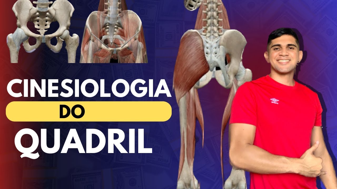
Cinesiologia do Quadril- Ossos, ligamentos , músculos : origem , inserção e ação.

Overview of Muscles of the Lower Limbs 🦵 - Quick Review - Anatomy Series
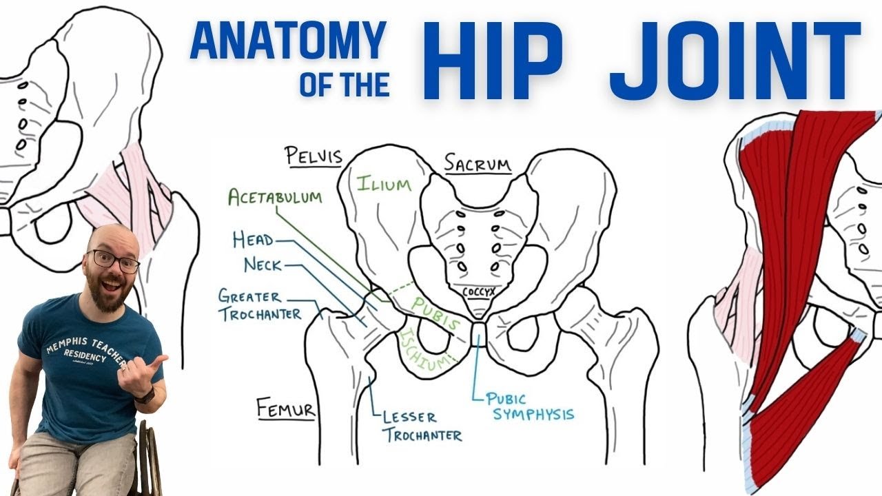
Anatomy of the Hip Joint | Bones, Ligaments, & Muscles

HIP JOINT DISTAL ARTICULATION (HIP JOINT COMPLEX BIOMECHANICS)Physiotherapy Tutorials
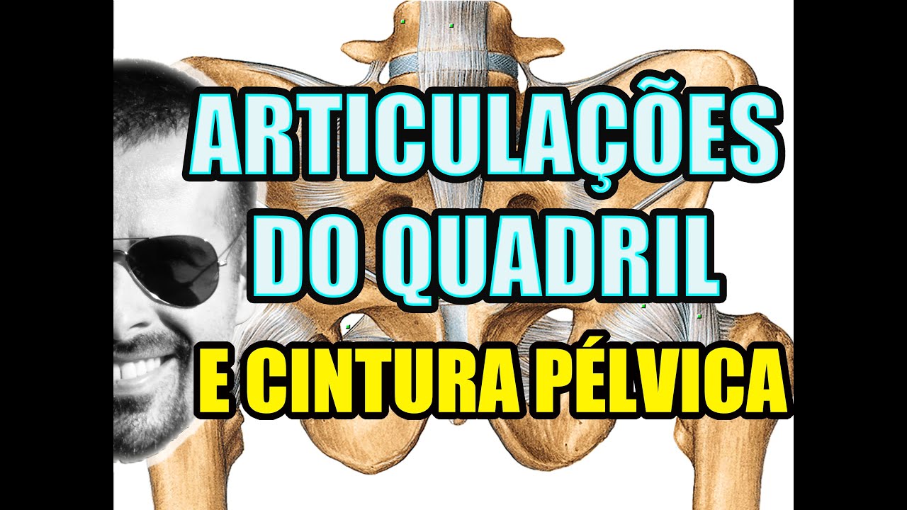
Vídeo Aula 115 - Anatomia Humana - Sistema Articular - Articulações do Quadril e da Cintura Pélvica

Ankle Joint - 3D Anatomy Tutorial
5.0 / 5 (0 votes)