Portosystemic anastomoses
Summary
TLDRThe video explains the liver’s regenerative capacity and its role in processing blood from the gastrointestinal system. It covers the anatomy of portal-systemic anastomoses, which are alternative blood routes when the liver becomes fibrous and blood flow is restricted. The script explores five key portal-systemic anastomoses: in the esophagus, anal canal, umbilicus, retroperitoneal area, and intrahepatic region. These anastomoses can lead to serious conditions like varices and ascites. The importance of understanding these alternate pathways is highlighted, especially in cases of liver disease, where they play a crucial role in bypassing the liver’s impaired circulation.
Takeaways
- 😀 The liver has an excellent regenerative capacity, but it can only repair itself so many times before it becomes fibrous, which limits blood flow.
- 😀 The liver is part of the gastrointestinal system, receiving blood from the gastrointestinal tract for processing before it enters the systemic circulation.
- 😀 The portal vein carries blood from the gastrointestinal tract to the liver, while the systemic veins return blood from the body to the heart.
- 😀 If the liver becomes fibrous, blood may struggle to pass through it, leading to the formation of alternate venous routes known as portosystemic anastomoses.
- 😀 Portosystemic anastomoses are connections between veins in different regions of the body that bypass the liver when blood flow is restricted.
- 😀 The most common portosystemic anastomosis is in the lower esophagus, where blood may bypass the liver by flowing into the azygos system and back to the heart.
- 😀 In the pelvic region, blood from the anal canal can take a detour through the inferior mesenteric vein and bypass the liver, returning to the heart.
- 😀 The umbilicus (belly button) has para-umbilical veins that connect to the liver and can link with epigastric veins, creating a route that bypasses the liver if it becomes fibrous.
- 😀 In rare cases, this can lead to caput medusae, where veins around the umbilicus become enlarged, resembling snakes, due to increased blood flow.
- 😀 Retroperitoneal portosystemic anastomoses are connections between veins in the posterior abdominal wall that help blood bypass the liver if it becomes congested.
- 😀 Intrahepatic portosystemic anastomoses occur within the liver itself, where blood can bypass the liver’s normal flow and enter the azygos system to reach the heart.
- 😀 Portal hypertension, caused by liver fibrosis, can lead to ascites, a condition where excess fluid builds up in the abdominal cavity, often due to retroperitoneal anastomoses.
Q & A
What is the regenerative capacity of the liver?
-The liver has a remarkable ability to regenerate itself. It can repair and grow new liver tissue when damaged. However, there is a limit to this regenerative capacity. Repeated injury and repair can lead to the liver becoming more fibrous, which affects its function.
What happens when the liver becomes more fibrous?
-When the liver becomes fibrous, it makes it harder for blood to pass through it. This condition can lead to the development of conditions like portal hypertension, as blood from the gastrointestinal (GI) tract finds alternate routes to return to the heart.
What are Porto-systemic anastomoses?
-Porto-systemic anastomoses are connections between the veins of the portal circulation (which normally drain into the liver) and the veins of the systemic circulation (which drain back to the heart). These anastomoses provide alternate pathways for blood to bypass the liver if it becomes obstructed.
How does blood flow through the gastrointestinal system to the liver?
-Blood from the gastrointestinal tract is absorbed and passed into the hepatic portal vein. This blood then travels to the liver, where the liver processes the nutrients before the blood continues through the inferior vena cava and returns to the heart.
Why is it a problem when blood can't pass through the liver?
-If blood cannot pass through the liver due to fibrosis or other obstructions, it will seek alternative routes. This may involve venous anastomoses that redirect the blood through other veins, which can lead to complications like varices and ascites.
What is the most common portal systemic anastomosis?
-The most common and clinically significant portal systemic anastomosis occurs in the lower esophagus. Blood from the lower esophagus can either flow through the portal vein to the liver or through alternative veins, such as the azygos system, bypassing the liver.
What is caput medusae?
-Caput medusae is a condition where veins around the umbilicus become distended and enlarged, resembling a ring of snakes. This occurs when blood is redirected through these veins due to portal hypertension, which is commonly seen in cirrhosis or liver disease.
What is the role of the para-umbilical veins in portal systemic anastomoses?
-Para-umbilical veins are remnants of the umbilical vein from fetal circulation. In adults, these veins can link the umbilicus to the liver's portal circulation. If the liver is fibrosed, blood can flow through these veins to the superficial epigastric veins, bypassing the liver.
What is the clinical significance of lower esophageal varices?
-Lower esophageal varices are dilated veins in the esophagus that can develop due to portal hypertension. They are dangerous because they can burst, leading to severe bleeding, shock, and potentially death. These varices are often caused by liver cirrhosis, typically due to chronic alcohol abuse.
What are the retroperitoneal portal systemic anastomoses?
-Retroperitoneal portal systemic anastomoses occur between veins draining retroperitoneal structures, like the kidneys and colon, and veins draining to the liver. If blood cannot pass through the liver, it may flow through these anastomoses to the inferior vena cava, bypassing the liver.
Outlines

This section is available to paid users only. Please upgrade to access this part.
Upgrade NowMindmap

This section is available to paid users only. Please upgrade to access this part.
Upgrade NowKeywords

This section is available to paid users only. Please upgrade to access this part.
Upgrade NowHighlights

This section is available to paid users only. Please upgrade to access this part.
Upgrade NowTranscripts

This section is available to paid users only. Please upgrade to access this part.
Upgrade Now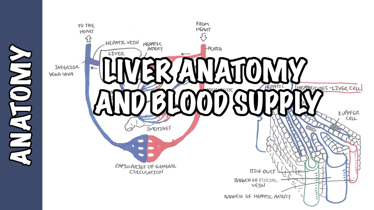


5.0 / 5 (0 votes)