Neuroanatomy S1 E2A: Introduction to the Spinal Cord #neuroanatomy #spinalcord #medicine
Summary
TLDRThis script explores the central nervous system, focusing on the spinal cord's structure and protection mechanisms. It details the meningeal layers: dura mater, arachnoid mater, and pia mater, and their roles. The script also discusses the spinal cord's termination at the conus medullaris and the cauda equina. It outlines the arrangement of spinal nerves in relation to the central nervous system, highlighting the cervical, thoracic, lumbar, sacral, and coccygeal regions, and their corresponding nerves. Additionally, it explains the spinal cord's enlargements related to the brachial and lumbosacral plexuses, which innervate the limbs.
Takeaways
- 🧠 The brain and spinal cord form the central nervous system, connecting the central and peripheral divisions of the nervous system.
- 🛡️ The spinal cord is protected by three meningeal layers: dura mater, arachnoid mater, and pia mater.
- 🌐 The arachnoid mater has a web-like appearance and forms the subarachnoid space filled with cerebrospinal fluid.
- 💧 Cerebrospinal fluid cushions and protects the brain and spinal cord both physically and chemically.
- ⚓ The pia mater has outgrowths like denticulate ligaments and the filum terminale that anchor and stabilize the spinal cord.
- 🏇 The cauda equina, resembling a horse's tail, consists of nerve roots at the end of the spinal cord.
- 🦴 The spinal cord is divided into regions (cervical, thoracic, lumbar, sacral) corresponding to different sets of vertebrae and nerves.
- 💪 The cervical and lumbosacral enlargements of the spinal cord correspond to areas that control the upper and lower limbs.
- 📍 The spinal cord ends at the L1-L2 vertebrae level, but the dural sac and cerebrospinal fluid continue to S2.
- 🦴 The vertebral column consists of cervical, thoracic, lumbar, sacral, and coccygeal vertebrae, with nerves emerging from each region.
Q & A
What structures make up the central nervous system?
-The central nervous system is made up of the brain and the spinal cord, which work together to connect the central and peripheral divisions of the nervous system.
What are the meningeal coverings, and what role do they play?
-The meningeal coverings are layers that surround and protect the spinal cord and brain. They include the dura mater, arachnoid mater, and pia mater, which help stabilize the spinal cord and brain within the skull and vertebral column.
What is the function of the dura mater?
-The dura mater is the outermost meningeal layer that forms the dural sac, surrounding the entire spinal cord and providing protection and stabilization.
What is the subarachnoid space, and why is it important?
-The subarachnoid space is the area between the arachnoid mater and the pia mater. It is filled with cerebrospinal fluid, which cushions and protects the brain and spinal cord physically and chemically.
What are arachnoid trabeculae, and what is their function?
-Arachnoid trabeculae are small strands of tissue within the subarachnoid space that suspend spinal blood vessels and connect to the pia mater, providing support and stability.
What is the role of the pia mater in spinal cord stabilization?
-The pia mater is the innermost meningeal layer that adheres tightly to the spinal cord. It gives rise to outgrowths like the denticulate ligaments and filum terminale, which anchor and stabilize the spinal cord.
What is the cauda equina, and where is it located?
-The cauda equina is a bundle of long lumbosacral nerve roots located at the end of the spinal cord, resembling a horse's tail. It surrounds the conus medullaris, the tapered end of the spinal cord.
How many cranial nerves does the brain give rise to, and how are they numbered?
-The brain gives rise to 12 cranial nerves, which are numbered using Roman numerals from I to XII.
How is the spinal cord divided based on body regions, and how many nerves are associated with each region?
-The spinal cord is divided into cervical, thoracic, lumbar, sacral, and coccygeal regions. There are 8 cervical nerves (C1-C8), 12 thoracic nerves (T1-T12), 5 lumbar nerves (L1-L5), 5 sacral nerves (S1-S5), and typically 1 coccygeal nerve.
Where does the spinal cord end, and how is this position identified on the body?
-The spinal cord ends at the intervertebral disc between L1 and L2. This position can be identified by using the iliac crest (top of the pelvis) to mark L4, and then counting up to L2 and L1.
Outlines

This section is available to paid users only. Please upgrade to access this part.
Upgrade NowMindmap

This section is available to paid users only. Please upgrade to access this part.
Upgrade NowKeywords

This section is available to paid users only. Please upgrade to access this part.
Upgrade NowHighlights

This section is available to paid users only. Please upgrade to access this part.
Upgrade NowTranscripts

This section is available to paid users only. Please upgrade to access this part.
Upgrade NowBrowse More Related Video

External Spinal Cord (Surface, Segments, Spinal Nerve, Enlargements, Reflex Arch) - Anatomy

vidio kelompok 1 mata kuliah ilmu biomedik dasar
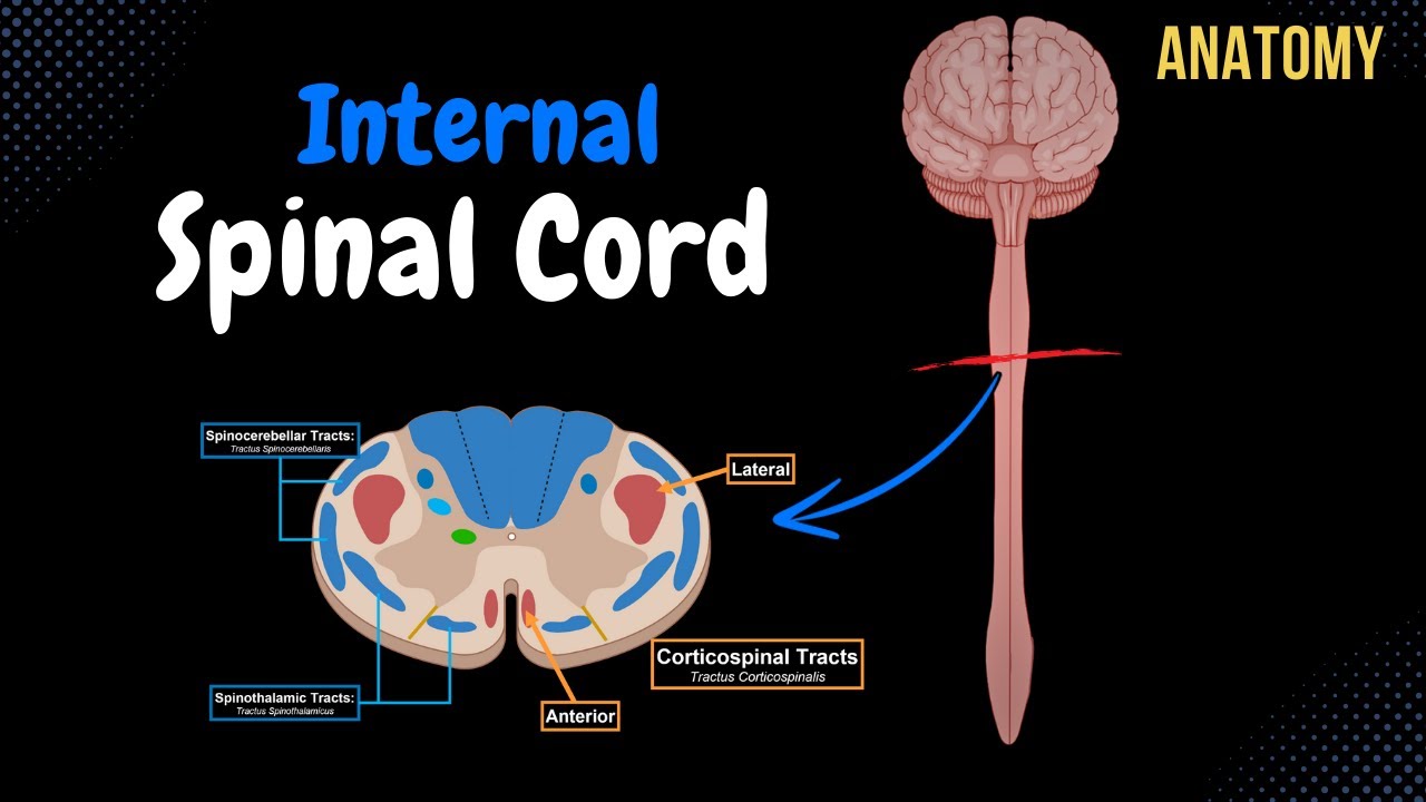
Internal Spinal Cord (Gray Matter, White Matter, Funiculus) - Anatomy
Neurology | Spinal Cord: White Matter Structure & Function

Anatomia nervoso1

Sistem Saraf - Part 2 : Sistem Saraf pusat (Otak dan STB)
5.0 / 5 (0 votes)