Pathogenesis of Entamoeba histolytica | parasitology | Basic Science Series
Summary
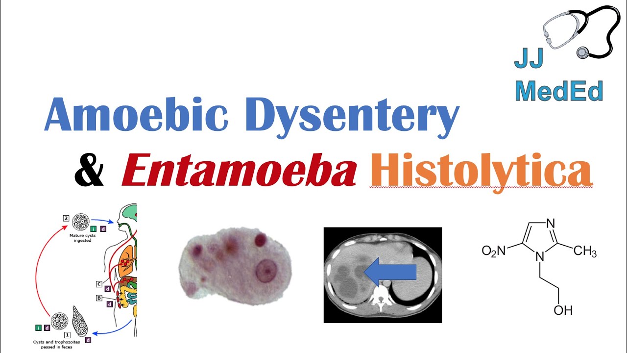
TLDRIn this video, the pathogenesis of *Entamoeba histolytica* infection is explored in detail. The narrator discusses the different layers of the intestinal wall and how the parasite damages these structures, causing issues like flask-shaped ulcers, perforation, and inflammation. The video highlights the importance of understanding the intestinal anatomy, with an in-depth explanation of various layers such as the mucosa, muscularis mucosa, submucosa, and the outermost serosa. It also covers the impact of the infection on the blood vessels and the complications that arise from the parasite's invasion, including extraintestinal spread and severe tissue damage.
Takeaways
- 😀 The video discusses the pathogenesis of Entamoeba histolytica infection, focusing on how it damages the intestinal wall.
- 😀 The first key damage caused by Entamoeba histolytica is the formation of flask-shaped ulcers in the colon.
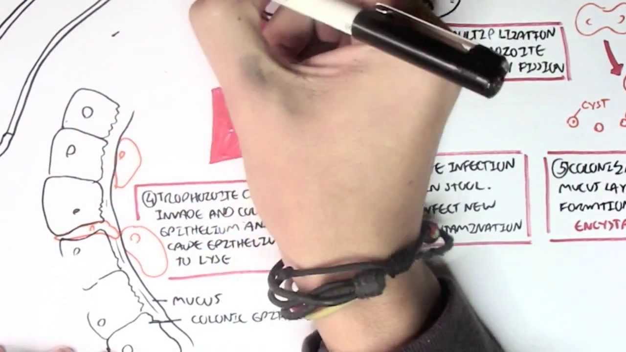
- 😀 Trophozoites, the active form of the parasite, cause significant damage to the intestine, leading to complications.
- 😀 The intestine has multiple layers: mucosa, muscularis mucosa, submucosa, circular muscles, longitudinal muscles, and serosa, each playing a specific role in digestion and protection.
- 😀 The mucosa layer of the intestine increases surface area for nutrient absorption and acts as a barrier against pathogens.
- 😀 Muscularis mucosa helps with the movement of the mucosa layer, facilitating digestion and waste transport.
- 😀 The submucosa, found below the mucosa, contains blood vessels, nerves, and lymphatics, aiding in nutrient and water absorption.
- 😀 Circular muscles in the intestine control peristaltic movement, helping with food and waste propulsion.
- 😀 Longitudinal muscles work with circular muscles for coordinated contraction, moving content through the digestive tract.
- 😀 The serosa is the outermost protective layer of the intestine and helps reduce friction and produces lubricating fluid for smoother digestion.
- 😀 Severe damage from Entamoeba histolytica infection can cause perforation, where a hole forms in the intestinal wall, leading to serious complications and potentially spreading to other organs.
Q & A
What is the difference between the cyst and trophozoite forms of *Entamoeba histolytica*?
-The cyst form is the inactive stage that helps *Entamoeba histolytica* survive in the environment, while the trophozoite form is active inside the host and causes damage to the intestine.
What is the role of the intestinal mucosa in digestion?
-The intestinal mucosa plays a critical role in digestion and nutrient absorption. It increases surface area through folds and acts as a barrier against harmful substances, pathogens, while secreting protective mucus and enzymes.
Why is the muscularis mucosa important in the digestive tract?
-The muscularis mucosa is a thin layer of smooth muscles that helps in the movement of the mucosa, allowing for the movement of food and waste through the intestine, and plays a role in digestion and transport.
What is the function of the submucosa layer in the intestine?
-The submucosa layer contains blood vessels, nerves, and lymphatics, which are essential for nutrient and water absorption, as well as digestion.
What are the circular muscles' roles in the intestine?
-The circular muscles are arranged in rings and their contraction narrows the inner space of the digestive tract. They play a key role in peristalsis, which is the coordinated wave-like motion that moves food through the intestine.
How do longitudinal muscles assist in digestion?
-Longitudinal muscles run along the length of the digestive tract and, in coordination with circular muscles, help propel the content forward, aiding in the movement of food and waste.
What is the function of the serosa layer in the intestine?
-The serosa is the outermost layer of the gut wall, offering protection and reducing friction during digestion. It also produces lubricating fluid, known as serous fluid, to facilitate smoother movements.
What is a flask-shaped ulcer, and how is it related to *Entamoeba histolytica* infection?
-A flask-shaped ulcer is a unique lesion caused by *Entamoeba histolytica* infection, typically found in the ascending colon. These ulcers penetrate deep into tissues, leading to inflammation and surrounding tissue damage.
What happens during perforation in the intestine due to *Entamoeba histolytica*?
-Perforation occurs when the ulcer caused by *Entamoeba histolytica* erodes through the entire intestinal wall, creating a hole. This is a severe complication that can lead to additional infections and requires immediate medical attention.
How does *Entamoeba histolytica* invade blood vessels and why is it significant?
-*Entamoeba histolytica* can invade blood vessels, allowing it to spread beyond the intestines to other organs, such as the liver. This can lead to extra-intestinal abscesses and further complicate the infection.
Outlines

This section is available to paid users only. Please upgrade to access this part.
Upgrade NowMindmap

This section is available to paid users only. Please upgrade to access this part.
Upgrade NowKeywords

This section is available to paid users only. Please upgrade to access this part.
Upgrade NowHighlights

This section is available to paid users only. Please upgrade to access this part.
Upgrade NowTranscripts

This section is available to paid users only. Please upgrade to access this part.
Upgrade NowBrowse More Related Video

AMEBÍASE - ENTAMOEBA HISTOLYTICA - PARASITOLOGIA| INFECTOLOGIA
Amebiasis (Amoebic Dysentery) | Entamoeba histolytica, Pathogenesis, Signs & Symptoms, Treatment
Amoebic dysentery - Entamoeba Histolytica

What is Dysentery? Causes, Signs and symptoms, Diagnosis and treatment.

SHOCK: Types, Pathogenesis of Septic Shock

Dengue Fever | Pathophysiology, Symptoms, Diagnosis & Treatment
5.0 / 5 (0 votes)