A 10-Minute Walkthrough of Grundium Ocus®20 and Ocus®40 Scanners – See how easy they are to use!
Summary
TLDRIn this video, Tuomo from Grundium introduces the Ocus scanners, highlighting their exceptional image quality, user-friendly interface, and affordability for remote pathology applications. Focusing on the Ocus®20 and Ocus®40 models, he demonstrates the scanning process, including slide insertion, selecting scan areas, and exporting images. The scanners feature a 500 GB storage capacity, enabling users to store numerous images. Tuomo also discusses additional functionalities such as live view, annotation tools, and advanced scanning modes. Overall, the Ocus scanners provide an accessible and efficient solution for remote pathology, with ease of use that can be learned quickly.
Takeaways
- 😀 The Ocus scanners provide high image quality, ease of use, and affordability for remote pathology applications.
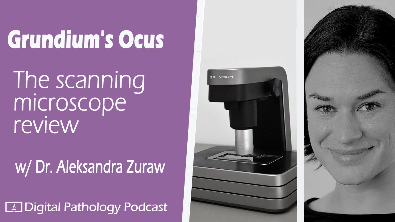
- 📦 There are three models available: Ocus®20, Ocus®40, and the new Ocus® M40.
- 🔌 The Ocus scanners feature essential components such as an overview camera, main objective, slide stage, USB port, and Ethernet port.
- 🖥️ Users can connect to the scanner via the Grundium website, allowing for easy access and control.
- 🔍 The scanning process starts with taking an overview image, which allows users to select a specific scan area.
- ✏️ The user interface includes tools for adjusting, adding, or removing scan areas with intuitive drag-and-drop functionality.
- ⏱️ The Ocus®20 scans at 20x magnification and optimally focuses on various fields of view for high-quality stitching.
- 💾 Each scanner has 500 GB of internal storage, enabling the storage of hundreds of images.
- 📤 Images can be exported in various formats (SVS, TIFF) to different destinations, including local downloads and network drives.
- 🖊️ The system supports live view and annotation features, allowing users to collaborate effectively without needing to scan.
Q & A
What are the main features of the Ocus scanners?
-The Ocus scanners offer high image quality, ease of use, and affordability, making them suitable for low to mid-throughput remote pathology applications.
Which models are currently available in the Ocus scanner lineup?
-The current lineup includes the Ocus®20, Ocus®40, and the new Ocus® M40 multi-slide scanner.
How do users initiate a scan with the Ocus scanners?
-Users log into the device through their web browser, select the scanner they wish to use, insert a slide, and then click 'new scan' to start the process.
What components make up the Ocus®20 scanner?
-The Ocus®20 includes an overview camera, main objective, slide stage, a USB port for exporting images, and an Ethernet port for internet connectivity.
How does the scanner determine the optimal focus for imaging?
-The Ocus scanners automatically detect the optimal layer in focus for each field of view and seamlessly stitch the images together during scanning.
What is the storage capacity of the Ocus®20 and Ocus®40?
-Both the Ocus®20 and Ocus®40 have 500 GB of internal storage, allowing them to store hundreds of images.
What export formats are available for images scanned by the Ocus scanners?
-The scanners support various export formats, with SVS and TIFF being the most common options.
What additional scanning features do the Ocus scanners provide?
-Additional features include extended depth of field, optical sections (Z stacking), live view of samples, and annotation tools for collaboration.
How can annotations be added to images in the Ocus scanner?
-Users can add annotations by selecting the area for annotation and entering text, which is stored with the image in the scan archive.
What is the learning curve for using the Ocus scanners?
-Users can typically learn to operate the Ocus scanners within 15 to 20 minutes, making them accessible for anyone.
Outlines

This section is available to paid users only. Please upgrade to access this part.
Upgrade NowMindmap

This section is available to paid users only. Please upgrade to access this part.
Upgrade NowKeywords

This section is available to paid users only. Please upgrade to access this part.
Upgrade NowHighlights

This section is available to paid users only. Please upgrade to access this part.
Upgrade NowTranscripts

This section is available to paid users only. Please upgrade to access this part.
Upgrade NowBrowse More Related Video
Ocus Grundium WSI Microscope review

The Secret To Designing PERFECT LOGO SHAPES (6 Things You Need)

A Stunning New AI Image Generator & New FREE AI Video!

Cara Membuat Cara Membuat Latar Belakang Skripsi | @Transfez

UI/UX Design, Skill IT Paling Dicari di Masa Depan (Eps 01)

What is Hugging Face? - Machine Learning Hub Explained
5.0 / 5 (0 votes)