Excitation Contraction Coupling | Skeletal Muscle Contraction | Cross Bridge Cycling | Myology
Summary
TLDRThis video explains the process of Excitation-Contraction coupling in skeletal muscles. It begins with a review of the structure of muscles, sarcomeres, and the neuromuscular junction. When a motor neuron triggers an action potential, it travels along the muscle membrane and T-tubules, activating calcium channels. Calcium is released from the sarcoplasmic reticulum, binds to troponin, and allows actin and myosin to form cross-bridges. This initiates muscle contraction through the sliding filament model, powered by ATP. The video also discusses how calcium levels regulate this process and how the muscle relaxes when action potentials cease.
Takeaways
- 💪 Excitation-Contraction coupling refers to the process that links muscle membrane excitation to muscle contraction.
- 🧬 Skeletal muscles are composed of fascicles, muscle fibers, myofibrils, and myofilaments (thick and thin filaments).
- ⚡ The neuromuscular junction is where the motor neuron triggers an action potential in the muscle membrane.
- 🧪 Action potentials travel along the membrane and into the T-tubules, which are vital for transmitting the signal deep into the muscle cell.
- 🧬 The Dihydropyridine receptor, a voltage-sensitive calcium channel, acts as a sensor for muscle contraction.
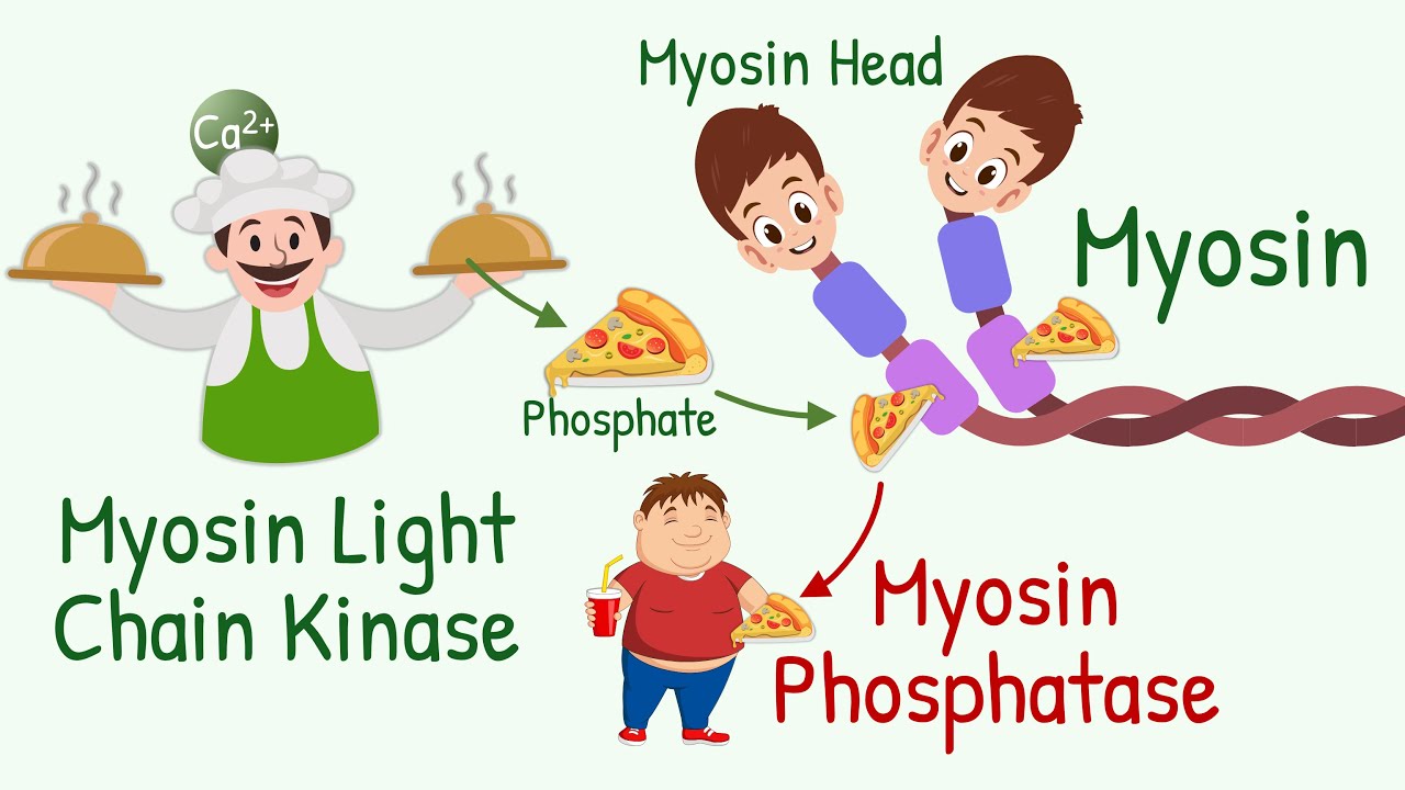
- 🔓 The Ryanodine receptor on the sarcoplasmic reticulum releases calcium into the sarcoplasm when activated, initiating contraction.
- 🧲 Calcium binds to Troponin C, which moves tropomyosin and allows myosin to bind to actin, forming cross-bridges.
- 🔄 The ATP-powered sliding filament mechanism involves myosin 'walking' along actin, creating the power stroke that contracts the muscle.
- 🔁 Continuous contraction requires repeated action potentials to keep calcium levels elevated in the sarcoplasm.
- 📉 Calcium is reabsorbed into the sarcoplasmic reticulum via the SERCA pump, leading to muscle relaxation when action potentials cease.
Q & A
What is the structure of a skeletal muscle at the microscopic level?
-Skeletal muscles are made up of fascicles, which are bundles of muscle fibers. Each muscle fiber contains myofibrils made up of thick and thin myofilaments. The arrangement of these myofilaments forms the sarcomere, the basic unit of muscle contraction.
What is the role of the sarcoplasmic reticulum in muscle contraction?
-The sarcoplasmic reticulum stores calcium, which is released during muscle contraction. The release of calcium into the sarcoplasm triggers the interaction between actin and myosin, leading to muscle contraction.
How does the neuromuscular junction initiate muscle contraction?
-At the neuromuscular junction, a motor neuron releases a neurotransmitter that triggers an action potential on the muscle membrane. This electrical signal is the first step in the process of muscle contraction.
What are T-tubules, and what role do they play in muscle contraction?
-T-tubules are invaginations of the sarcolemma (muscle cell membrane) that carry action potentials deep into the muscle cell. They help transmit the signal to the sarcoplasmic reticulum, ensuring calcium release for muscle contraction.
What is Excitation-Contraction coupling?
-Excitation-Contraction coupling refers to the process where an action potential on the muscle membrane leads to calcium release, which in turn allows actin and myosin to form cross-bridges, ultimately causing muscle contraction.
How do Dihydropyridine receptors and Ryanodine receptors function during muscle contraction?
-Dihydropyridine receptors on the T-tubules act as voltage sensors that detect the action potential and trigger the Ryanodine receptors on the sarcoplasmic reticulum to release calcium into the sarcoplasm, facilitating muscle contraction.
What happens when calcium binds to Troponin C?
-When calcium binds to Troponin C, it causes a conformational change that moves tropomyosin away from actin's binding sites, allowing actin and myosin to form cross-bridges and initiate muscle contraction.
What is the role of ATP in muscle contraction?
-ATP binds to the myosin head, causing it to detach from actin. The breakdown of ATP to ADP and phosphate provides energy for the myosin head to perform a power stroke, moving actin and causing muscle contraction.
What is the sliding filament model of muscle contraction?
-The sliding filament model describes how thin filaments (actin) slide over thick filaments (myosin) during muscle contraction, shortening the sarcomere and causing the muscle to contract.
How does the muscle relax after contraction?
-Muscle relaxation occurs when calcium is pumped back into the sarcoplasmic reticulum by the SERCA pump, lowering calcium levels in the sarcoplasm and stopping the formation of cross-bridges between actin and myosin.
Outlines

This section is available to paid users only. Please upgrade to access this part.
Upgrade NowMindmap

This section is available to paid users only. Please upgrade to access this part.
Upgrade NowKeywords

This section is available to paid users only. Please upgrade to access this part.
Upgrade NowHighlights

This section is available to paid users only. Please upgrade to access this part.
Upgrade NowTranscripts

This section is available to paid users only. Please upgrade to access this part.
Upgrade NowBrowse More Related Video

[#1] Fisiologia do Músculo Esquelético: CONTRAÇÃO MUSCULAR | MK Fisiologia

Skeletal muscle contraction : Muscle physiology Animations

Skeletal Muscle Contraction and Relaxation Physiology Animation / Excitation Contraction Coupling 💪

Guyton and Hall Medical Physiology (Chapter 7) Muscle excitation-contraction coupling || Study This!
Excitation Contraction Coupling in SMOOTH Muscles || Its DIFFERENT than in Skeletal Muscle

How Your Muscles Work
5.0 / 5 (0 votes)