Neurofacile 1 : La Voie Pyramidale.
Summary
TLDRThis video explains the voluntary motor pathways in the human nervous system, focusing on the two-neuron structure of motor transmission. It details the function of the first motor neuron (N1), which connects the brain to the spinal cord, forming the pyramidal tract. The video also covers the topographic organization of motor functions in the brain, the effects of cortical lesions, and clinical implications for motor recovery. Additionally, it touches on the phenomenon of motor plasticity and the role of the corticospinal tract in controlling muscle movements. Future videos will delve into related topics like spasticity and motor rehabilitation.
Takeaways
- 🧠 Human voluntary motor control is transmitted through a fast neural pathway involving two neurons: N1 (central pathway) and N2 (peripheral pathway).
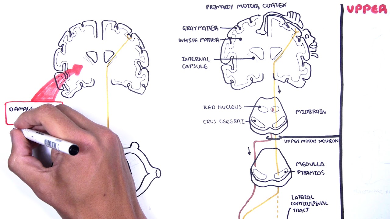
- 🦴 N1, or upper motor neurons, transmit signals from the brain to the spinal cord, forming part of the pyramidal tract, while N2 transmits from the spinal cord to muscles.
- 🔺 The pyramidal tract gets its name from the triangular shape of the cell bodies located in the motor cortex.
- 🌍 N1 axons descend through the white matter, crossing at the base of the medulla, and synapse in the anterior horn of the spinal cord with N2.
- 🖐️ The motor cortex is organized topographically, with each area corresponding to specific body parts, forming the motor homunculus.
- 💡 Topographical organization helps understand vascular hemiplegias, with specific arteries (middle cerebral and anterior cerebral) supplying different regions of the motor cortex.
- 🩸 A stroke in the middle cerebral artery affects the upper body and face, while one in the anterior cerebral artery affects the lower body.
- ♻️ Cortical plasticity allows adjacent areas to compensate for motor function loss after damage, aiding in post-injury motor recovery.
- 🔀 90% of pyramidal tract fibers cross at the medulla (decussation), controlling the contralateral side of the body, while 10% remain uncrossed, innervating the ipsilateral side.
- 🤸 The direct corticospinal tract ensures rapid recovery of axial muscle control due to double innervation from both hemispheres.
Q & A
What are the two types of motoneurons involved in voluntary motor activity?
-The two types of motoneurons involved are upper motoneurons (N1), which transmit signals from the brain to the spinal cord, and lower motoneurons (N2), which transmit signals from the spinal cord to the muscles.
Where are the cell bodies of upper motoneurons located?
-The cell bodies of upper motoneurons (N1) are located in the grey matter of the brain, specifically in the motor cortex.
What is the pyramidal tract and how is it named?
-The pyramidal tract is a collection of axons that transmit motor signals from the motor cortex to the spinal cord. It is named after the triangular shape of the cell bodies that form it.
What is the role of the internal capsule in the transmission of motor signals?
-The internal capsule is a region in the brain where the axons of the upper motoneurons (N1) gather and concentrate before descending to the spinal cord. It plays a crucial role in transmitting motor signals to the body.
How does the somatotopic organization of the motor cortex influence motor control?
-The motor cortex is organized somatotopically, meaning that specific areas of the cortex control specific parts of the body. This organization creates a topographic map, where larger areas control more finely controlled parts like the hands and face.
What happens when there is an interruption of blood flow to the middle cerebral artery?
-An interruption of blood flow to the middle cerebral artery can cause hemiplegia, particularly affecting the upper limbs and face, as this artery supplies the lateral part of the motor cortex responsible for these areas.
What is the difference between proportional and non-proportional hemiplegia?
-Proportional hemiplegia affects the entire half of the body equally and typically results from lesions in the subcortical regions, whereas non-proportional hemiplegia varies in intensity across different body parts and indicates a cortical lesion.
How does brain plasticity aid in motor recovery after a lesion?
-Brain plasticity allows adjacent areas of the cortex to take over the function of damaged regions, such as when a hand's motor function is partially restored by nearby areas following a lesion.
What is the decussation of the pyramids, and why is it important?
-The decussation of the pyramids refers to the crossing of the motor fibers at the base of the brainstem, which ensures that each cerebral hemisphere controls the opposite side of the body. It is crucial for understanding the location and effects of motor impairments.
How does the corticospinal tract ensure balance control after hemisphere lesions?
-Balance control is maintained after lesions in one hemisphere because the axial muscles receive dual innervation from both the crossed and uncrossed corticospinal fibers, allowing rapid recovery of postural control.
Outlines
🧠 Human Voluntary Motor Control Pathways
The human voluntary motor system is transmitted via a fast, two-neuron pathway. The first motor neuron, called the upper motor neuron (N1), sends signals from the brain to the spinal cord through the central pathway, while the second motor neuron, called the peripheral motor neuron (N2), transmits signals from the spinal cord to the skeletal muscles. The N1 neurons form the pyramidal tract, originating from the motor cortex and extending through the spinal cord. Each region of the motor cortex corresponds to specific body parts, creating a somatotopic map known as the motor homunculus. This map explains how different areas of the body are represented in the brain, with larger representations for parts like the hands. Understanding this mapping aids in diagnosing conditions like hemiplegia, where a stroke affecting the middle cerebral artery impacts motor function of the face and upper limbs, while anterior cerebral artery involvement leads to leg-related issues.
🔄 Motor Control Pathway Decussation and Clinical Implications
The pyramidal tract's crossing, or decussation, at the medulla allows each hemisphere to control the opposite side of the body (contralateral innervation). This crossing is significant for diagnosing central paralysis. Lesions above the decussation cause contralateral paralysis, while lesions below lead to ipsilateral paralysis. Although 90% of pyramidal fibers cross, 10% form an uncrossed corticospinal tract for ipsilateral innervation. These uncrossed fibers mainly control muscles that maintain posture and balance, explaining why axial muscles recover quickly after brain damage. Additionally, the termination of N1 fibers in the spinal cord involves synapses that both excite target muscles and inhibit antagonist muscles, preventing counterproductive contractions. Disruptions in this inhibitory control can lead to spasticity.
Mindmap
Keywords
💡Motor Neuron
💡Pyramidal Tract
💡Cortex Motor
💡Somatotopy
💡Hemiplegia
💡Decussation of the Pyramids
💡Corticospinal Tract
💡Capsule Internal
💡Synapse
💡Cerebral Plasticity
Highlights
The human voluntary motor control is transmitted through the nervous system by a rapid pathway consisting of only two neurons: the upper motor neuron (N1) and the lower motor neuron (N2).
The upper motor neuron (N1) transmits information from the brain to the spinal cord, while the lower motor neuron (N2) transmits it from the spinal cord to the striated muscles.
N1 neurons form the pyramidal tract, with their cell bodies located in the motor cortex of the brain, specifically in the frontal cortex.
The term 'pyramidal tract' comes from the triangular shape of the cell bodies of N1 neurons located in the motor cortex.
The motor cortex is organized topographically, meaning different regions control specific parts of the body. This organization is known as the motor homunculus.
The motor homunculus illustrates that areas such as the hands and face have a larger representation in the motor cortex due to their higher complexity of movement.
Damage to different arteries supplying the motor cortex can result in distinct types of hemiplegia (paralysis), depending on which artery is affected.
An interruption of blood flow to the middle cerebral artery causes hemiplegia with a predominance of facial and upper limb paralysis.
Damage to the anterior cerebral artery results in hemiplegia with more prominent paralysis in the lower limbs.
In cases of cortical lesions, the brain's plasticity allows adjacent areas to take over some of the motor functions, aiding in recovery.
Pyramidal fibers cross at the base of the medulla oblongata, ensuring that each hemisphere of the brain controls the opposite side of the body.
Lesions occurring above the pyramidal decussation (crossing) result in contralateral paralysis, while lesions below the decussation cause ipsilateral paralysis.
Approximately 90% of pyramidal fibers cross over, while the remaining 10% continue uncrossed and control muscles bilaterally.
The synapse between N1 and N2 occurs in the anterior horn of the spinal cord, where excitatory signals cause muscle contraction.
Inhibitory signals are also sent to the antagonist muscle to prevent opposition during movement, a key mechanism for coordinated motion.
Transcripts
la motricité volontaires humaine est
transmise dans le système nerveux par
une voie rapide à seulement deux
neurones
le premier moto neurones encore appelée
motoneurones supérieur transmet
l'information du cerveau vers la moelle
épinière
c'est la voie centrale que l'on va
appeler n 1 le deuxième moto neurones
motoneurones périphériques transmet
l'information de la moelle épinière vers
le muscles striés c'est la voie
périphérique que l'on va appeler n 2 et
qui fera l'objet de la prochaine vidéo
maintenant concentrons nous sur les n 1
qui réunit tous ensemble constitue le
faisceau pyramidale
comme tout le run and bike constituée
d'un corps cellulaire qui se situe dans
la substance grise du cerveau est
spécifiquement dans le cortex moteur
c'est la forme triangulaire de ce corps
cellulaire qui est à l'origine de la
dénomination du faisceau pyramidale
l'accent de n1 quant à lui va traverser
la substance blanche sous corticales du
cerveau celle du trans cérébrale puis
descendre dans un cordon de la moelle
épinière jusqu'au point de liens avec et
deux au niveau de la corne antérieures
les corps cellulaire de tous les n1 se
répartissent dans le cortex frontal dans
une zone bien identifiés appelé et
remontrer ce primaire cette zone s'étend
sur une bande verticale juste en avant
de la cie sur de rolando qui correspond
dans le jargon anatomique à la
circonvolution frontale ascendante
chaque n1 partant du cortex frontal va
transmettre l'ordre au moteur
motoneurones périphériques destinés à un
muscle précis qui peut être située au
niveau de la face par exemple ou bien au
niveau du bras ou dans une autre partie
du corps ainsi chaque petite zone du
cortex moteur correspond à une zone
spécifique de l'organisme dans la
projection topographiques sur le cortex
réalise ce qu'on appelle le manque ulus
moteur si on s'amusait à faire une
modélisation du corps selon la taille de
projection de chaque membre sur le
cortex moteur on obtiendrait ce genre de
petit bonhomme dont les mains sont
géante car très mou
cette organisation topographiques très
précise et connu sous le nom de sommets
tout opi soma est un terme qui renvoie
au corps et au pis pour topographie
cette somme est au tapis à deux
applications cliniques très intéressante
la première application nous sert à
comprendre la sémiologie de l'hémiplégie
vasculaires en effet la vascularisation
du cerveau est issu d'un système
vasculaire bien alimenté partant d'un
réseau située à la base du cerveau
appelée polygone de willis il donne
naissance à plusieurs branches terminale
dans l'artère cérébrale moyenne qui va
assurer la vascularisation de toute la
partie latérale du cortex moteur destiné
essentiellement à l'innervation motrice
du membre supérieur et de la face il
donne aussi naissance à l'artère
cérébrale antérieures qui vascularisé la
partie médiane du cortex moteur
innervant essentiellement le membre
inférieur en cas d'interruption de la
vascularisation corticale de la
cérébrale moyenne on va avoir une
hémiplégie à prédominance braqueurs
faciale alors qu'une atteinte de la
cérébrale antérieures entraînerait
bientôt plus tôt une hémiplégie à
prédominance rural c'est à dire plus
marquée au niveau du membre inférieur
c'est ce qu'on appelle en sémiologie une
hémiplégie nos proportionnelle car elle
n'a pas la même intensité sur l'ensemble
du corps et sous-entend une lésion
corticale la deuxième application
clinique de cette somme est au pis nous
permet de comprendre le mécanisme de
récupération post lésionnel supposons
que la partie du cortex destiné à
l'innervation de la main a subi une
lésion qu'elles soient d'origine
vasculaire tumorales autres il a été
démontré que la fonction motrice de la
main pourrait être partiellement supplée
y est paru des zones adjacentes grâce à
la plasticité cérébrale qui sera abordée
plus en détail dans une autre vidéo afin
de comprendre son implication dans les
processus de récupération motrice
une fois les axones dn1 en kit et le
cortex moteur
ils vont se réunir au niveau de la
substance blanche sous corticales pour
fermer pour former un faisceau de petits
voulu concentrant toutes les
fibres nerveuses au niveau d'une zone
appelée capsule interne une lésion
vasculaire sous corticales notamment au
niveau de cette zone va être responsable
d'une hémiplégie massive touchant de
façon semblable l'ensemble de l'emi
corps c'est ce qu'on appelle en
sémiologie l'hémiplégie proportionnelle
qui renvoie à une lésion pyramidale sous
corticales le faisceau pyramidale se
caractérise par son croisement au niveau
de la base du bulbe
c'est ce qu'on appelle la dégustation
pyramidale qui fait que chaque
hémisphère cérébral assure mme
innervation motrice de la partie contre
latéral de l'emi corps ce croisement et
sa localisation une importance majeure
dans le diagnostic topographiques des
paralysies centrale toute lésion du
faisceau pyramidale survenant au dessus
du croisement qu'elle soit au niveau du
cerveau ou du transfert hébral va
s'exprimer par une paralysie dit contrat
lésionnel alors que toute lésion
survenant après croisement engendrera
une paralysie dit ipsi lésionnel c'est à
dire du même côté de la lésion il est
important de savoir que ce croisement
concerne uniquement 90% des fibres
pyramidale réalisant le faisceau
corticaux spinale croisé destinés à des
nikkor controlatéral les 10% des fibres
qui restent ne vont pas croisé la ligne
médiane et réalise le faisceau corticaux
spinale directe destinée à l'ennemi
corps ipsi latéral ce faisceau direct
est essentiellement destinée aux muscles
action qui reçoivent une double
innervation aussi bien par le faisceau
direct que par le fait se croiser et
donc par les deux hémisphères
c'est ce qui explique la conservation ou
récupération très rapide de l'equilibre
axial assurée par ses muscles après
lésion d'un seul hémisphère la dernière
notion que j'aimerais aborder dans cette
vidéo cancer la particularité du lien
entre la terminaison de n1 et le corps
cellulaire de n2 c'est la synapse
médullaire qui a lieu dans la coque
l'intérieur de la moelle épinière
lorsqu'une fibre du faisceau corticaux
spinale harry
au niveau des laitages médullaire
correspondant à sa terminaison elle va
rentrer dans la corne antérieures pour
stimuler par une synapse excitatrice le
motoneurones périphériques qui va faire
contracter le muscle cible cette même
fibre va aussi donner une branche
terminal qui va inhiber le moto neurones
du muscle antagonistes au muscle cible
afin d'éviter qu'ils s'opposent au
mouvement
par exemple pour contracter le biceps
brachial il faut une iv sont
antagonistes c'est à dire le triceps
pour assurer son allongement c'est le
contrôle inhibiteur descendant dans
l'altération est à la base de la
spasticité qui sera abordée plus en
détail dans une prochaine vidéo
voilà c'est tout pour la voix pyramidale
pour rester informé des prochaines
vidéos
n'oubliez pas de vous abonner à la
chaîne et de suivre nos activités sur
leur contenu à très bientôt
[Musique]
Browse More Related Video
¡Vía CORTICOESPINAL y Síndromes de Motoneurona! PARA ESTUDIANTES
Neurology | Spinal Cord: White Matter Structure & Function
Gray and white matter | Organ Systems | MCAT | Khan Academy
Organizzazione del Sistema Nervoso | NEUROSCIENZE - Lezione 9
BAB 2 Sistem Koordinasi Manusia || Sistem Saraf Manusia || IPA SMP/MTs Kelas 9 Kurikulum Merdeka
NEUROFISIOLOGIA - AS GRANDES VIAS AFERENTES E EFERENTES - Prof Mara Ribeiro - Aula 13
5.0 / 5 (0 votes)