Cardiac Action Potential, Animation.

Alila Medical Media
24 Jan 201707:50

Summary

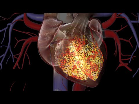
TLDRThe heart is a self-regulating muscular organ that pumps blood through a series of electrical impulses. Its specialized cardiac myocytes initiate contractions via action potentials, which start at the SA node and spread via the conduction system. The SA node's pacemaker cells generate action potentials that synchronize atrial and ventricular contractions. The unique ion channels and calcium handling in cardiac cells ensure a prolonged plateau phase, allowing for efficient blood expulsion, while a longer refractory period prevents uncontrolled muscle responses.

Takeaways

  • 💓 The heart is a muscle that pumps blood through contractions initiated by electrical impulses called action potentials.
  • 🔌 Unlike skeletal muscles, the heart generates its own electrical stimulation, allowing it to beat independently of the nervous system.
  • 🌀 The cardiac conduction system, starting with the SA node, controls the heart's rhythm and can continue to function even if the SA node is damaged.
  • 🔥 The SA node's spontaneous firing of action potentials at about 80 per minute results in an average heart rate of 80 beats per minute.
  • 🔄 The electrical signals propagate through the atria and ventricles via gap junctions, ensuring synchronized contractions.
  • 🔮 Pacemaker cells have a unique action potential generation process involving 'funny' currents that lead to the pacemaker potential.
  • 💥 The action potential in cardiac myocytes involves a rapid influx of sodium followed by a plateau phase due to a balance of calcium and potassium ions.
  • 🏋️‍♂️ The influx of calcium ions is crucial for muscle contraction, with a significant amount released from the sarcoplasmic reticulum.
  • 🔙 The repolarization phase involves the closing of calcium channels and the predominance of potassium efflux, returning the cell to its resting state.
  • ⏱️ The cardiac muscle's absolute refractory period is much longer than skeletal muscle, preventing unwanted summation and tetanus that could disrupt the heartbeat.

Q & A

  • What is the primary function of the heart?

    -The primary function of the heart is to contract and pump blood throughout the body.

  • What are cardiac myocytes and how are they involved in the heart's function?

    -Cardiac myocytes are specialized muscle cells that contract to pump blood. They initiate contraction through electrical impulses known as action potentials.

  • How does the heart generate its own electrical stimulation?

    -The heart generates its own electrical stimulation through a group of specialized myocytes called pacemaker cells, which are part of the cardiac conduction system.

  • What is the role of the SA node in the heart?

    -The SA node, or sinoatrial node, is the primary pacemaker of the heart. It initiates all heartbeats and controls the heart rate.

  • How do the impulses from the SA node spread through the heart?

    -The impulses from the SA node spread through the conduction system and to the contractile myocytes via gap junctions, which allow ions to flow from one cell to another.

  • What is the significance of the resting membrane potential in cardiac cells?

    -The resting membrane potential, usually negative, indicates the cell's electrical state at rest. It is crucial for the generation of action potentials.

  • How does the action potential in cardiac cells differ from that in skeletal muscle cells?

    -Cardiac cells have a plateau phase in their action potentials, which allows them to stay contracted longer than skeletal muscle cells, necessary for the expulsion of blood from the heart.

  • What is the role of the sarcoplasmic reticulum (SR) in cardiac muscle cells?

    -The SR in cardiac muscle cells stores a large amount of calcium, which is crucial for the 'calcium-induced calcium release' process that triggers muscle contraction.

  • What is the significance of the absolute refractory period in cardiac muscle?

    -The absolute refractory period in cardiac muscle is much longer than in skeletal muscle, ensuring that the muscle has relaxed before it can respond to a new stimulus, preventing summation and tetanus.

  • How does the nervous system influence the heart's beating?

    -The nervous system can influence the heart's beating by making the heartbeats go faster or slower but cannot generate the heartbeats itself.

  • What happens if the SA node is damaged?

    -If the SA node is damaged, other parts of the conduction system may take over the role of initiating heartbeats.

Outlines

00:00

🫀 Heart's Electrical Conduction System

The heart is a muscular organ that contracts to pump blood, composed of specialized cells known as cardiac myocytes. These cells contract due to electrical impulses called action potentials. Unlike skeletal muscles, the heart generates its own electrical stimulation, allowing it to continue beating even when removed from the body. The nervous system can influence the heart rate but does not create the heartbeats. The cardiac conduction system, starting with the pacemaker cells, initiates these impulses. The SA node, as the primary pacemaker, controls the heart rate. If damaged, other parts of the system can take over. The action potentials spread through gap junctions, electrically coupling neighboring cells. The conduction system ensures synchronized contraction of all myocytes. The SA node cells have a unique way of generating action potentials, with no true resting potential and a spontaneous depolarization due to 'funny' currents. The action potential involves the flow of ions through voltage-gated channels, leading to the contraction of the heart muscle.

05:03

🔬 Cardiac Action Potential and Muscle Contraction

The second paragraph delves into the specifics of the cardiac action potential and how it differs from that of skeletal muscles. The contractile myocytes have a stable resting potential and depolarize only upon stimulation. The depolarization leads to the opening of fast sodium channels, causing a rapid rise in voltage. L-type calcium channels open subsequently, contributing to the depolarization. The action potential reaches a plateau phase, unique to cardiac cells, where calcium influx from the extracellular fluid and the sarcoplasmic reticulum triggers muscle contraction. The plateau phase allows for a longer contraction time, essential for blood expulsion from the heart. The refractory period in cardiac muscle is significantly longer than in skeletal muscle, preventing unwanted summation and tetanus, which could halt heart function. The restoration of ionic balance is facilitated by pumps and the sodium/potassium pump, ensuring the muscle can relax and prepare for the next contraction.

Mindmap

Keywords

💡Cardiac Myocytes

Cardiac myocytes are specialized muscle cells that make up the heart. They are responsible for the heart's ability to contract and pump blood. Unlike skeletal muscles, cardiac myocytes can initiate their own contractions without the need for nervous system stimulation, as highlighted in the script with the phrase 'the heart generates its own electrical stimulation.' This autonomy is crucial for the heart's continuous function, even when it is outside the body.

💡Action Potentials

Action potentials are electrical impulses that trigger the contraction of cardiac myocytes. They are initiated by a change in the electrical potential across the cell membrane. The script explains that these impulses are essential for the synchronized beating of the heart, with the statement 'Action potential generation and conduction are essential for all myocytes to act in synchrony.' The process involves the movement of ions across the cell membrane, leading to the cell's depolarization and repolarization.

💡Pacemaker Cells

Pacemaker cells are a group of specialized myocytes that initiate the electrical impulses for heartbeats. The script mentions 'The SA node is the primary pacemaker of the heart,' indicating that these cells are located in the sinoatrial (SA) node. They are unique because they can spontaneously generate action potentials, which then spread throughout the heart to cause it to beat, as described in 'The cells of the SA node fire spontaneously, generating action potentials that spread though the contractile myocytes of the atria.'

💡Cardiac Conduction System

The cardiac conduction system is a network of cells responsible for the generation and transmission of electrical impulses that regulate the heartbeat. The script describes it as starting from the pacemaker cells and includes the 'SA node,' 'AV node,' and the 'ventricular myocytes.' This system ensures that the electrical signals are coordinated, allowing the heart to contract in a synchronized manner.

💡Resting Membrane Potential

Resting membrane potential refers to the electrical voltage across the cell membrane when the cell is at rest. In the context of the script, it is mentioned that 'In a resting cell, the membrane voltage, known as the resting membrane potential, is usually negative.' This negative potential is due to the concentration gradients of ions, with more sodium and calcium outside the cell and more potassium inside. This potential is crucial for the generation of action potentials.

💡Depolarization

Depolarization is the process by which the cell membrane potential becomes less negative, moving towards a more positive value. The script explains this as 'When membrane voltage INCREASES and becomes LESS negative, the cell is LESS polarized, and is said to be depolarized.' This is a critical step in the generation of an action potential, as it leads to the opening of voltage-gated ion channels and the initiation of an electrical impulse.

💡Repolarization

Repolarization is the return of the cell membrane potential to its resting state after an action potential. As described in the script, 'At the peak of depolarization, potassium channels open, calcium channels inactivate, potassium ions leave the cell and the voltage returns to -60mV.' This process is essential for resetting the cell's electrical state so that it can respond to new stimuli and continue to function properly.

💡Threshold

The threshold is the critical value of membrane potential at which an action potential is generated. The script states that 'For an action potential to be generated, the membrane voltage must depolarize to a critical value called the THRESHOLD.' When the membrane potential reaches this threshold, it triggers the opening of ion channels, leading to the rapid depolarization phase of the action potential.

💡Voltage-gated Ion Channels

Voltage-gated ion channels are proteins in the cell membrane that open or close in response to changes in the membrane voltage. As mentioned in the script, 'These channels are passageways for ions in and out of the cell, and as their names suggest, are regulated by membrane voltage.' They play a crucial role in the generation and propagation of action potentials by allowing the flow of ions such as sodium, potassium, and calcium across the cell membrane.

💡Calcium-induced Calcium Release

Calcium-induced calcium release is a process where the influx of calcium ions into the cell triggers the release of more calcium from the sarcoplasmic reticulum (SR). The script explains this as 'Instead, it triggers a MUCH greater calcium release from the SR, in a process known as, “calcium-induced calcium release”.' This process is essential for coupling the electrical excitation of the cell to its physical contraction, as it provides the necessary calcium ions to initiate muscle contraction.

💡Refractory Period

The refractory period is a time after an action potential during which the cell cannot respond to another stimulus. The script mentions that 'The absolute refractory period is also much longer - 250 msec compared to 1 msec in skeletal muscle.' This period ensures that the cell has returned to its resting state and is ready to receive a new stimulus, preventing unwanted summation of signals or tetanus, which could disrupt the regular beating of the heart.

Highlights

The heart is a muscle that pumps blood through contraction.

Heart contractions are initiated by electrical impulses called action potentials.

The heart generates its own electrical stimulation independently of the nervous system.

Pacemaker cells are specialized myocytes that initiate and conduct action potentials.

The SA node is the primary pacemaker, controlling the heart rate.

If the SA node is damaged, other parts of the conduction system can take over.

Action potentials propagate through gap junctions connecting myocytes.

The AV node slows impulses to allow atrial contraction before ventricular contraction.

Action potential generation and conduction synchronize all myocytes.

Pacemaker cells and contractile myocytes have different action potential forms.

Resting membrane potential is negative, indicating the cell is more negative inside.

Ion concentration gradients are maintained by pumps moving sodium and calcium out, potassium in.

An action potential is a brief reversal of the cell membrane's electric polarity.

Pacemaker cells of the SA node fire about 80 times per minute, correlating to an average heart rate.

Pacemaker cells lack a true resting potential and spontaneously depolarize to threshold.

The influx of calcium ions is crucial for the rising phase of the action potential.

The plateau phase of the cardiac action potential is unique to heart muscle cells.

Calcium-induced calcium release from the sarcoplasmic reticulum triggers muscle contraction.

The absolute refractory period in cardiac muscle is much longer than in skeletal muscle.

Transcripts

play00:03

The heart is essentially a muscle that contracts and pumps blood.

play00:07

It consists of specialized muscle cells called cardiac myocytes.

play00:12

The contraction of these cells is initiated by electrical impulses, known as action potentials.

play00:18

Unlike skeletal muscles, which have to be stimulated by the nervous system, the heart

play00:23

generates its own electrical stimulation.

play00:27

In fact, a heart can keep on beating even when taken out of the body.

play00:31

The nervous system can make the heartbeats go faster or slower, but cannot generate them.

play00:37

The impulses start from a small group of myocytes called the pacemaker cells, which constitute

play00:43

the cardiac conduction system.

play00:46

These are modified myocytes that lose the ability to contract and become specialized

play00:50

for initiating and conducting action potentials.

play00:54

The SA node is the primary pacemaker of the heart.

play00:58

It initiates all heartbeats and controls heart rate.

play01:01

If the SA node is damaged, other parts of the conduction system may take over this role.

play01:07

The cells of the SA node fire spontaneously, generating action potentials that spread though

play01:13

the contractile myocytes of the atria.

play01:15

The myocytes are connected by gap junctions, which form channels that allow ions to flow

play01:21

from one cell to another.

play01:23

This enables electrical coupling of neighboring cells.

play01:27

An action potential in one cell triggers another action potential in its neighbor and the signals

play01:32

propagate rapidly.

play01:34

The impulses reach the AV node, slow down a little to allow the atria to contract, then

play01:39

follow the conduction pathway and spread though the ventricular myocytes.

play01:44

Action potential generation and conduction are essential for all myocytes to act in synchrony.

play01:51

Pacemaker cells and contractile myocytes exhibit different forms of action potentials.

play01:58

Cells are polarized, meaning there is an electrical voltage across the cell membrane.

play02:03

In a resting cell, the membrane voltage, known as the resting membrane potential, is usually

play02:09

negative.

play02:10

This means the cell is more negative on the inside.

play02:13

At this resting state, there are concentration gradients of several ions across the cell

play02:18

membrane: more sodium and calcium outside the cell, and more potassium inside the cell.

play02:25

These gradients are maintained by several pumps that bring sodium and calcium OUT, and

play02:30

potassium IN.

play02:31

An action potential is essentially a brief REVERSAL of electric polarity of the cell

play02:36

membrane and is produced by voltage-gated ion channels.

play02:41

These channels are passageways for ions in and out of the cell, and as their names suggest,

play02:46

are regulated by membrane voltage.

play02:49

They open at some values of membrane potential and close at others.

play02:53

When membrane voltage INCREASES and becomes LESS negative, the cell is LESS polarized,

play02:59

and is said to be depolarized.

play03:01

Reversely, when membrane potential becomes MORE negative, the cell is repolarized.

play03:07

For an action potential to be generated, the membrane voltage must depolarize to a critical

play03:13

value called the THRESHOLD.

play03:15

The pacemaker cells of the SA node SPONTANEOUSLY fire about 80 action potentials per minute,

play03:22

each of which sets off a heartbeat, resulting in an average heart rate of 80 beats per minute.

play03:28

Pacemaker cells do NOT have a TRUE RESTING potential.

play03:31

The voltage starts at about -60mV and SPONTANEOUSLY moves upward until it reaches the threshold

play03:38

of -40mV.

play03:40

This is due to action of so-called “FUNNY” currents present ONLY in pacemaker cells.

play03:46

Funny channels open when membrane voltage becomes lower than -40mV and allow slow influx

play03:53

of sodium.

play03:54

The resulting depolarization is known as “pacemaker potential”.

play03:58

At threshold, calcium channels open, calcium ions flow into the cell further depolarizing

play04:05

the membrane.

play04:06

This results in the rising phase of the action potential.

play04:10

At the peak of depolarization, potassium channels open, calcium channels inactivate, potassium

play04:17

ions leave the cell and the voltage returns to -60mV.

play04:22

This corresponds to the falling phase of the action potential.

play04:26

The original ionic gradients are restored thanks to several ionic pumps, and the cycle

play04:31

starts over.

play04:34

Electrical impulses from the SA node spread through the conduction system and to the contractile

play04:39

myocytes.

play04:41

These myocytes have a different set of ion channels.

play04:44

In addition, their sarcoplasmic reticulum, the SR, stores a large amount of calcium.

play04:50

They also contain myofibrils.

play04:52

The contractile cells have a stable resting potential of -90mV and depolarize ONLY when

play04:59

stimulated, usually by a neighboring myocyte.

play05:02

When a cell is depolarized, it has more sodium and calcium inside the cell.

play05:08

These positive ions leak through the gap junctions to the adjacent cell and bring the membrane

play05:13

voltage of this cell up to the threshold of -70mV.

play05:18

At threshold, fast sodium channels open creating a rapid sodium influx and a sharp rise in

play05:24

voltage.

play05:25

This is the depolarizing phase.

play05:28

L-type, or slow, calcium channels also open at -40mV, causing a slow but steady influx.

play05:36

As the action potential nears its peak, sodium channels close quickly, voltage-gated potassium

play05:42

channels open and these result in a small decrease in membrane potential, known as early

play05:49

repolarization phase.

play05:51

The calcium channels, however, remain open and the potassium efflux is eventually balanced

play05:57

by the calcium influx.

play05:59

This keeps the membrane potential relatively stable for about 200 msec resulting in the

play06:05

PLATEAU phase, characteristic of cardiac action potentials.

play06:09

Calcium is crucial in coupling electrical excitation to physical muscle contraction.

play06:15

The influx of calcium from the extracellular fluid, however, is NOT enough to induce contraction.

play06:21

Instead, it triggers a MUCH greater calcium release from the SR, in a process known as

play06:28

“calcium-induced calcium release".

play06:30

Calcium THEN sets off muscle contraction by the same “sliding filament mechanism”

play06:35

described for skeletal muscle.

play06:37

The contraction starts about half way through the plateau phase and lasts till the end of

play06:42

this phase.

play06:43

As calcium channels slowly close, potassium efflux predominates and membrane voltage returns

play06:49

to its resting value.

play06:51

Calcium is actively transported out of the cell and also back to the SR.

play06:56

The sodium/potassium pump then restores the ionic balance across the membrane.

play07:02

Because of the plateau phase, cardiac muscle stays contracted longer than skeletal muscle.

play07:08

This is necessary for expulsion of blood from the heart chambers.

play07:12

The absolute refractory period is also much longer - 250 msec compared to 1 msec in skeletal

play07:20

muscle.

play07:21

This long refractory period is to make sure the muscle has relaxed before it can respond

play07:26

to a new stimulus and is essential in preventing summation and tetanus, which would stop the

play07:31

heart from beating.

Rate This

5.0 / 5 (0 votes)

Related Tags
Cardiac MuscleHeart FunctionElectrical ImpulseMyocyte ActionPacemaker CellsConduction SystemAction PotentialHeart Rate ControlMuscle ContractionIonic ChannelsCardiac Physiology