Skeletal System (Part 3) - Vertebrae, ribs and sternum

Pinoy Vet Anatomist
15 Jan 202113:19

Summary

TLDRThis module discusses the vertebral formula, structure, and classification of the vertebrae, ribs, and sternum in animals, with a focus on dogs. It explains the arrangement of vertebrae (cervical, thoracic, lumbar, sacral, caudal), the features of each type, and their role in supporting the body. The rib structure and their articulation with the thoracic vertebrae and sternum are also covered. Notable sections include descriptions of the atlas, axis, anticlinal vertebra, and the differences between sternal and floating ribs. A quiz is offered at the end of the module.

Takeaways

  • πŸ“š The vertebral formula of a dog is C7, T13, L7, S3, and Ca 20-24, meaning 7 cervical, 13 thoracic, 7 lumbar, 3 sacral, and 20-24 caudal vertebrae.
  • 🦴 Vertebrae are irregular bones that form the vertebral column, extending from the skull to the tail.
  • πŸ¦“ The vertebrae are grouped by location: cervical (neck), thoracic (chest), lumbar (loin), sacral (pelvic), and caudal (tail).
  • πŸ•β€πŸ¦Ί A typical vertebra has a body, an arc, and several processes; the sixth cervical vertebra is used as an example.
  • πŸ—οΈ The first cervical vertebra, the atlas, supports the head, and the second cervical vertebra, the axis, forms a pivot joint with the atlas.
  • πŸ”§ The thoracic vertebrae are characterized by tall spinous processes and articulate with the ribs, with T11 serving as the anticlinal vertebra in dogs.
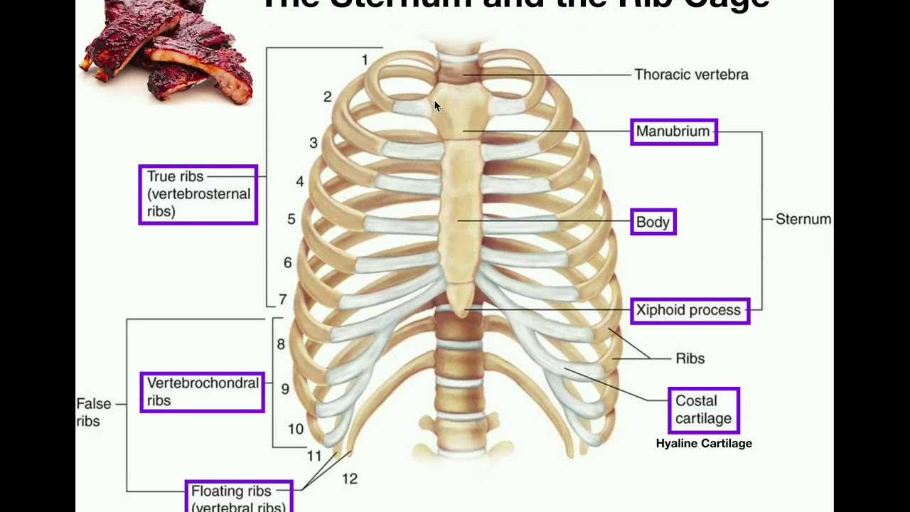
  • 🐾 There are 13 pairs of ribs in dogs, corresponding to the number of thoracic vertebrae, with 9 true ribs, 3 false ribs, and 1 floating rib.
  • 🧩 The sternum is composed of 8 fused bones called sternebrae and is divided into the manubrium, body, and xiphoid process.
  • πŸ”© The ribs are divided into different types based on their attachment to the sternum: true ribs, false ribs, and floating ribs.
  • πŸ” Key processes on vertebrae include spinous, transverse, and articular processes, with additional features like transverse foramina in cervical vertebrae.

Q & A

  • What is the vertebral formula of a dog?

    -The vertebral formula of a dog is C7, T13, L7, S3, Ca20-24. This means there are 7 cervical, 13 thoracic, 7 lumbar, 3 sacral, and 20-24 caudal vertebrae.

  • Where are the cervical vertebrae located, and how many are there in a dog?

    -The cervical vertebrae are located in the neck region, and there are 7 cervical vertebrae in a dog.

  • What are the key characteristics of thoracic vertebrae?

    -Thoracic vertebrae are characterized by tall spinous processes, short bodies, and transverse processes. They also articulate with the ribs.

  • What is the function of the atlas (C1) in the vertebral column?

    -The atlas, or C1, supports the head and connects it with the rest of the body. It is morphologically unique, lacking a spinous process and having wing-like transverse processes.

  • How do the ribs articulate with the vertebral column in dogs?

    -Ribs articulate with the thoracic vertebrae. The head of the rib connects with the costal fovea on the body of the vertebra, while the tubercle of the rib connects with the transverse fovea on the transverse process of the vertebra.

  • How are sternal and asternal ribs different in dogs?

    -Sternal ribs, also called true ribs, directly articulate with the sternum via costal cartilages, while asternal ribs, or false ribs, do not directly attach to the sternum and are instead fused into the costal arch.

  • What is the role of the sacrum in the vertebral column?

    -The sacrum is formed by the fusion of sacral vertebrae and articulates with the hip bones to form the sacroiliac joint, providing support to the pelvis.

  • What are the typical components of a vertebra?

    -A typical vertebra consists of a body, an arc (composed of the lamina and pedicle), and various processes, including spinous, transverse, and articular processes.

  • What is the anticlinal vertebra, and where is it located in a dog?

    -The anticlinal vertebra in a dog is T11, characterized by a vertically oriented spinous process, in contrast to the adjacent thoracic vertebrae that have spinous processes angled cranially or caudally.

  • What is the purpose of the hemal arches in the caudal vertebrae of dogs?

    -Hemal arches are bony structures found on the ventral surface of the 4th to 6th caudal vertebrae in dogs. They protect the median caudal artery.

Outlines

00:00

🦴 Introduction to Vertebrae and Skeletal Anatomy

This paragraph introduces the vertebral column, ribs, and sternum, explaining their location and function in the body. It describes how the vertebrae are classified into different groups based on their location (cervical, thoracic, lumbar, sacral, and caudal) and highlights the vertebral formula for a dog: C7, T13, L7, S3, and Ca20-24. The paragraph also mentions that vertebrae are classified as irregular bones, sharing a common structure with a body, arc, and processes. It concludes by describing the basic parts of a typical vertebra and their respective roles.

05:02

πŸ“ Anatomical Characteristics of the Atlas and Axis

This paragraph focuses on the first two cervical vertebraeβ€”the atlas and axis. The atlas holds the head and is unique in its structure, lacking a spinous process and featuring modified wings. The axis, on the other hand, has a tooth-like projection called the 'dens,' which forms a pivot joint with the atlas. Additionally, the thoracic vertebrae are discussed, characterized by their tall spinous processes and articulation with the ribs, with specialized structures for rib attachment, such as the transverse fovea and costal fovea.

10:04

πŸ• Overview of Rib Types and Sternum Structure

This paragraph explains the structure and function of the ribs and sternum, highlighting that there are 13 pairs of ribs corresponding to the number of thoracic vertebrae. Ribs are classified as sternal (true ribs), asternal (false ribs), and floating ribs, with dogs having 9 pairs of sternal ribs and 3 pairs of asternal ribs. The sternum is described as consisting of eight individual bones (sternebrae), with the manubrium as the first segment and the xiphoid process as the last. The paragraph ends with a brief mention of a quiz on this section and an introduction to the next module covering the bones of the thoracic limb.

Mindmap

Keywords

πŸ’‘Vertebrae

Vertebrae are the individual bones that form the vertebral column or backbone. In the context of the video, they are classified based on their location: cervical, thoracic, lumbar, sacral, and caudal. These bones protect the spinal cord and provide structural support for the body. Examples include the cervical vertebrae in the neck and the thoracic vertebrae in the chest region.

πŸ’‘Vertebral Formula

The vertebral formula is a shorthand used to represent the number of each type of vertebra in an animal. For dogs, the formula is C7, T13, L7, S3, Ca 20-24, meaning they have 7 cervical, 13 thoracic, 7 lumbar, 3 sacral, and 20 to 24 caudal vertebrae. This formula helps identify the anatomical structure of the vertebral column.

πŸ’‘Cervical Vertebrae

Cervical vertebrae are the vertebrae located in the neck region. In dogs, there are seven cervical vertebrae (C1 to C7), with C1 (atlas) and C2 (axis) being the most distinct. The cervical vertebrae support the skull and allow head movement, with unique structures like the transverse foramen in mammals.

πŸ’‘Atlas and Axis

The atlas (C1) and axis (C2) are the first two cervical vertebrae. The atlas holds the head and allows it to nod, while the axis has a tooth-like projection called the dens that forms a pivot joint with the atlas, enabling head rotation. These bones are vital for the mobility and support of the head.

πŸ’‘Thoracic Vertebrae

Thoracic vertebrae are found in the chest area and articulate with the ribs. In dogs, there are 13 thoracic vertebrae, and their spinous processes are tall and oriented caudally. They support the rib cage, protecting vital organs like the heart and lungs. The T11 vertebra is known as the anticlinal vertebra, marking a change in spinous process orientation.

πŸ’‘Lumbar Vertebrae

Lumbar vertebrae are located in the lower back or loin region, characterized by large, flat transverse processes that project laterally. In dogs, there are 7 lumbar vertebrae, and they provide support for the abdominal organs while also allowing for flexible movement in the lower back.

πŸ’‘Sacral Vertebrae

Sacral vertebrae are fused bones found in the pelvic region. In dogs, there are three sacral vertebrae, which form the sacrum. The sacrum articulates with the hip bones to form the sacroiliac joint, which is important for stability and movement of the hind limbs.

πŸ’‘Ribs

Ribs are long, curved bones that form the lateral walls of the thorax, protecting organs such as the heart and lungs. In dogs, there are 13 pairs of ribs, which correspond to the thoracic vertebrae. They are classified into sternal (true ribs), asternal (false ribs), and floating ribs based on their connection to the sternum.

πŸ’‘Sternum

The sternum is a series of bones located at the ventral midline of the thorax, providing attachment for the costal cartilages of the ribs and the pectoral muscles. It consists of eight bones called sternebrae, which fuse with age. The first sternebra is the manubrium, and the last is the xiphoid process.

πŸ’‘Caudal Vertebrae

Caudal vertebrae are found in the tail of the dog. Their number varies from 20 to 24, becoming progressively smaller toward the tail's tip. These bones support the tail's structure and movement, and in some vertebrae, hemal arches protect the median caudal artery.

Highlights

Introduction to the vertebrae, ribs, and sternum in the skeleton.

The vertebral formula of a dog includes seven cervical, 13 thoracic, seven lumbar, and three sacral vertebrae, with 20 to 24 caudal vertebrae.

Vertebrae are classified as irregular bones due to their shape, but share common structural parts.

The cervical vertebrae, located in the neck region, include C1 (atlas) and C2 (axis), which are atypical compared to the others.

C1, or the atlas, supports the head and lacks a spinous process, with transverse processes modified into wings.

C2, or the axis, has a tooth-like projection called the 'dens' or 'odontoid process,' which articulates with the atlas to form a pivot joint.

Thoracic vertebrae are characterized by tall spinous processes, short bodies, and articulate with the ribs.

The anticlinal vertebra in a dog is T11, where the orientation of the spinous process changes direction.

Lumbar vertebrae, located in the loin area, have large, flat transverse processes and include accessory processes.

The sacrum, composed of fused sacral vertebrae, forms the sacroiliac joint by articulating with the hip bones.

Caudal vertebrae, forming the tail, range from 6 to 24 in number and become rod-like distally.

Ribs are long, curved bones that form the lateral walls of the thorax, and the number of ribs corresponds with the thoracic vertebrae.

In dogs, there are nine pairs of sternal ribs and three pairs of asternal (false) ribs.

The sternum, made up of eight segments called sternebrae, forms the ventral part of the thorax and supports the ribs.

The manubrium is the first and longest segment of the sternum, while the xiphoid process is the last segment, holding the xiphoid cartilage.

Transcripts

play00:00

welcome to part three of module two

play00:03

after identifying the bones of the head

play00:05

let us now continue discussing other

play00:07

parts of the actual skeleton

play00:09

like the vertebrae the ribs and the

play00:12

sternum

play00:13

at the end of this presentation you

play00:16

should be able to write the vertebral

play00:17

formula of dog

play00:19

identify the total number of free pairs

play00:21

present

play00:22

and its relation to the number of sterne

play00:24

braid

play00:25

first let us see the general arrangement

play00:28

of the vertebrae

play00:29

ribs and sternum in the body of an

play00:32

animal

play00:33

the vertebrae are located at the dorsal

play00:36

midline

play00:37

the rib forms a lateral bony structure

play00:40

supporting the thorax

play00:42

while the sternum are located at the

play00:44

ventral midline

play00:47

the vertebral column is composed of a

play00:49

number of unpaired bones

play00:51

called vertebrae arranged in a series

play00:54

along the midline of the body

play00:56

and extending from the base of the skull

play00:58

to the tip of the tail

play01:00

the vertebrae can be grouped based on

play01:02

their location

play01:03

cervical vertebrae are located at the

play01:06

neck region

play01:07

the thoracic vertebrae are located at

play01:09

the level of the chest

play01:12

lumbar vertebrae are those present at

play01:14

the loin area

play01:16

sacral vertebrae are the fused vertebrae

play01:19

at the level of the pelvic area

play01:21

and finally the caudal vertebrae which

play01:24

supports the tail

play01:27

vertebral formula represents the number

play01:29

of specific

play01:30

vertebrae in an animal the number is

play01:33

constant except for the caudal vertebrae

play01:36

take note that the number varies per

play01:38

species

play01:39

in dog the vertebral formula is c7

play01:43

t13 l7 s3

play01:47

ca 20-24 this means

play01:50

that the dog has seven cervical

play01:52

vertebrae

play01:54

13 thoracic vertebrae 7 lumbar vertebrae

play01:58

3 sacral vertebrae and around 20 to 24

play02:01

caudal vertebrae

play02:04

remember that vertebrae are classified

play02:07

as irregular bones because of their

play02:09

irregular shape

play02:10

however most of the vertebrae have the

play02:12

basic parts in common

play02:15

a typical vertebra has a body an arc

play02:18

and number of processes let's identify

play02:22

the parts of a vertebra

play02:24

this is the sixth cervical vertebra

play02:28

the body is a cylindrical mass forming

play02:30

the ventral part of the vertebra

play02:32

and the floor of the vertebral foramen

play02:35

it is

play02:36

convex cranially as shown here and

play02:38

concave

play02:39

caudally the vertebral art completes the

play02:43

vertebral foramen dorsally

play02:45

the arc is composed of a lamina and

play02:48

pedicle

play02:50

the lamina forms the roof of the ark

play02:53

while the pedicle forms the wall of the

play02:55

arc

play02:57

on the average there are seven processes

play03:00

present at the vertebral arc

play03:02

there is a single spinous process that

play03:05

projects

play03:06

dorsally from the arc of the vertebra

play03:09

the paired transverse processes project

play03:12

laterally from the arc

play03:14

there are also four articular processes

play03:18

two cranial and two codal articular

play03:20

processes

play03:22

they form joints between adjacent

play03:24

vertebrae

play03:26

the vertebral foramen is the space

play03:29

formed by the vertebral arc and the body

play03:32

the vertebral foramina of all vertebrae

play03:34

form the vertebral canal

play03:38

the paired transverse foramen is an

play03:40

opening

play03:41

present at the transverse process

play03:43

through which vertebral

play03:44

artery passes there are seven

play03:48

cervical vertebrae in mammals these are

play03:51

the vertebrae at the neck region

play03:54

they are characterized by a massive

play03:56

appearance and by the presence of

play03:58

transverse foramen as discussed

play04:00

previously

play04:01

this slide shows examples of the

play04:04

cervical vertebrae

play04:06

note that they have different views of

play04:08

presentation

play04:09

c1 or atlas and c2 or axis

play04:12

are considered as atypical meaning they

play04:15

are not

play04:16

similar in appearance with the rest of

play04:18

the cervical vertebrae

play04:20

third to fifth cervical vertebrae are

play04:22

almost the same with the sixth cervical

play04:24

vertebra

play04:25

the seventh cervical vertebra locks the

play04:28

transverse foramen

play04:29

and has a tall spinous process we will

play04:33

just

play04:33

focus on the first and second cervical

play04:35

vertebrae

play04:37

the first cervical vertebra is the atlas

play04:41

the name of this bone was derived from

play04:43

the greek mythology character atlas

play04:46

which was given the task of holding up

play04:48

the heavens

play04:49

the same with the first cervical

play04:51

vertebra it is holding the head in place

play04:53

with the rest of the body

play04:56

morphologically the atlas is a typical

play04:59

it does not follow the same structures

play05:02

present in most vertebrae

play05:05

some books considered the body of atlas

play05:08

to be absent

play05:09

but in some it is described as a thin

play05:11

structure

play05:12

forming the ventrum of the bone this is

play05:15

in contrast to the bulky body of other

play05:17

vertebrae

play05:19

here is a dorsal and caudal view of the

play05:21

atlas

play05:22

take note that it lacks spinous process

play05:26

the transverse processes are modified

play05:28

into wings

play05:30

it has a transverse foramen and lateral

play05:33

vertebral foramen

play05:35

the alar foramen is modified into our

play05:38

notch

play05:39

the presence of allah notch is

play05:41

characteristic of a dog atlas

play05:45

the axis or epistrophuse is the second

play05:48

cervical vertebra

play05:50

it has a tooth like bony projection at

play05:52

the cranial end

play05:53

called dense or odontoid process which

play05:57

articulate with the atlas forming a

play05:59

pivot joint

play06:01

take note that it has a large ridge-like

play06:04

spinous process

play06:06

the thoracic vertebra is characterized

play06:09

by the tall spinous process short body

play06:14

and transverse processes the thoracic

play06:17

vertebrae articulate with the rib

play06:19

thus it has structures specialized for

play06:22

such

play06:23

the transverse fovea is a plane surface

play06:26

at the transverse process of the

play06:27

vertebra

play06:28

that articulates with the tubercle of

play06:30

the rib

play06:31

on the other hand the costal fovea is a

play06:34

plane surface that articulates with the

play06:37

head of the ribs

play06:38

they are located at the body of the

play06:40

vertebra

play06:41

in dogs there are 13 number of repairs

play06:45

look at this schematic drawing of the

play06:47

thoracic vertebrae of dog

play06:50

observe that the spinous processes are

play06:52

oriented caudally

play06:54

until one spinous process is vertically

play06:56

inclined

play06:58

this vertebra is called the anticlinal

play07:00

vertebra

play07:02

in contrast the spinous processes of the

play07:04

vertebrae cuddle to the anticlinal

play07:06

vertebra

play07:07

is oriented cranially the anticlinal

play07:10

vertebra of dog is t11

play07:15

as you recall the lumbar vertebrae are

play07:17

located at the loin area

play07:20

the lumbar vertebrae can be

play07:22

distinguished from the rest of the

play07:23

vertebrae

play07:24

because of its large flat or plate-like

play07:27

transverse

play07:28

processes that project laterally

play07:31

in addition to other processes present

play07:33

in other vertebrae

play07:35

the lumbar vertebrae has an accessory

play07:37

process

play07:38

note that this are present from the mid

play07:40

thoracic vertebrae to the lumbar

play07:42

vertebrae

play07:44

this should not be confused with a

play07:46

ruptured intervertebral disc

play07:49

they also have mammiliary process which

play07:52

are present at the cranial articular

play07:56

processes

play07:58

and the processes of the three sacral

play08:00

vertebrae fuse to form the sacrum

play08:03

here is a lateral dorsal

play08:07

and ventral view of the sacrum

play08:10

the median sacral crest is the fused

play08:13

spinous processes

play08:14

present at the dorsal surface of the

play08:16

suck room

play08:17

the wing forms the lateral parts of the

play08:20

sacrum

play08:20

that articulates with the hip bone to

play08:22

form the sacroiliac joint

play08:25

you can also observe paired openings

play08:27

seen at the dorsal and ventral views

play08:30

this are the dorsal and ventral sacral

play08:33

foramina

play08:34

these are for the passage of the spinal

play08:37

nerves

play08:38

the promontory is a transverse ridge at

play08:41

the cranioventral part of the base

play08:44

this can be best seen on a cranial or

play08:47

ventral view

play08:50

the vertebrae of the tail has an average

play08:52

number of 20

play08:54

but may range between 6 to 24.

play08:57

they became smaller distally until it

play09:00

become a rod-like structure

play09:03

hemal arches are present as a separate

play09:06

bone that articulates with the ventral

play09:08

surface of the fourth

play09:10

fifth and sixth caudal vertebrae in life

play09:13

this structure is protecting the median

play09:16

caudal artery

play09:19

again here are the segments of the

play09:21

vertebral column

play09:23

cervical thoracic lumbar

play09:27

sacral and caudal vertebrae

play09:31

take note that in writing the vertebral

play09:33

formula

play09:34

it must be written in chronological

play09:36

order

play09:38

after finishing the bones of the

play09:39

vertebral column

play09:41

let us now discuss the bones of the

play09:43

thorax

play09:44

this is a bony cavity formed by the

play09:47

sternum

play09:48

ribs costal cartilages

play09:51

and the bodies of the thoracic vertebrae

play09:55

this bones support and protect the soft

play09:57

tissues and organs of the thoracic

play09:59

cavity

play10:00

like the lungs and the heart ribs

play10:04

are longed curved bone forming the

play10:06

lateral walls of the thorax

play10:08

usually the number of pairs of ribs is

play10:12

the same with the number of the thoracic

play10:14

vertebrae

play10:15

remember that the thoracic vertebrae is

play10:17

13 in number

play10:19

thus the number of rib bears is also 13.

play10:23

ribs can be classified depending on

play10:26

their articulation with the sternum

play10:29

sternal or true ribs truribs articulates

play10:32

with the sternum by means of its costal

play10:34

cartilage

play10:36

the costal cartilages are cartilaginous

play10:38

cylindrical

play10:39

distal continuation of the bony rib

play10:42

sternal ribs are also known as immobile

play10:45

ribs

play10:46

there are nine pairs of sternal ribs in

play10:49

dog

play10:51

asternal or false ribs have no direct

play10:54

attachment with the sternum

play10:56

however their cartilages are fused to

play10:59

form the costal arc

play11:02

they are also called the respiratory rib

play11:04

because they are mobile

play11:06

there are three pairs of external ribs

play11:09

in dog

play11:11

here is the coastal ark it is formed by

play11:14

the costal cartilages

play11:15

of the 10 11 and 12 rib

play11:19

with the last segment of the sternum

play11:23

the last is the floating rib it has no

play11:25

connection with the sternum

play11:27

and no cartilage is present it is

play11:30

usually the last stream

play11:31

and found only in dog and man

play11:35

let us then discuss the parts of a rib

play11:38

the head of the rib articulates with the

play11:41

costal fovea of the thoracic vertebrae

play11:45

the neck of the rib connects the head to

play11:48

the body

play11:49

or shaft of the rib the tubercle of the

play11:53

rib

play11:53

is a bony small prominence lateral to

play11:56

the head

play11:57

that articulates with the transverse

play11:59

fovea of the thoracic vertebrae

play12:03

the sternum forms the ventrum of the

play12:06

bony thorax

play12:07

and gives attachment to the coastal

play12:09

cartilages

play12:10

of the ribs as well as providing a bony

play12:13

origin for the pectoral muscles

play12:15

the sternum is consists of eight

play12:17

individual bones called sterne bray that

play12:20

tend to fuse as age advances

play12:24

manubrium is the first segment of the

play12:26

sternum

play12:27

it is longer than the other sternobray

play12:31

the body is the middle portion composed

play12:33

of several sterner brain

play12:36

and finally the siphoid process is the

play12:39

last segment of the sternum

play12:41

it is wide and flat and it holds the

play12:43

siphoid cartilage

play12:47

that ends our lecture on the remaining

play12:49

parts of the actual skeleton

play12:51

i hope you are now able to differentiate

play12:53

vertebrae from one another

play12:55

familiar with the vertebral formula of

play12:57

dog the number of rib pairs

play13:00

and segments of the sternum a short quiz

play13:03

on this part module is prepared for you

play13:05

to answer

play13:06

after that you may now proceed to part 4

play13:09

of module 2

play13:10

which is the bones of the thoracic limb

Rate This
β˜…
β˜…
β˜…
β˜…
β˜…

5.0 / 5 (0 votes)

Related Tags
Dog AnatomyVertebraeRibsSternumSkeleton StructureVeterinary ScienceVertebral FormulaBone StudyThoracic BonesAnimal Physiology