BMS 1 Anatomy: Musculoskeletal system for dentistry
Summary
TLDRThis lecture focuses on the musculoskeletal system, particularly the skull and mandible, using 3D reconstructions from CT scans to illustrate the importance of understanding normal anatomy for clinical practice. It covers the skull's structure, including the cranial cavity, orbits, and nasal cavity, and highlights the significance of cranial foramina for nerves and blood vessels. The lecture also discusses the mandible's anatomy and its role in holding teeth, and delves into facial expression muscles and their clinical relevance. Additionally, it touches on the vertebral column's function in supporting the head and protecting the spinal cord, with an emphasis on the cervical vertebrae's unique features.
Takeaways
- π The class focuses on the musculoskeletal system, specifically the anatomy of the skull and mandible, emphasizing the importance of understanding these structures for clinical practice.
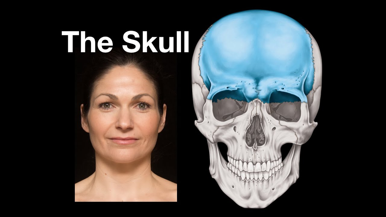
- π The lecture includes a 3D reconstruction from a CT scan, highlighting the visualization of soft tissues and skeletal structures, which is crucial for identifying pathological lesions.
- π₯ A significant pathological lesion in the mandible is presented to illustrate the importance of understanding normal musculoskeletal anatomy to detect abnormalities.
- π The course is structured into four main parts: studying the skull and mandible, discussing facial expression and masticatory muscle groups, and exploring the bounce of the next topic.
- π¨βπ« The lecture includes a motivational quote to inspire students to take anatomy seriously, as it can be life-saving in clinical practice.
- π‘ The skull's functions are highlighted, including housing the brain, cranial nerves, and special sense organs, as well as being the entry point for air and food.
- 𦴠Detailed descriptions of the skull's bones, sutures, and foramina are provided, with an emphasis on their clinical relevance for identifying fractures and other abnormalities.
- π The lecture mentions the importance of learning cranial landmarks and sutures for future studies in volumetric analysis and forensic odontology.
- π¦· The mandible's structure is discussed in detail, including the alveolar process, body, and mental protuberance, which are essential for dental and facial surgeries.
- π€ The facial expression muscles are explored, explaining how they contribute to non-verbal communication and the potential issues that arise when they are not functioning properly, such as facial paralysis.
Q & A
What is the main topic of the BMW 1 class on anatomy?
-The main topic of the BMW 1 class is the muskuloskeletal system, focusing on the anatomy of the skull and mandible.
What is the significance of the 3D reconstruction shown in the class?
-The 3D reconstruction from CT Scan images is significant as it helps visualize soft tissues in transparent blue and skeletal structures of the skull, which is crucial for understanding clinical applications.
Why is it important for dentists to understand the normal structure of the muskuloskeletal system?
-Understanding the normal structure is essential for dentists to identify abnormalities or pathologies, such as lesions or fractures in the facial bones, which is vital for proper diagnosis and treatment.
What is the role of the skull in the human body as described in the script?
-The skull serves as a protective housing for the brain and cranial nerves, contains the special sense organs for sight, hearing, and olfaction, and forms the entryway for air and food into the respiratory and digestive systems.
What are the key components of the cranial cavity mentioned in the script?
-The key components include the cranial cavity itself, which houses the brain, the orbital cavity for the eyes, the nasal cavity for olfaction, and various foramina for the passage of nerves and blood vessels.
What is the importance of knowing the cranial foramina and their contents?
-Knowing the cranial foramina and their contents is important for medical professionals to understand the pathways of nerves and blood vessels, which is crucial for diagnosing and treating conditions related to the skull.
How many cranial bones are there, and what are they called?
-There are 8 cranial bones, which include the frontal, ethmoid, sphenoid, and parietal bones, with the sphenoid and temporal bones being paired.
What is the significance of the mandible in dentistry?
-The mandible is significant in dentistry as it holds the lower teeth and is involved in mastication. Understanding its structure is essential for diagnosing and treating oral health issues.
What are the facial expression muscles, and why are they important?
-Facial expression muscles are responsible for the various facial expressions and are important for non-verbal communication. They include muscles like the frontalis, orbicularis oculi, and others that allow for a range of emotional expressions.
What is the role of the mastication muscles in the human body?
-The mastication muscles, including the temporalis and masseter, are responsible for the movement of the jaw, which is essential for chewing and processing food.
What are the typical and atypical vertebrae in the cervical spine, and how do they differ?
-The typical vertebrae in the cervical spine are C3, C4, C5, and C6, characterized by a small vertebral body and large vertebral foramen. The atypical vertebrae are C1 (atlas), which lacks a body, and C2 (axis), which has an odontoid process. C7 is also atypical due to its long spinous process, known as the vertebra prominens.
Outlines
𦴠Introduction to Musculoskeletal Anatomy
The lecture begins with a focus on the musculoskeletal system, emphasizing the importance of understanding the normal structure of the human body for clinical practice. A three-dimensional reconstruction from a CT scan is showcased, highlighting the soft tissues in blue and the skeletal structure of the skull. The lecturer points out a pathological lesion in the mandible, stressing the need for dentists to comprehend the musculoskeletal system to identify abnormalities. The class structure includes practical tasks, tutorials, presentations, quizzes, and final projects. The session then transitions into a detailed study of the skull and mandible, with an emphasis on fractures and the need for dental professionals to recognize such conditions.
ποΈβπ¨οΈ Skull and Mandible Structures
The lecture delves into the anatomy of the skull, discussing its various components such as the cranial cavity, orbital and nasal cavities, and their functions. The skull's role in housing the brain, special sense organs, and serving as a pathway for air and food is highlighted. The lecturer describes the lateral view of the skull, noting the respiratory passage and the anterior part of the neck. The neurocranium, or the cranial part of the skull, is detailed, including its eight bones and sutures. The importance of understanding the foramina, the holes in the skull that allow nerves and blood vessels to pass through, is emphasized for future studies in volumetric analysis and forensic odontology.
π§ Detailed Cranial Anatomy and Landmarks
This section focuses on the detailed anatomy of the skull, including the sphenoid bone and its separation from other bones. The lecture mentions the need to understand the cranial nerves and blood vessels that exit through the foramina. The importance of learning craniometric points, or landmarks, for future courses in volumetric analysis and radiology is stressed. The lecture also covers the inferior view of the skull, highlighting various foramina and their contents, as well as the significance of these structures for clinical practice.
π¦· Mandible and Muscles of the Head and Neck
The lecture shifts to the mandible, detailing its structure and its importance in housing the lower teeth. The alveolar part of the mandible, which holds the teeth, and other parts like the body, mental foramen, and condylar process are discussed. The muscles of the head and neck are categorized into groups, with a focus on the facial expression muscles that enable humans to express a range of emotions. The lecture also touches on the clinical implications of facial muscle dysfunction, such as asymmetry resulting from facial paralysis.
π£οΈ Facial Expression Muscles and Mastication
This part of the lecture explores the facial expression muscles in detail, explaining how they allow for a variety of emotional expressions. The muscles are categorized and their functions, origins, insertions, and innervations are discussed. The clinical aspects of these muscles are highlighted, emphasizing the importance of understanding their functions for non-verbal communication. The lecture also covers the muscles of mastication, including the temporalis and masseter, and their roles in the chewing process.
π Vertebral Column and Clinical Relevance
The final section of the lecture addresses the vertebral column, explaining its function in supporting the weight of the head and trunk and protecting the spinal cord. The typical and atypical vertebrae are differentiated, with a focus on the unique features of C1 (atlas), C2 (axis), and C7. The lecture concludes with a brief overview of the clinical relevance of understanding the vertebral column, setting the stage for further exploration in practical sessions.
Mindmap
Keywords
π‘Anatomy
π‘Mandible
π‘Musculoskeletal System
π‘CT Scan
π‘Skull
π‘Facial Expression Muscles
π‘Mastication Muscles
π‘Foramen
π‘Vertebral Column
π‘Cranial Fossa
π‘Sphenoid Bone
Highlights
Introduction to the muskuloskeletal system using a 3D reconstruction from a CT Scan.
Importance of understanding soft tissue and skeletal structures for clinical practice.
Identification of a pathological lesion affecting the mandible and its clinical implications.
Emphasis on the significance of normal anatomical structures for detecting abnormalities.
The role of anatomy in preventing clinical errors and the importance of continuous learning.
Overview of the course structure including practical tasks, presentations, and assessments.
Detailed examination of the skull and mandible structures in 3D.
Discussion on the functions of the skull, including housing the brain and special sense organs.
Identification of cranial and facial bones from a CT Scan image.
Explanation of the sphenoid bone's features and its role in the skull's structure.
Importance of knowing cranial foramina and their contents for clinical practice.
Clinical relevance of understanding the muscles of facial expression and mastication.
Description of the vertebral column's anatomy and its clinical significance.
Differentiation between typical and atypical vertebrae in the cervical spine.
Clinical application of understanding the vertebral column's anatomy for head and neck support.
Summary of the key points covered in the lecture andι’ε of further practical exploration.
Transcripts
Assalamualaikum warahmatullahi
wabarakatuh
Hari ini kita ada pada kelas BMW 1
anatomi dengan topik muskuloskeletal
system
pada gambar di samping slide ini saya
Tampilkan
rekonstruksi tiga dimensi dari CT Scan
images yang mulanya dua dimensi dengan
menggunakan software kemudian bisa
direkonstruksi seperti ini dan disetting
bahwa soft tissue atau jaringan lunak ya
terlihat pada warna biru transparan ya
bisa kita lihat profilnya dan di
dalamnya terlihat
struktur skeletal atau tulang dari
tengkorak
saya jadikan cover pada topik kuliah
hari ini karena merupakan
representasi bahwa pentingnya topik hari
ini untuk
kegiatan klinik nanti sehari-hari ya
bisa kita lihat pada gambar ini ada Lesi
patologis yang cukup besar ya merusak
Ramos dari mandible ya Nah bayangkan
kalau dokter gigi tidak memahami
struktur normal ya dari
muskuloskeletal sistem pada manusia ini
ada rongga disangka Lesi ini ada rongga
di Sangkala isi atau Bahkan sebaliknya
ini rongga normal untuk mata ini juga
rongga normal padahal berbeda ada yang
normal ada yang patologis Oleh karena
itu kita di sini untuk belajar
bersama-sama tentang muskuloskeletal
sistem
quadly dan saya Tampilkan juga sebuah
quotes yang
bisa membuat mahasiswa lebih semangat
untuk mempelajari
damage satu anatomi itu yaitu
knowledge of anatomi can save your
passions jadi lebih semangat lebih
serius lagi dalam membaca
boleh kita hari ini akan dibagi menjadi
4 bagian besar pertama kita lihat
struktur tengkorak dan mandibula
Kemudian ada beberapa grup dari seluruh
grup otot yang ada di head and back
untuk topik ini kita hanya bahas dua
grup yaitu facial expression dan
masticase
kemudian terakhir kita akan bahas
tentang Bounce of the next
Nah untuk secara umum ya kegiatan kuliah
di BMW 1 terdiri dari tugas praktikum
gitu ya Dan nanti ada Kegiatan praktikum
yang harus hadir dengan presensi minimal
80% kemudian ada tutorial presentasi
makalah kemudian ada Kuis UTS us dan
juga tugas akhir
Nah
kita sekarang beralih ke bagian pertama
yaitu school and mandable ini adalah
struktur 3 dimensi dari tulang saja
tanpa jaringan lunak dari
tengkorak pasien dan ini adalah
penampakan frontal atau anterior View
bisa kita lihat di sini terdapat
fraktur yang cukup parah pada tulang apa
saja Nah inilah yang harus kita ketahui
supaya nanti dokter gigi bisa melihat
bisa mengidentifikasi
kelainan atau misalnya fraktur pada
tulang-tulang wajah dan kepala
Nah ini salah satu contoh juga yang saya
screenshot dari
presentasi Prof assaumi visiting lecture
beberapa waktu yang lalu di fkg4 ya
Mi University menampilkan
MRI scan dari pasiennya
bisa kita lihat nah saya ingin tanya
adik-adik harus bisa jawab ini potongan
apa kemarin sudah belajar di
bab 1 introduction ini bidangnya bidang
apa sehingga menghasilkan Gambaran
seperti ini harus bisa jawab nah pada
gambaran kamera ini
terlihat ada Lesi patologis yang cukup
besar
nah kembali lagi bahwa pentingnya
ilmu anatomi yang mempelajari struktur
manusia normal sehingga nanti Ketika
menemukan struktur yang abnormal atau
patologis itu klinicy bisa mendeteksinya
tengkorak ya tulang tengkorak itu
menjadi rumah bagi beberapa fungsi
pertama di dalam tengkorak ada otak dan
kranials kemudian di dalam kepala juga
ada special sense organs seperti
penglihatan penghidup pendengaran dan
juga ada pengecatan
dan juga kepala merupakan jalur masuk
bagi udara untuk pernafasan dan juga
bagi makanan dan minuman untuk
sebagai pintu masuk saluran pencernaan
nah kemudian kita lihat di sini struktur
tengkorak pertama ada kranial
di mana di dalamnya ada cranial cavity
yang diisi oleh otak ya Jadi ini
kemudian ada rongga orbit tempat
duduknya bola mata kemudian ada rongga
nasal tempat
apa penghidup Indra penghidupan
du apabila dilihat dari lateral ya
ada respiratory passage yang bisa di apa
sebagai pintu masuknya adalah hidung
ataupun mulut ya kemudian lanjut ke
bagian anterior dari leher jadi di depan
tulang belakang ya oke yang elementary
passage atau saluran pencernaan ya pintu
masuknya adalah rongga mulut yang
kemudian nanti berlanjut ke bagian leher
ya kemudian ada yang ini lubang namanya
external akustik mattes tempat atau
pintu masuk ya dari
hearing and balance
Kemudian pada leher ada 7 servival
yang menopang kepala
nah bila dilihat dari lateral ya kita
bisa melihat bagian besar nih datang
kerak yang disebut namanya
neurocranium atau kranial volt ya
kemudian bila kita tarik Garis khayal
dari titik di atas rongga orbit dan di
bawah telinga di bawah eksternal akustik
netes Garis khayal ini akan membagi
tengkorak menjadi dua bagian yaitu
kranial pot tadi yang tempat otak
di dalamnya kemudian ada tulang wajah
disebut juga Fisher
kranial volt ini terdiri dari 8 tulang
dimana 4 adalah singular letaknya di
tengah collinsing Killer Ya udah frontal
edmoid venoid
ini di sini kurang terlihat kecil karena
agak dalam posisinya
kemudian ada dua tulang yang berpasangan
seperti temporal dan pariental
ya
jadi total untuk kranial foad ada 8
tulang yang dihubungkan dengan
sutura-sutura sutura adalah fibers
yang menghubungkan dua tulang nah
bagaimana dengan fishrocranium pembentuk
wajah ada 15 airgular Bounce yang
terdiri dari tiga singular ya Yang di
tengah itu
terdiri dari
6 tulang yang berpasangan membentuk
wajah yaitu maksila inferior nasal
kontrakan
nah Apabila ada gambaran round synografi
seperti ini ya kemudian terlihat ada
fraktur-fraktur
dokter gigi harus bisa mengidentifikasi
ini fraktur di tulang apa
tulang frontal
variety
nah ini saya terlihatkan gambar-gambar
dari berbagai
sisi ya bahwa tulang tengkorak kita
merupakan gabungan dari beberapa tulang
yang menempel melalui sutora dan tidak
hanya bentuknya yang berbeda-beda tetapi
juga ada struktur yang namanya
foramen-fornamen yang harus mahasiswa
ketahui satu persatu
ada gambar-gambar yang di color coded ya
jadi memudahkan untuk pembelajaran ya
mana tulang mana beda warnanya
ini anterior view Tetapi ada juga nanti
gambar yang tidak color coded ya untuk
melatih adik-adik mahasiswa karena
memang nanti ketika melihat misalnya
tengkoraknya langsung itu tidak ada
warnanya ya warnanya sama putih seperti
ini
nah ini yang lateral view dilihat satu
persatu
kemudian yang posterior view apabila
tulang Oxy vitalnya kita lepaskan maka
akan terlihat di dalamnya kranial cabity
yang memiliki
struktur-struktur foramen di dalamnya ya
dan masing-masing foramen ada namanya
Kenapa dinamakan satu-satu karena supaya
membedakan dan isinya biasanya nama
mencerminkan isinya isi atau konten yang
melalui foramen tersebut apa
oke nah Ini apabila dilihat dari
Superior ya bisa kita lihat suturanya
dan ada titik-titik ya
ini adalah
craniometric point atau Landmark atau
antropometrik points ya Kenapa sih harus
belajar titik-titik ini nanti akan
diulang lagi dipakai lagi pada mata
kuliah selanjutnya misalnya pada
volumetric analysis atau pada
forensik odontologi nah tulang
titik-titik Lemak ini akan dipakai lagi
akan muncul kembali dan akan juga muncul
pada mata kuliah radiologi
ini adalah gambaran dari tulang sfenoid
secara sendiri dipisahkan ya dari
tulang-tulang lain di sekitar
Nah kalau kita potong kranial volt atau
Tempurung lalu kita pindahkan isinya
yaitu otaknya dan otaknya dibalik ya
dilihat dari bawah akan banyak
syarat-syarat dan pembuluh darah seperti
ini ini adalah kranial nanti kita akan
belajar lebih lanjut di slide berikutnya
dan pada topik 3 ya
nah nervus nervus ini dan pembuluh darah
ini akan keluar dari kranial kbt menuju
tempat-tempat yang
ditargetkannya melalui foramen-fornamen
ini
ya jadi harus mengetahui
foramen Apa isinya apa nervus apa ya
inilah struktur
tengkorak apabila dilihat dari inferior
view ya Jadi tadi kan ada
fornamen-forman kalau dilihat dari
internal dan interior nah
maka setelah masuk ke foramen keluarnya
di sini nanti sama namanya ya yang kayak
yang tadi ya
dilihat dari inferior view Tadi dilihat
dari dalam ada juga namanya
dibaca satu persatu agar bisa
diidentifikasi dan kemudian dihafalkan
ini juga inferior view tapi tanpa warna
color coded
nah ini khusus untuk foramennya saja
banyak sekali foramennya
dan nanti dihafalkan juga dipahami juga
bahwa misalnya
foramen rotundum ya dan foramen
spinosum
Superior orbital visure dan optik karena
dan foramen ovalet ini ada di tulang
sfenoid
ya jadi jangan sampai ketika saya tanya
ada di mana kalian jawab di sphenoid itu
salah oke
nah ini adalah rangkuman dari kranial
foramina yang tadi
sudah ditunjukkan strukturnya lokasinya
di mana dan isinya karena masing-masing
mayoritas isinya adalah kranials tapi
ada juga pembuluh darahnya
nah ini masih sama hanya saja ada
struktur-struktur tulang yang lain
selain
kita sudah memahami dua tadi ya dari
gambar sebelumnya
dilihat dari lateral ada juga
titik-titik lain yang ada ion
asterion
vertex terion labela Nation saya
jelaskan Coba satu-satu ya lamda ini
titik apabila dilihat dari lateral ini
pada garis tengah berarti ini semua pada
garis tengah yang ini
landak itu pertemuan antara tulang
osipital dan
tulang pariental pada midline vertex
adalah titik tertinggi pada posisi
Frankfurt horizontal print seperti ini
adalah pertemuan antara tulang frontal
dan pariental ya pada garis tengah
pada garis tengah juga
yaitu titik
pada tulang frontak Nation pertemuan
antara tulang frontal dan tulang nasal
pada garis tengah asterion adalah
pertemuan sutura dua sutura yaitu sutura
squamosa dengan sutura lamproid nah ini
titiknya disebut asterium sedangkan
terion adalah bukan suatu titik tapi
suatu garislah garis yang mempertemukan
dengan
ya dan
Spanyol frontause venoidalis future yang
ini jadi ada pertemuan tiga sudut
disebut period ya
adalah titik ada di sini eksternal
opsipital
Oke kita sekarang beralih ke mendible
dulu mendible ini strukturnya sangat
spesifik ya Jadi ini harus
dipelajari dengan baik karena ini
merupakan rumah dari gigi-gigi rahang
bawah yang merupakan nanti pekerjaan
kita sehari-hari
pertama selain gigi geligi ada bagian
tulang yang memegang gigi-gigi tersebut
menyangga gigi tersebut disebut alveolar
part of the mendyble kemudian ada body
dari mendible bagian tengah ada mendidih
kemudian ada mental frame tempat
keluarnya apa Nanti kita lihat ya
Kemudian dilihat dari sisi sebelah sini
ya nanti Tempat masuknya saraf apa kita
pelajari nanti terus kemudian ujungnya
keluar di mental
koramidele
yang vertikal ini ramus of
kemudian ada koronoid proses yang di
anterior nah yang ini adalah kondiloid
proses ada haidnya ada netnya dan
tertukar ya mana yang konoid mana yang
Nah kita berlanjut sekarang ke otot
otot-otot pada kepala dan leher itu
dibagi menjadi beberapa grup kita lihat
dulu yang di kepala ada ekstrak untuk
menggerakkan bola mata dan juga membuka
kelopak mata
ada otot-otot pada telinga tengah ya
kemudian ada otot facial expression yang
kita pelajari hari ini
kemudian ada masaus of mastication untuk
pengunyahan membuka mulut nutup mulut ya
ini kita pelajari hari ini ada otot soft
pelet itu nanti di bbs2 Mas source of
the town ini untuk di BMS 2
kemudian ada pada lehernya ada otot
faring dan lari ya yang laring kita
pelajari di topik berikutnya yang faring
di bns2 kemudian ada strap
pada leher ada otot dari outer Survival
color pada postural massos ini nanti di
BNS 3
Nah kita pelajari grup yang pertama
yaitu facials expressions Kenapa manusia
bisa berekspresi dengan berbagai jenis
emosinya
itu karena ada otot-otot wajah yang
bekerja ya ketika otot-otot wajah tidak
berfungsi dengan baik persarapannya
contohnya misalnya pada kejadian
kelumpuhan sebagian otot wajah cari itu
kelainan apa namanya itu bisa terlihat
pada gambar yang sebelah kiri terlihat
ada asimetris pada
otot wajahnya pada wajahnya gitu ya
Nah itu karena ada gangguan pada
otot-otot wajahnya
bisa kita lihat di sini dari tampak
depan ya Ada oksip itu frontalis massals
proceus
supersiliae Ada orbicularis Of Your Life
nasalis lavator
lebia superioris
dan depresor septinasi ada orbiculation
ada vaksinator kemudian ada levator ini
angler Oris tapi ini yang di sudut ya
berbeda dengan labiay superioris
kemudian ini levator
ini selalu engkel posisinya di Angle of
Nah itulah otot-otot yang membentuk
wajah kita kenapa wajah kita
berbeda-beda karena pertama struktur
tulang wajahnya dulu berbeda ketika
struktur tulangnya berbeda maka
otot-otot ini akan menempelnya sama di
area itu tetapi karena strukturnya itu
sendiri struktur tulangnya itu berbeda
menghasilkan wajah yang berbeda
nah ini facial expression Masters bila
dilihat dari later ya dibaca lagi satu
persatu dan saya ingin Perlihatkan di
sini bahwa semua facial expression
massal dipersarapi oleh nervus
yang bercabang menjadi 6 ada posterior
auricular ada temporal ada
bukel ada Marginal
dasar bike
nah kemudian kita lanjut ke facial
expression massal di bagian rongga mulut
Ada apa saja nih di bagian rongga mulut
nih
ada orbicularis okula ya dan ada
depresor nilai Oris nanti di sini juga
ada depresornya
inferiors
lavatornya juga ada dua ada yang Angela
ada yang
ini berkompleks atau membentuk suatu
anyaman lah ya dengan pembuluh darah dan
juga nanti di bawahnya ada
struktur yang akan kita bekerja
sehari-hari jadi harus diketahui mulut
itu terdiri dari komposisinya apa saja
nah ini ya
ini adalah detail dari tadi nama
musself ototnya apa saja Origin dan
insentions dan fungsinya
nah ini adalah gambaran klinis dari
fungsi-fungsi otot-otot tersebut satu
persatu ya Jadi mohon nanti bisa
diketahui
otot apa fungsinya untuk menghasilkan
ekskresi wajah yang mana Inilah grup
Mass inilah yang
meng-inible manusia untuk berkomunikasi
tanpa kata-kata tanpa verbal
nah grup selanjutnya yaitu massage of
mystication ada 4 ya yang dikuningan ini
yang besar adalah temporalis dan maseter
Oke apabila mansetter dan sebagian dari
mendiblenya kita
remove akan terlihat di dalamnya letral
dari guide yang horizontal dan medil
very good yang agak komplit agak miring
ini adalah satu persatu dijelaskan
Origin inversion innervation dan
fungsinya
fungsinya ya Ini penting
Oke kita lanjut ke bagian terakhir yaitu
Bounce of the next ya terdiri dari
servival vertebray kemudian ada
highlight
Nah untuk vertebral kolom ya tulang
belakang terdiri dari 7 vertikal 12 yang
bagian terakhir
fungsi dari vertebral kolom pertama
adalah untuk support beratnya dari
kepala dan apa Batang tubuh pada saat
berdiri sehingga
vertebral kolom itu ada ligamen untuk
menopangnya dan juga fungsi yang kedua
untuk memproteksi dari Spanyol chord ya
karena di dalamnya ada
foramen atau seperti terowongan yang
panjang isinya Spanyol
dan juga karena ada otot-otot yang
attach pada vertebral kolom tersebut dan
ada join joinnya sehingga
batang tubuh
nah tulang dari vertebral kolom tadi kan
Ya ada
yang bagian survivalnya ada 7 nah ini
yang bagian kita yang kita pelajari
hanya unsur ada 7
C1 C2 C3 456 dan C7
yang 3456 disebut tipikal Survival
vertebrate jadi bentuknya
dan nanti kita bahas slide selanjutnya
yang C1 C2
C7 itu bentuknya beda sendiri Makanya
disebut atipikal
kalau yang C1 atau namanya atlas seperti
cincin
ya yang C2 itu ada odontoid prosesnya
namanya atlas ya si tulang
C1 atlas C2 itu Axis nah Axis ini ada
odontoid proses atau dance nama lainnya
yang ini beda sendiri
yang C7 ini dia punya
proses yang paling panjang diantara
semuanya sehingga disebut vertebrate
prominence karena teraba pada tengkuk
proses pertama yang teraba itu adalah
batik C7
Nah untuk yang tipikal vertebray yaitu
c3456 ya dia punya karakteristiknya sama
semuanya ada vertebral bodynya kecil
gitu ya dan ada vertebral foramennya
besar transfer prosesnya punya Superior
passesnya ada juga kemudian spiness
prosesnya agak pendek dibandingkan
dengan yang 7 ya
Oke kemudian apabila kita rekap seperti
ini ya Jadi c3456 ini tipikal
C1 nya atlas ya dia tidak ada bodynya
seperti cincin fungsinya untuk loading
yang C2 dia punya odontoid proses atau
dance ya
namanya Axis nih c2-nya nama lainnya
adalah Axis fungsinya untuk shaking head
atau rotasi ya kemudian tadi C7 yang
antipikal juga ya dia punya proses yang
panjang disebut vertebrata
nah sekian kuliah singkat yang saya
sampaikan hari ini akan kita explore
lebih lanjut nanti di sesi praktikum
Untuk itu saya sampaikan sekian terima
kasih wassalamualaikum
warahmatullahi wabarakatuh
5.0 / 5 (0 votes)