What is a Protein? (from PDB-101)
Summary
TLDRProteins, composed of 21 amino acids, are essential for various biological functions, from transporting nutrients to facilitating chemical reactions. Their primary structure is a sequence of amino acids encoded by DNA, forming peptide bonds. Proteins fold into secondary structures like alpha helices and beta sheets, which contribute to their three-dimensional tertiary structures. These shapes are crucial for their roles, as seen in hemoglobin's oxygen binding or antibodies' pathogen recognition. The RCSB Protein Data Bank offers more insights into proteins' functions and structures.
Takeaways
- π Proteins are essential in the biological world, serving a variety of functions including transport, catalysis, and structural support.
- π All proteins are composed of the same 21 amino acids, which are the building blocks of proteins.
- π§ Amino acids consist of carbon, oxygen, nitrogen, and hydrogen, with some also containing sulfur or selenium.
- π The side chain of an amino acid is what varies and determines its properties, such as being hydrophobic, hydrophilic, or charged.
- 𧬠The primary structure of a protein is its linear sequence of amino acids as determined by DNA.
- π Peptide bonds link amino acids together, forming the protein backbone and releasing a water molecule in the process.
- π Proteins can fold into secondary structures like alpha helices or beta sheets, stabilized by hydrogen bonds.
- 𧬠The tertiary structure is the three-dimensional shape of the protein, influenced by the amino acids' characteristics.
- π Globular proteins often have hydrophobic side chains on the inside, while membrane-bound proteins have them on the outside for lipid interaction.
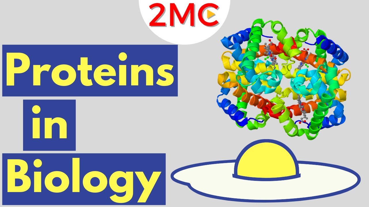
- π The function of many proteins is reliant on their three-dimensional shape, such as hemoglobin's ability to bind oxygen.
- π Visual representations of proteins, like space-filling, ribbon, and surface diagrams, provide insights into their structure and function.
- π‘ The size of proteins, exemplified by hemoglobin at 6.5 nanometers, is significant for their function and interaction within the body.
- π₯ The RCSB Protein Data Bank is a resource for learning more about the functions and 3D structures of proteins and other molecular machines.
Q & A
What are the primary functions of proteins in the biological world?
-Proteins serve various functions such as transporting nutrients, facilitating chemical reactions, and building the structures of living organisms.
How many building blocks are there for proteins, and what are they called?
-There are 21 building blocks for proteins, known as amino acids.
What elements make up amino acids, and is there any exception with a different element?
-Amino acids are composed of carbon, oxygen, nitrogen, and hydrogen, with the exception of selenocysteine, which also contains a selenium atom.
What are the three main components of an amino acid?
-An amino acid consists of an amino group, a carboxyl group, and a side chain attached to a central carbon atom.
How does the side chain of an amino acid affect its properties?
-The side chain varies from one amino acid to another and determines its properties, such as being hydrophobic, hydrophilic, or charged.
What is the primary structure of a protein, and how is it formed?
-The primary structure is the linear sequence of amino acids as encoded by DNA, with amino acids joined by peptide bonds, releasing a water molecule each time.
What are the two types of secondary structures that protein chains can fold into?
-Protein chains can fold into alpha helices or beta sheets, both stabilized by hydrogen bonds.
How does the tertiary structure of a protein differ from its secondary structure?
-The tertiary structure refers to the three-dimensional shape of the entire protein chain, determined by the characteristics of the amino acids within it.
What role does the shape of a protein play in its function?
-The three-dimensional shape of a protein is crucial for its function, allowing it to interact with other molecules or perform specific tasks within the body.
Can you provide an example of a protein that relies on its three-dimensional shape for function?
-Hemoglobin is an example of a protein that relies on its shape to form a pocket that holds heme and binds oxygen.
What are some different visual representations of proteins, and what do they show?
-Visual representations include space filling diagrams that show all atoms, ribbon or cartoon diagrams that show the protein backbone and alpha helices, and surface representations that highlight areas accessible to water molecules.
Where can one find more information about the functions and 3D structures of proteins?
-The RCSB Protein Data Bank is a resource where one can learn more about the functions and 3D structures of proteins and other molecular machines.
Outlines
𧬠Protein Structure and Function
Proteins are essential to life, with roles ranging from nutrient transport to structural support. They are composed of 21 amino acids, which vary in their side chains, affecting their interactions with water and other molecules. The primary structure of a protein is its sequence of amino acids, determined by DNA. This sequence folds into secondary structures like alpha helices and beta sheets, stabilized by hydrogen bonds. The tertiary structure is the three-dimensional shape of the protein, influenced by the amino acids' properties. This shape is crucial for function, as seen in hemoglobin's ability to bind oxygen. Some proteins, like antibodies and insulin, rely on their shape for immune function and glucose regulation, respectively. Proteins can also be represented visually through space filling, ribbon, and surface diagrams to understand their structure and function better.
π οΈ Specific Proteins and Their Roles
The calcium pump, powered by ATP and aided by magnesium, moves calcium ions to the sarcoplasmic reticulum, essential for muscle contraction. Ferritin is a spherical protein that stores iron in a non-toxic form, with channels for iron entry and exit based on the organism's needs. Inside, ferritin has a hollow space with atoms attached to the interior wall. Collagen is another structural protein, forming a strong triple helix that supports the body, particularly in skin and tendons, by forming elongated fibrils into collagen fibers. The RCSB Protein Data Bank offers more insights into the functions and 3D structures of proteins and other molecular machines, highlighting the complexity and diversity of these biological molecules.
Mindmap
Keywords
π‘Proteins
π‘Amino Acids
π‘Peptide Bonds
π‘Primary Structure
π‘Secondary Structures
π‘Tertiary Structure
π‘Globular Proteins
π‘Membrane-Bound Proteins
π‘Hemoglobin
π‘Antibodies
π‘Collagen
Highlights
Proteins have a wide range of functions in the biological world, including transport, catalysis, and structural support.
All proteins are composed of the same 21 amino acids, which vary in their side chains.
Selenocysteine is the only standard amino acid containing a selenium atom.
Amino acids are categorized as hydrophobic, hydrophilic, or charged based on their side chain properties.
The primary structure of a protein is its linear sequence of amino acids as encoded by DNA.
Peptide bonds link amino acids together in a protein, forming the protein backbone.
Proteins can fold into secondary structures like alpha helices and beta sheets, stabilized by hydrogen bonds.
The tertiary structure is the three-dimensional shape of a protein, determined by amino acid characteristics.
Many proteins form globular shapes with hydrophobic side chains sheltered inside.
Membrane-bound proteins have hydrophobic residues on the outside to interact with lipids in the membrane.
Charged amino acids allow proteins to interact with molecules with complementary charges.
The three-dimensional shape of a protein is crucial for its function, as seen in hemoglobin's ability to bind oxygen.
Hemoglobin's four subunits cooperate to pick up and release oxygen in the body.
Different visual representations of proteins, like space filling and ribbon diagrams, provide insights into protein structure and function.
Hemoglobin molecules are about 6.5 nanometers in size and are found in high concentration in red blood cells.
Antibodies have flexible arms that recognize and bind to pathogens, targeting them for immune system destruction.
Insulin is a small, stable protein that regulates blood glucose levels while traveling through the blood.
Alpha Amylase is an enzyme that initiates starch digestion in saliva.
The calcium pump moves calcium ions back to the sarcoplasmic reticulum after muscle contraction, aided by magnesium and powered by ATP.
Ferritin is a spherical protein that stores iron in a non-toxic form and allows iron atoms to enter and exit depending on the organism's needs.
Collagen forms a strong triple helix for structural support in the body, and collagen molecules can aggregate to form collagen fibers found in skin and tendons.
The RCSB Protein Data Bank provides more information on protein functions and 3D structures.
Transcripts
Proteins play countless roles throughout the biological world.
Some transport nutrients throughout the body.
Some help the chemical reactions happen at a faster rate.
Others build the structures of the living things.
Despite this wide range of functions, all proteins are made out of the same 21 building
blocks, called amino acids.
Amino acids are made of carbon, oxygen, nitrogen, and hydrogen, and some contain sulfur atoms.
Selenocysteine is the only standard amino acid that contains a selenium atom.
These atoms form an amino group, a carboxyl group, and a side chain attached to a central
carbon atom.
The side chain is the only part that varies from amino acid to amino acid and determines
its properties.
Hydrophobic amino acids have carbon-rich side chains, which donβt interact well with water.
Hydrophilic or polar amino acids interact well with water.
Charged amino acids interact with oppositely charged amino acids or other molecules.
Primary Structure The primary structure of a protein is the
linear sequence of amino acids as encoded by DNA.
The amino acids in a protein are joined by peptide bonds, which link the amino group
of one amino acid to the carboxyl group of another.
A water molecule is released each time a bond is formed.
The linked series of carbon, nitrogen, and oxygen atoms make up the protein backbone.
These protein chains often fold into two types of secondary structures: alpha helices, or
beta sheets.
An alpha helix is a right-handed coil stabilized by hydrogen bonds between the amine and carboxyl
groups of nearby amino acids.
Beta-sheets are formed when hydrogen bonds stabilize two or more adjacent strands.
The tertiary structure of a protein is the three-dimensional shape of the protein chain.
This shape is determined by the characteristics of the amino acids making up the chain.
Many proteins form globular shapes with hydrophobic side chains sheltered inside, away from the
surrounding water.
Membrane-bound proteins have hydrophobic residues clustered together on the outside, so that
they can interact with the lipids in the membrane.
Charged amino acids allow proteins to interact with molecules that have complementary charges.
The functions of many proteins rely on their three-dimensional shapes.
For example, hemoglobin forms a pocket to hold heme, a small molecule with an iron atom
in the center that binds oxygen.
Two or more polypeptide chains can come together to form one functional molecule with several
subunits.
The four subunits of hemoglobin cooperate so that the complex can pick up more oxygen
in the lungs and release it in the body.
Different visual representations of proteins can give us visual clues about the protein
structure and function.
This space filling diagram shows all atoms that are making up this protein.
This representation, called ribbon or cartoon diagram shows the organization of the protein
backbone and highlights the alpha helices.
This surface representation shows the areas that are accessible to water molecules.
Most proteins are smaller than the wavelength of light.
For example, the hemoglobin molecule is about 6.5 nanometers in size.
Hemoglobin is found in high concentration in red blood cells.
A typical red blood cell contains about 280 million hemoglobin molecules.
The three-dimensional shapes of proteins determine their function.
The flexible arms of antibodies protect us from disease by recognizing and binding to
pathogens and targeting them for destruction by the immune system.
The hormone insulin is a small, stable protein that can easily maintain its shape while traveling
through the blood to regulate the blood glucose level.
Alpha Amylase is an enzyme that begins digestion of starches in our saliva.
The calcium pump is aided by magnesium and powered by ATP
to move calcium ions back to the sarcoplasmic reticulum after each muscle contraction.
Ferritin is a spherical protein with channels that allow the iron atoms to enter and exit
depending upon organismβs needs.
On the inside ferritin forms a hollow space interior with atoms attached to the inner
wall.
Ferritin stores iron in the non-toxic form.
Collagen forms a strong triple helix that is used throughout the body for structural support
Collagen molecules can form elongated fibrils which aggregate to form collagen fibers.
This type of collagen is found in skin and tendons.
Learn more about the functions and 3D structures of proteins and other molecular machines at
the RCSB Protein Data Bank.
Browse More Related Video
Protein structure | Primary | Secondary | Tertiary | Quaternary
What is a Protein? Learn about the 3D shape and function of macromolecules
Protein Structure - Primary, Secondary, Tertiary, & Quarternary - Biology
Chem 1307 Ch 19.3 Proteins - Secondary Structure
Proteins | Biological Molecules Simplified #2
Proteins AP Biology
5.0 / 5 (0 votes)