Anatomy and Physiology of the Integumentary System
Summary
TLDRThis video script delves into the integumentary system, highlighting its role in skin and accessory structures like nails, hair, and glands. It underscores the skin's multifaceted functions, including temperature regulation, protection, sensory information, and vitamin D synthesis. The script explores skin layers, from the epidermis to the dermis, detailing their composition and functions. It also examines hair and nail structures, the role of melanocytes in pigmentation, and the significance of sweat and sebaceous glands in maintaining skin health and body temperature.
Takeaways
- 📚 The integumentary system derives from 'in' (inward) and 'tegere' (to cover), highlighting its role in covering the body with skin and accessory structures like nails, hairs, glands, and receptors.
- 🌡️ It plays a crucial role in maintaining body temperature, protecting the body, providing sensory information, reflecting emotions, and aiding in physiological processes like sweating.
- 💊 The skin synthesizes vitamin D, which is vital for calcium and phosphate absorption, starting with the molecule 7-dihydrocholesterol that converts to active vitamin D3 upon UV exposure.
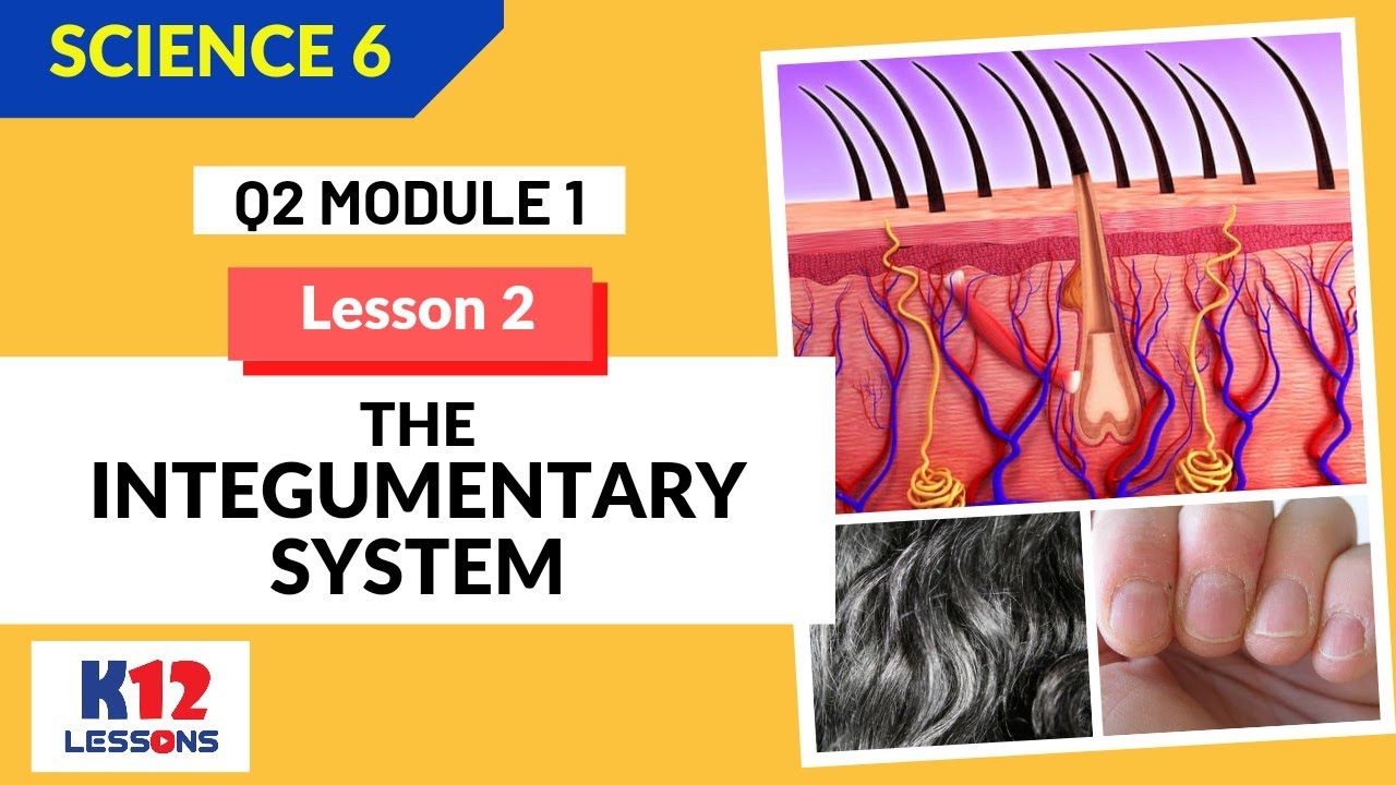
- 🧬 The skin, being the largest organ, varies in thickness and is composed of the epidermis and dermis, with the epidermis having 4-5 layers of epithelial cells and no blood supply of its own.
- 🔬 The epidermis consists of keratinocytes that produce keratin for protection, melanocytes for pigmentation, Langerhans cells for immune response, and Merkel cells for light touch and pressure detection.
- 🌞 Melanocytes are responsible for skin color and protection against UV damage, with increased melanin production in response to sun exposure, leading to tanning and freckle formation.
- 🌱 The stratum basale is the epidermal layer where new cells are formed through mitosis, containing stem cells that differentiate into keratinocytes.
- 🌀 The dermis is denser and contains blood vessels, nerves, and appendages like hair follicles and sweat glands, providing strength, elasticity, and hydration to the skin.
- 👁️ Dermal papillae increase friction for grip and contain sensory receptors for touch, pressure, and temperature, with unique fingerprint patterns due to genetic variation.
- 🦴 Collagen and elastic fibers in the dermis contribute to skin strength and elasticity, with a decrease in these fibers leading to aging effects like sagging and wrinkles.
- 🌡️ Blood vessels in the dermis help regulate body temperature by dilating to release heat or constricting to conserve it, affecting skin color and sensation.
Q & A
What is the integumentary system?
-The integumentary system is the body system that includes the skin and its accessory structures such as nails, hairs, glands, and sensory receptors. It plays a crucial role in covering the body, maintaining body temperature, protection, sensory information, and reflecting emotions.
What is the role of vitamin D in the integumentary system?
-Vitamin D is synthesized in the skin and functions as a hormone that stimulates the absorption of calcium and phosphate from the intestines. The molecule 7-dihydrocholesterol, a precursor of vitamin D, is converted into cholecalciferol when exposed to ultraviolet light from the sun and is later modified by the liver and kidneys to form active vitamin D3 or calcitriol.
What are the two main layers of the skin?
-The two main layers of the skin are the epidermis and the dermis. The epidermis is the outermost layer composed of epithelial cells, while the dermis is the thicker layer beneath, made up of dense irregular connective tissue containing collagen and elastic fibers.
What are the different types of cells found in the epidermis?
-The epidermis contains various cells including keratinocytes, melanocytes, Langerhans cells, and Merkel or tactile cells. Keratinocytes produce keratin, melanocytes produce melanin for skin color, Langerhans cells are part of the immune system, and Merkel cells are associated with detecting light touch and pressure.
How does the skin help in maintaining body temperature?
-The skin helps maintain body temperature through the blood vessels in the dermis. When the body temperature is high, the capillaries become engorged, allowing heat to radiate from the skin surface. In a cool environment, blood bypasses the capillaries to conserve body heat.
What is the function of the stratum corneum in the skin?
-The stratum corneum is the outermost layer of the epidermis and consists of dead keratinized cells. It serves as a barrier to prevent the penetration of microbes and dehydration of underlying tissues, providing mechanical protection for the delicate underlying layers.
What is the structure of hair and what are its functions?
-Hair consists of a shaft that projects above the skin surface and a root below the surface. It is composed of layers including the medulla, cortex, and cuticle. Hair provides protective functions such as shielding the eyes through eyelashes and preventing foreign particles from entering the respiratory tract through nasal hairs.
How does the nail structure differ from hair?
-Nails are thin plates made of layers of dead stratum corneum cells containing a very hard type of keratin. Unlike hair, nails have a free edge, a body, and a root. The nail matrix is responsible for nail growth, and nails are mostly non-living materials that appear pink due to the underlying blood supply.
What are the two types of sweat glands and their functions?
-There are two types of sweat glands: eccrine and apocrine. Eccrine glands are widespread and produce sweat for temperature regulation. Apocrine glands are found in areas like the armpits and genitalia and produce secretions that, when metabolized by bacteria, contribute to body odor.
What is the role of sebaceous glands in the skin?
-Sebaceous glands produce sebum, an oily substance that prevents the skin from drying and provides protection against some bacteria. Sebum also helps keep the skin and hair moisturized and soft.
What is the significance of the dermis in terms of skin strength and elasticity?
-The dermis, composed of dense irregular connective tissue with collagen and elastic fibers, provides the skin with tensile strength, allowing it to stretch without breaking. As we age, the decrease in collagen and elastic fibers leads to a loss of skin elasticity, causing sagging and wrinkles.
Outlines
🧬 Overview of the Integumentary System
The integumentary system, derived from Latin roots meaning 'inward' and 'to cover', encompasses the skin and its accessory structures such as nails, hair, glands, and sensory receptors. This system is crucial for maintaining body temperature, protection, sensory information, and emotional expression. It also plays a vital role in vitamin D synthesis, which aids in calcium and phosphate absorption. The skin, being the body's largest organ, varies in thickness and is composed of the epidermis and dermis, with the epidermis being avascular and consisting of cells like keratinocytes, melanocytes, and Merkel cells, responsible for skin hardness, color, and light touch sensation, respectively.
🔍 Structure and Function of Skin Layers
The skin's structure is detailed with various layers serving distinct functions. The epidermis has 4 to 5 layers, including the stratum basale for cell production, stratum spinosum for cell division, stratum granulosum for cell apoptosis, and the stratum corneum for barrier protection. Thick skin, found on palms and soles, includes an additional stratum lucidum. Dendritic cells in the epidermis play a role in immune response, while the dermis, composed of the papillary and reticular layers, contains blood vessels, nerves, and appendages like hair follicles and sweat glands, contributing to the skin's strength, elasticity, and sensory reception.
🦠 Immunity and Sensory Reception in the Skin
The skin's immune function is facilitated by dendritic cells that engulf pathogens and alert the immune system. Merkel cells and tactile corpuscles in the dermis are associated with nerve endings for light touch and pressure detection. The skin also contains unique fingerprint patterns due to dermal papillae arrangements, which increase grip and produce sweat for temperature regulation. The dermis's blood vessels help maintain body temperature by dilating or constricting in response to thermal changes.
💇♀️ Hair and Nail Structure and Growth Cycles
Hair and nails are accessory structures of the skin with specific protective roles. Hair shields the eyes and respiratory tract, while its structure consists of the medulla, cortex, and cuticle made of keratinized cells. Hair growth occurs in cycles with a growth stage followed by a resting stage, leading to hair replacement. Nails are composed of layers of keratinized cells and have a matrix for nail growth, appearing pink due to underlying blood supply. The visible part of the nail, the lunula, is a crescent-shaped area at the base.
💧 Glands and Their Functions in the Skin
Sebaceous and sweat glands are integral to the skin's function. Sebaceous glands produce sebum for skin protection and hydration, while sweat glands regulate body temperature and produce sweat, which also contributes to body odor when metabolized by bacteria. The apocrine glands, found in armpits and genital areas, secrete substances that become odoriferous post-bacterial action. Cerumen glands in the ear produce earwax, a protective barrier against foreign bodies and microorganisms.
Mindmap
Keywords
💡Integumentary System
💡Keratin
💡Melanocytes
💡Dermis
💡Epidermis
💡Vitamin D
💡Sebaceous Glands
💡Sweat Glands
💡Hair Follicle
💡Nail
💡Cerumen
Highlights
The integumentary system includes the skin and its accessory structures like nails, hairs, glands, and sensory receptors.
The system plays a crucial role in maintaining body temperature, protection, sensory information, and reflecting emotions.
Skin is capable of synthesizing vitamin D, which is essential for calcium and phosphate absorption.
7-dihydrocholesterol in the skin converts to cholecalciferol upon UV exposure, forming active vitamin D3.
Human skin is the largest organ, covering about 2 square meters and weighing 4.5 to 5 kilograms.
The skin varies in thickness from 0.5 millimeters on eyelids to 4 millimeters on heels.
The skin is composed of the epidermis and dermis, with the epidermis being the outermost and thinner layer.
Keratinocytes in the epidermis produce keratin, providing hardness and water resistance to the skin.
Melanocytes produce melanin, influencing skin color and protecting against UV damage.
Langerhans cells in the epidermis are part of the immune system, responding to threats like bacteria and viruses.
Merkel cells are associated with nerve endings, detecting light touch and pressure.
The stratum basal is responsible for cell production and is the deepest layer of the epidermis.
The dermis contains blood vessels, nerves, and glands, and is much thicker than the epidermis.
Collagen and elastic fibers in the dermis provide strength, toughness, and elasticity to the skin.
Blood vessels in the dermis help regulate body temperature by dilating or constricting.
Hair serves protective functions and is composed of a shaft and root with a complex structure.
Hair growth occurs in cycles with a growth stage followed by a resting stage.
Hair color is determined by the amount and type of melanin produced by melanocytes in the hair follicle.
Nails are made of layers of keratinized cells and protect the sensitive tips of fingers and toes.
Sebaceous glands produce sebum for skin protection, while sweat glands help regulate body temperature.
Cerumen, produced by cerumenos glands, provides a protective barrier in the ear canal.
Transcripts
in this video you will be learning about
the integumentary system
the word in documentary was actually
derived from the latin words in
which means inward and tigera which
means to cover
this makes sense because the
integumentary system
refers to the skin and its accessory
structures
like the nails hairs glands
and sensory receptors
aside from covering our body the
integumentary system
also plays several important functions
like helping maintain a constant body
temperature
protecting the body providing sensory
information
reflecting our emotions and other
aspects of normal physiology
such as sweating we can fully understand
how these functions are performed
by the integumentary system as we study
its different
structures another important function of
the skin
is to synthesize vitamin d vitamin d
functions as a hormone that stimulates
the absorption
of calcium and phosphate from the
intestines
the molecule 7-dihydrocholesterol
which is a precursor of vitamin d is
stored in the skin
when exposed to ultraviolet light of the
sun
this molecule is converted into
cholecalciferol
which is released into the blood and
modified by the liver
and later by the kidneys to form active
vitamin d3
or calcitriol we will now
look into the structures of our skin
our skin is also known as as accutaneous
membrane
well obviously our skin covers the
surface of our body
and it is the largest organ in fact
it covers an area of about 2 square
meters and weighs about 4.5
to 5 kilograms which is about 7
of our total body weight
it also varies in thickness from 0.5
millimeters in our eyelids
up to four millimeters in our heels
in most parts of the body it is about
one to two
millimeters thick
the skin is made of multiple layers of
cells and tissues which are held to the
underlying structures by connective
tissues
it is composed of two main layers in the
epidermis and
the dermis these can be seen in this
micrograph
which shows a sectional view of
the skin the epidermis is located here
in the outermost layer as you can see
it is a thinner xanthodermis
constituting of only four or five layers
of epithelial cells
depending on its location in the body
like other epithelial tissues the
epidermis is a vascular
which means that it has no blood supply
of its own
this explains why a man can shave daily
and not
bleed even though he cuts off many cell
layers each time he shaves
the cells that make up the epidermis
include
the carotenoid sites melanocytes
dendritic cells and merkel or tactile
cells
these keratinocytes are cells that
produce and store keratin
keratin is a protein that gives our
hairs
nails and skin their hardness and water
resistant properties
it also helps protect the skin and
underlying tissues from
injuries pathogens heat and chemicals
the carotenoid sites in the uppermost
layer of the skin
known as the stratum corneum are dead
and
regularly slough away being replaced by
cells from the deeper layers
about eight percent of the epidermal
cells are these
spider-shaped cells known as melanocytes
these cells produce the pigment melanin
which is transferred
into the keratinocytes and contributes
to skin
color dark skinned individuals produce
more melanin
than those with pale skin also
exposure to the uv rays of the sun
causes melanin to be manufactured and
built up
in keratinocytes which results in the
darkening of the skin
in fact freckles and moles are seen
where melanin is concentrated in one
spot
this increased melanin accumulation is
important in protecting the dna
of epidermal cells from uv ray damage
and the breakdown of folic acid
longer hand cells are dendritic cells
or cells that have tree like structures
they engulf bacteria foreign particles
and damaged
cells that occur in the epidermis
these cells arise from red bone marrow
and
migrate to the epidermis
they also alert and activate immune
system cells
to a threat such as bacterial or viral
invasion
the merkel or tactile cells are the
least numerous epidermal cells
which are located in the deepest layer
of the epidermis
or in the epithermal dermal junction
they are associated with the nerve
endings
responsible for detecting light touch
and superficial pressure
now it was mentioned a while ago that
the skin may have four to five layers of
these cells
skeins that has four layers of cells
is referred to as thin skin
from deep to superficial these layers
are the stratum basal
stratum spinosum stratum granulosum
and stratum corneum most of the skin can
be classified as a thin skin
thick skin is found only on the palms of
the hands and the soles of the feet
it has a fifth layer called the stratum
lucidum which is located between the
stratum corneum and the stratum
granulosum
the deepest layer of the epidermis is
the stratum basal or also known as
stratum germinativum
to indicate its role in forming new
cells
it attaches the epidermis to the basal
lamina
below which lie as the layers of the
dermis
stratum basal is a single layer of cells
primarily made of basal cells
basal cells are cube-shaped stem cells
that will soon become keratino sites of
the epidermis all of the keratinocytes
are produced from the single layer of
cells
which are constantly going through
mitosis or cell division
to produce new cells as new cells are
formed as the existing cells are pushed
upwards
away from the stratum basal
merkel cells and melanocytes are also
found in the stratum basal
superficial to or just above the stratum
basil
is the stratum spinosum as the name
implies
this layer is spiny in appearance
it consists of numerous keratinocytes
arranged in eight to ten layers
these keratinocytes formed as a result
of cell division
in the stratum basal they also retained
their ability to divide aside from zechy
retinol sites
langerhans cells are also located in
this layer
as new keratinocytes are produced above
the stratum basal
the carotenoid sites of the stratum
spinozum
are pushed into the stratum granulosum
cells flatten as as they move upwards
cells in this layer undergo apoptosis or
programmed cell deaths as as they move
farther away
from the source of nutrition which is
the thermal blood vessels
also their cell membranes thicken and as
they generate large amounts of protein
keratin
and keratohyalin the nuclei
and other cell organelles disintegrate
as the cells die
leaving behind the carotene keratohyalin
and cell membranes that will form the
stratum lucidum
stratum corneum and the accessory
structures of hairs and
nails the stratum lucitum consists of
four to six
layers of flat and clear dead cave
retinol sites that contain large amounts
of keratin
and second plasma membranes it is
present only in the thick skin of
areas such as fingertips palms and soles
this provides an additional level of
toughness in these regions of thick skin
the stratum corneum is the most exposed
and superficial layer of
the epidermis it usually consists of 15
to 30 layers of cells
this dry dead layer helps prevent the
penetration of microbes
and the dehydration of underlying
tissues and provides
a mechanical protection for the more
delicate
underlying layers cells in this layer
are shed periodically and are
replaced by cells pushed up from the
stratum granulosum
or stratum lucidum in the case of palms
and soles
the entire layer is replaced during a
period of about four weeks
if the skin is constantly exposed to
friction
there is an increased cell and keratin
production
that results in the formation of a
callus and abnormal thickening of the
stratum cranium
below the epidermis is the dermis which
is
much thicker it is composed of dense
irregular connective tissue
containing collagen and elastic fibers
which contribute to its great tensile
strength
when we say tensile strength we refer to
the ability to stretch without breaking
in fact leathers used in making belts
bags and shoes are dried and treated
terminals of other animals
unlike the epidermis which is largely
made of cells
the dermis contains blood and leaf
vessels
nerves sweat glands and other structures
such as hair follicles
also portions of them project up into
and through the epidermis
the dermis also has varying thickness
depending on the location
for instance it is a thicker on the
palms and soles and thinner
in the eyelids the dermis
is made of two layers the papillary
and reticular
the superficial or upper portion is the
popular layer
which lies immediately deep to the
stratum basal
we can see in this is that the papillary
layer has finger-like projections called
dermal patella
many of these thermal papillae contain
capillary loops
that furnish nutrients to the epidermis
in the palms and soles this papillae are
arranged in reaches as that increase
friction and improves the grip of the
hands and feet
the ridges of the fingertips are well
provided with sweat pores
that produce sweat which may leave
fingerprints and footprints
on surfaces they touch because popular
patterns are genetically determined
everyone even an identical twin has
unique fingerprints and footprints
other dermal papillae contain misner or
tactile corpuscles that respond to light
touch
and free nerve endings that function as
pain
or temperature receptors the reticular
layer is the deepest
skin layer which accounts for
approximately eighty percent of dermal
thickness
it contains dense irregular connective
tissues
blood vessels sweat and oil glands and
deep pressure receptors
called lamellar or pacinian corpuscles
sensory receptors which are actually
parts of the nervous system
are also located in the skin these
include touch
pressure temperature and pain receptors
that provides us with information about
our external environment
in addition phagocytes are also found
here
these are cells that protect the body by
ingesting harmful foreign particles and
microbes
that have managed to get through the
epidermis
collagen and elastic fibers are found
throughout the dermis
collagen fibers are responsible for the
toughness and strength of the dermis
they also attract and bind water to help
keep the skin hydrated
elastic fibers give the skin its
elasticity
but as we age the number of collagen and
elastic fibers decreases
and the subcutaneous tissue losses fat
as a result the skin losses its
elasticity and begins to sag
and wrinkle the blood vessels in our
dermis play a role in maintaining body
temperature
when body temperature is high the
capillaries of the dermis become
engorged or
swollen with heat applied and as the
skin becomes
reddened and warm this allows body heat
to radiate from the skin surface
if the environment is cool and body heat
must be conserved
blood bypasses as the term is
capillaries temporarily
allowing internal body temperature to
remain high
the epidermis and dermis are firmly
connected
however a burn or friction such as the
rubbing of the skin
and a poorly feeding shoe may cause
them to separate this causes
interstitial fluid or
the fluid that surrounds the body cells
to accumulate in the cavity between the
layers
which results in a blister
one of the accessory structures of the
skin is
the hair in humans it is found
everywhere on the skin
except the palms soles as a lips
nipples parts of the external genitalia
and the distal segments of the fingers
and toes
it has protective functions which
include shielding the eyes as through
the eyelashes
and helping to keep foreign particles
out of the respiratory tract
through the nasal hairs
we now look into the structure of our
hair
a hair is divided into the shaft which
projects above the surface of the skin
and the root which is located below the
surface
most of the root and the shaft are
composed of columns of dead
keratinized epithelial cells arranged in
three
layers the medulla is the cortex
and the cuticle the medulla is the
central axis of the hair
and it consists of two or three layers
of cells
containing soft keratin the cortex
surrounds the medulla and forms the bulk
of the hair
the cells of the cortex contain hard
keratin
the cortex is covered by the cuticle a
single layer of
cells which also contain hard keratin
the cuticle is the most heavily
keratinized region
it provides strength and helps keep the
inner layers
tightly compacted but because it is most
subject to abrasion
the cuticle tends to wear away at the
tip of the shaft
causing split ends the hair follicle is
a tube-like invacination of the
epidermis
that extends into the dermis a hair
develops and grows
within each hair follicle a hair
follicle consists
of a thermal brew cheese and an
epithelial blue cheese
the epithelial root sheath is divided
into external
and internal parts when a hair is pulled
out
the internal epithelial root cheese
usually comes
out as well and is plainly visible as
whitish tissue
around the root of the hair the base of
the root is expanded to form the hair
bulb
inside it is a mass of epithelial cells
called as a matrix which produces the
hair
and the internal epithelial will achieve
our hairs are produced in cycles that
involve a gross
stage and resting stage during
the growth stage a hair increases in
lens as
new matrix cells are produced
the hair grows longer as cells are added
at the base of the hair root
eventually hair growth stops and the
hair follicle shortens and holds
the hair in place this is the resting
period
after which a new cycle begins and a new
hair
replaces the old hair which falls out of
the hair follicle losing a hair
normally means that the hair is being
replaced
hair color is made by melanocytes in the
hair bubble
these cells produce melanin and passive
to keratinophytes in the hair cortex
and medulla varying amounts and types of
melanin
cause different shades of hair color for
instance
blonde hair has little black brown
melanin
while black hair has the most melanin
intermediate amounts of melanin account
for different shades of brown
and red hair is caused by varying
amounts of red type of melanin
the amount of melanin in hair can
decrease with age causing
hair color to fade or become white
if we look carefully at the structure of
the hair follicle
we will notice that it is slightly
slanted
small bands of smooth muscle cells
called
erector pili connect each side of the
hair follicle
to the dermal tissue when these muscles
contract as
one year cold or frightened the hair is
pulled upright
and causing goose bumps
another accessory structure of the skin
is the nail
which is a thin plate consisting of
layers of dead stratum corneum cells
that contain a very hard type of keratin
each nail has a free edge a body
which is the visible portion and the
root which is embedded in the skin
the borders of the nail are overlapped
by these nail folds
the edge of the thick proximal nail fold
is commonly called as a cuticle
the stratum basal of the epidermis
extends beneath
the nail as a nail bed this
second area in the nail bed called the
nail matrix is
responsible for nail curls as the matrix
produces nail cells
they become heavily curetonized and die
thus nails like hairs are mostly
non-living materials
nails are transparent and nearly
colorless but they look pink because of
the rich blood supply
in the underlying dermis the exception
to this
is the region over the second nail
matrix
that appears as a white crescent and is
called the linole
as mentioned earlier sebaceous and sweat
glands are also located within the
dermis of the skin
the sebaceous gland produces sebum which
is an oily substance that prevents
drying
and protects against some bacteria there
are two types of sweat glands
the acream and apple cream the aquarine
which produces mostly water some salts
and
small amounts of ammonia urea uric acid
and lactic acid
is the most common type this is
important in regulating temperature
the apocrine is a simple coiled tubular
gland that usually opens into hair
follicles
it is found in the armpit and genitalia
secretions from the apocrine gland
contain organic substances
that are essentially odorless when first
released
however they are quickly metabolized by
bacteria
to cause what is commonly known as body
odor
cerumenos glands are modified sweat
glands in the external ear
that produce a waxy lubricating
secretion
the combined secretion of the cerumenos
and sebaceous glands
is a yellowish material called cerumen
or earwax
together with the hairs in the external
auditory canal
they provide a sticky barrier that
impedes the entrance of foreign bodies
and
insects cerumen also waterproofs
the canal and prevents bacteria and
fungi
from entering cells
5.0 / 5 (0 votes)