The Coyle Method
Summary
TLDRThis video demonstrates the coil method for elbow imaging, focusing on the radial head and coronoid process. The instructor guides a live patient through proper positioning, emphasizing the importance of shielding during X-rays, especially in trauma cases. Viewers learn how to direct the central ray toward the shoulder to elongate the radial head, and at a 45-degree angle away from the shoulder to visualize the coronoid process. The tutorial highlights precise table and arm adjustments, proper use of markers, and techniques to detect fractures or dislocations, providing a clear, step-by-step approach for effective and safe elbow imaging.
Takeaways
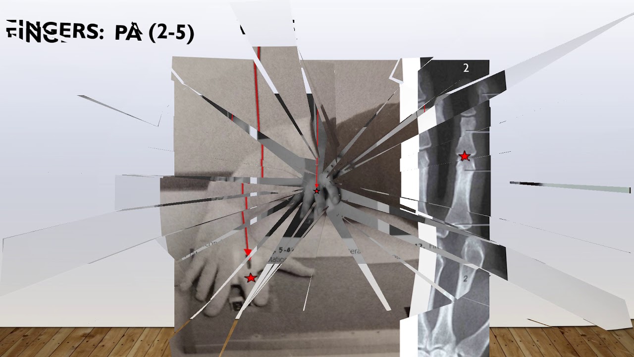
- 😀 The coil method involves positioning the central ray to either the radial head or the coronoid process depending on the desired view.
- 😀 When focusing on the radial head, the central ray is directed towards the shoulder, helping to identify fractures and dislocations.
- 😀 When focusing on the coronoid process, the central ray is directed away from the shoulder, also to look for fractures or dislocations.
- 😀 It’s essential to shield the patient during the procedure to ensure safety, especially for trauma patients.
- 😀 The coil method is typically used on trauma patients who may have injuries to the elbow region.
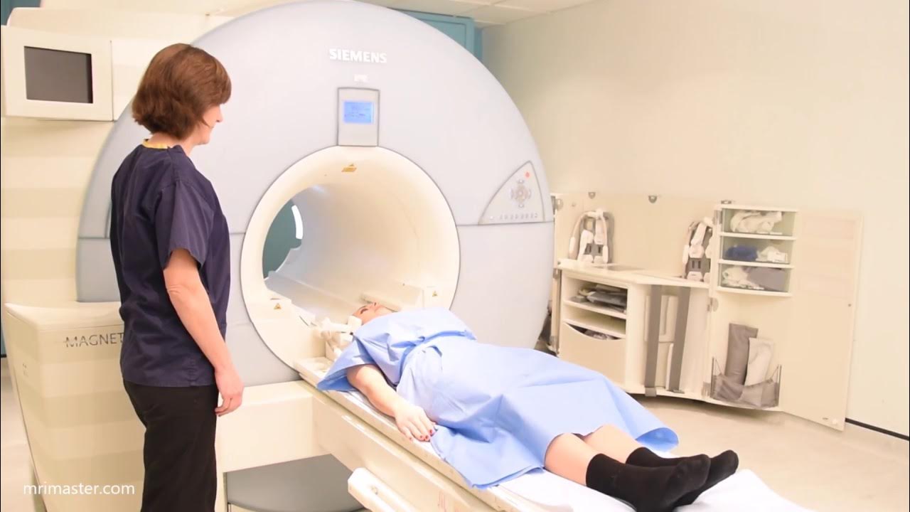
- 😀 The patient’s elbow should be properly positioned on the imaging table to align the arm in the same plane as the radiographic beam.
- 😀 The imaging procedure involves raising the table to ensure proper alignment for a lateral elbow view.
- 😀 Markers should be placed on the image to indicate the correct side of the body being imaged.
- 😀 Elongation of the radial head is achieved through the central ray's positioning to help visualize potential fractures and dislocations.
- 😀 For viewing the coronoid process, the central ray is angled 45 degrees to elongate the structure and assess for injuries.
Q & A
What is the primary focus of the coil method in this procedure?
-The coil method focuses on examining the radial head and the coronoid process of the elbow by adjusting the central ray towards or away from the shoulder.
Why is it important to shield the patient during this procedure?
-It is important to shield the patient to minimize radiation exposure, as there’s no reason not to use protection during elbow imaging.
What is the typical scenario where this procedure is performed?
-This procedure is typically performed on trauma patients to assess for fractures or dislocations in the elbow region.
How does the central ray direction affect the imaging outcome?
-When the central ray is directed towards the shoulder, it focuses on the radial head, and when directed away from the shoulder, it targets the coronoid process.
Why is it necessary to adjust the table height during the procedure?
-The table height is adjusted to ensure the patient's arm is in the correct position, similar to performing a lateral elbow view.
What are the key anatomical structures being assessed in this procedure?
-The key anatomical structures being assessed are the radial head and the coronoid process of the elbow joint.
How is the radial head assessed during the procedure?
-The radial head is assessed by elongating it with the central ray directed towards the shoulder to check for fractures or dislocations.
How is the coronoid process evaluated in this procedure?
-The coronoid process is evaluated by rotating the central ray 45 degrees away from the shoulder to elongate the process and identify potential fractures or dislocations.
What role does the marker play in this imaging process?
-The marker is placed on the image to identify the side of the body being imaged and to ensure the correct area is being assessed.
What are the potential abnormalities being checked for in this procedure?
-The procedure looks for fractures or dislocations in the radial head and coronoid process of the elbow joint.
Outlines

Этот раздел доступен только подписчикам платных тарифов. Пожалуйста, перейдите на платный тариф для доступа.
Перейти на платный тарифMindmap

Этот раздел доступен только подписчикам платных тарифов. Пожалуйста, перейдите на платный тариф для доступа.
Перейти на платный тарифKeywords

Этот раздел доступен только подписчикам платных тарифов. Пожалуйста, перейдите на платный тариф для доступа.
Перейти на платный тарифHighlights

Этот раздел доступен только подписчикам платных тарифов. Пожалуйста, перейдите на платный тариф для доступа.
Перейти на платный тарифTranscripts

Этот раздел доступен только подписчикам платных тарифов. Пожалуйста, перейдите на платный тариф для доступа.
Перейти на платный тарифПосмотреть больше похожих видео

Elbow Joint: Bones, Muscles & Movement - Human Anatomy | Kenhub

How To Grow The BIGGEST Tricep
Radiographic Upper Extremity Positioning
Brain MRI scan protocols, positioning and planning

Galvanometer | moving coil galvanometer 12th class explanation construction and working animation HD
This Simple Technique Improves Your Lightroom Skills by 99%
5.0 / 5 (0 votes)
