Muscles of Respiration | Breathing Mechanics | Respiratory Physiology
Summary
TLDRThis video from Bite Size Med explores the muscles involved in respiration and the mechanics of the chest wall during breathing. It explains how inspiration and expiration are facilitated by the diaphragm, ribs, and accessory muscles. The diaphragm is the primary muscle for inspiration, flattening to increase thoracic volume, while expiration is generally passive due to lung elasticity. The video also highlights the role of external intercostal muscles and the importance of the phrenic nerve in diaphragm function. It concludes with a mnemonic to help remember the respiratory muscles and their actions.
Takeaways
- 🌀 The breathing cycle consists of inspiration (inhaling air into the lungs) and expiration (exhaling air out of the lungs).
- 🏗️ Changes in the volume of the thoracic cavity during respiration are achieved by altering its dimensions vertically, anteroposteriorly, and transversely.
- 📐 The ribs are hinged at the back and angle downwards to attach to the sternum, allowing for changes in the thoracic cavity's diameters.
- 💪 The diaphragm is the primary muscle of respiration, contracting to flatten and increase the vertical dimension of the thoracic cavity during inspiration.
- 🔄 During quiet breathing, expiration is a passive process due to the lungs' elastic properties and does not require muscle contraction.
- 📉 The external intercostal muscles are involved in inspiration, elevating the ribs and moving the sternum forward to increase the thoracic cavity's volume.
- 🔄 Accessory muscles, such as the sternocleidomastoid, serratus anterior, and scalene muscles, assist in respiration when extra effort is needed.
- 📚 The diaphragm is innervated by the phrenic nerve, which originates from C3, C4, and C5, and damage to this nerve can lead to diaphragmatic palsy.
- 📉 The internal intercostal muscles and abdominal wall muscles are involved in expiration, pulling the ribs down and reducing the thoracic cavity's volume.
- 🔄 Quiet expiration is generally a passive process due to the lungs' elastic recoil, but active muscle involvement indicates the need for extra effort.
- 👍 Remembering the muscles of respiration can be aided by associating inspiration with 'E' for external intercostals and expiration with 'I' for internal intercostals.
Q & A
What is the primary function of the diaphragm in the respiratory process?
-The diaphragm is the primary muscle of respiration responsible for most of the work during inspiration. When it contracts, it flattens, increasing the vertical dimension of the thoracic cavity, allowing the lungs to expand and draw in air.
What are the two phases of the breathing cycle?
-The two phases of the breathing cycle are inspiration, which is the process of taking air into the lungs, and expiration, which is the process of breathing air out.
How do the ribs contribute to the changes in the thoracic cavity's dimensions during respiration?
-The ribs, which are hinged at the back and angle downwards, can move to change the diameters of the thoracic cavity from front to back and sideways. The upper ribs move with a pump handle movement, increasing the anteroposterior diameter, while the lower ribs move with a bucket handle movement, increasing the transverse diameter.
What is the difference between active and passive expiration?
-Active expiration requires the use of muscles to force air out of the lungs, often during conditions like respiratory distress. Passive expiration, on the other hand, is the result of the lungs' elastic properties causing them to recoil, and it does not require muscle action during quiet breathing.
Which muscles are considered accessory muscles of respiration and when do they come into play?
-Accessory muscles of respiration, such as the sternocleidomastoid, serratus anterior, and scalene muscles, come into play during situations that require extra effort, like in respiratory distress, when the primary muscles are not sufficient.
How do the external intercostal muscles assist in inspiration?
-The external intercostal muscles are attached to the ribs and are directed forwards and downwards. When they contract, they elevate the ribs, contributing to the increase in the thoracic cavity's volume during inspiration.
What is the role of the internal intercostal muscles during expiration?
-The internal intercostal muscles are attached to the ribs with fibers that run downwards and backwards. They contract during expiration to depress the ribs, reducing the thoracic volume and aiding in the passive expulsion of air from the lungs.
What muscles are involved in the abdominal wall that assist in forced expiration?
-The muscles of the abdominal wall that assist in forced expiration include the rectus abdominis, external and internal obliques, and the transversus abdominis. These muscles pull the ribs down and increase intra-abdominal pressure, which helps force air out of the lungs.
What nerve supplies the diaphragm and what condition can occur if it is damaged?
-The diaphragm is supplied by the phrenic nerve, which originates from the cervical spinal nerves C3, C4, and C5. Damage to this nerve can result in diaphragmatic palsy, a condition that makes breathing difficult.
How does the movement of the ribs and sternum during inspiration and expiration relate to the anteroposterior and transverse diameters of the thoracic cavity?
-During inspiration, the ribs move up and the sternum moves forward, increasing the anteroposterior diameter. The lower ribs' bucket handle movement increases the transverse diameter. During expiration, the ribs move down and the sternum falls back, reducing both the anteroposterior and transverse diameters.
What is the mnemonic provided in the script to help remember the muscles involved in inspiration and expiration?
-The mnemonic provided in the script is 'E for I and I for E' to help remember that the external intercostal muscles are involved in inspiration and the internal intercostal muscles are involved in expiration.
Outlines

Этот раздел доступен только подписчикам платных тарифов. Пожалуйста, перейдите на платный тариф для доступа.
Перейти на платный тарифMindmap

Этот раздел доступен только подписчикам платных тарифов. Пожалуйста, перейдите на платный тариф для доступа.
Перейти на платный тарифKeywords

Этот раздел доступен только подписчикам платных тарифов. Пожалуйста, перейдите на платный тариф для доступа.
Перейти на платный тарифHighlights

Этот раздел доступен только подписчикам платных тарифов. Пожалуйста, перейдите на платный тариф для доступа.
Перейти на платный тарифTranscripts

Этот раздел доступен только подписчикам платных тарифов. Пожалуйста, перейдите на платный тариф для доступа.
Перейти на платный тарифПосмотреть больше похожих видео

Surfactant and Surface Tension in Respiration | Breathing Mechanics | Respiratory Physiology
MEKANISME PERNAPASAN DADA DAN PERUT | SISTEM PERNAPASAN MANUSIA
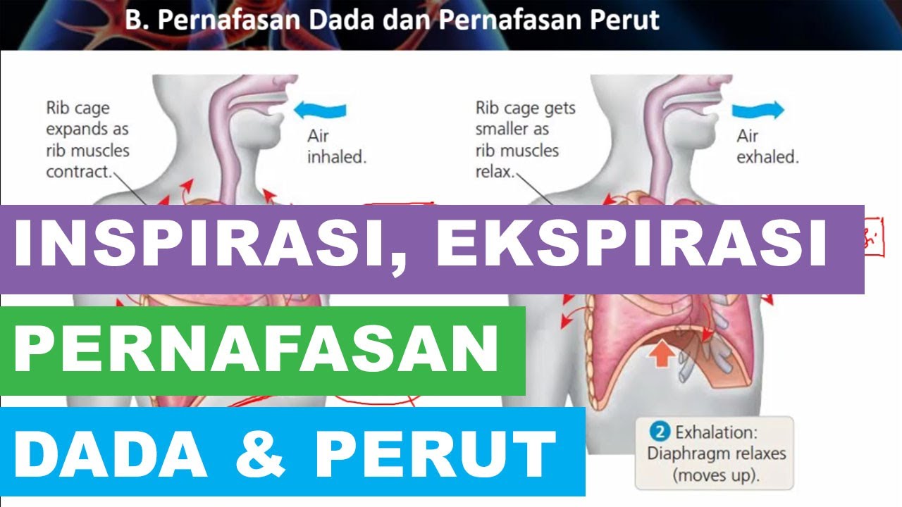
Mekanisme Inspirasi dan Ekspirasi Pernafasan Dada dan Pernafasan Perut

MEKANISME PERNAPASAN MANUSIA

Respiratory Response To Exercise | Respiratory Physiology

BIOLOGI SMA Kelas 11 - Sistem Pernapasan | GIA Academy
5.0 / 5 (0 votes)
