PET scan | How Does a PET Scan Work? | Clinical application of PET scan | #biomedicine series
Summary
TLDRThis video explains the mechanism and significance of a PET scan (Positron Emission Tomography), a technique used to reveal the metabolic function of tissues and organs. The video covers how PET scans detect cancer and other diseases by measuring metabolic activity through the use of 18-FDG, a radioactive glucose analog that emits positrons. The process involves annihilation reactions, gamma ray emissions, and coincidence imaging. The video also discusses the integration of PET with CT scans for more precise anatomical localization, enhancing cancer detection and treatment planning.
Takeaways
- 🧪 Positron Emission Tomography (PET) scan reveals the metabolic or biochemical function of tissues and organs.
- 🔬 PET scans use positrons, the antimatter counterpart of electrons, to create images based on metabolic activity.
- 🏥 PET scans are functional imaging techniques, whereas MRI and CT scans provide structural imaging.
- 🛠 The PET scan machine is located in a radiation-proof room, and the process involves scanning the patient while they lie down inside a tunnel.
- 💡 PET scans use 18-FDG, a glucose analog that emits positrons, to detect cells with high metabolic activity.
- 🌟 Cells with higher metabolic activity emit more gamma rays, which creates a 'hot spot' on the PET scan image.
- 🌑 Cells with lower metabolic activity emit fewer gamma rays, resulting in a 'cold spot' on the image.
- 🎯 PET scans are commonly used to detect cancer, as cancer cells have high metabolic activity and show up as hot spots.
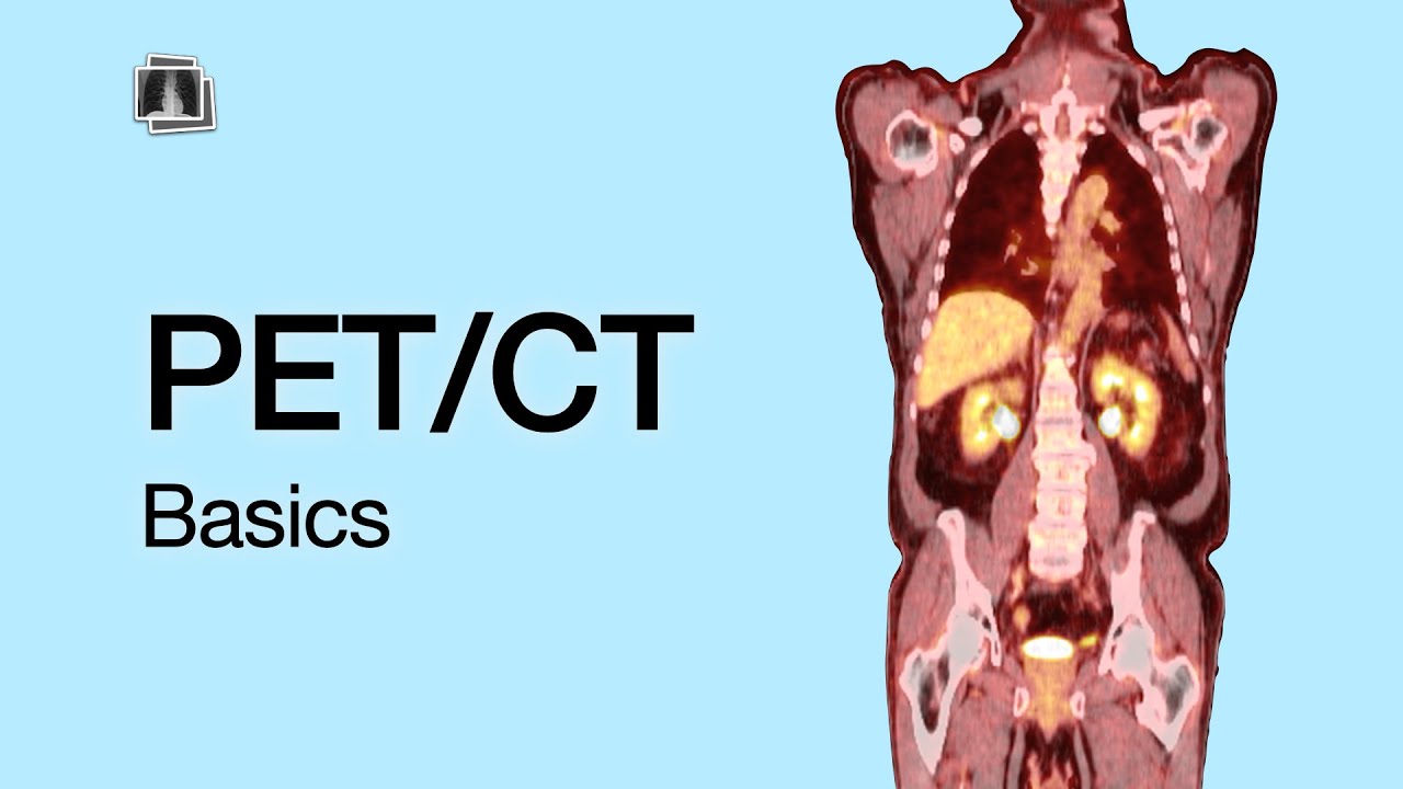
- 🖼 PET scans can be combined with CT or MRI scans (PET-CT) for higher resolution images, providing both metabolic and structural data.
- 📊 The combination of PET and CT images helps accurately localize lesions or tumors, improving diagnosis and treatment planning.
Q & A
What is a PET scan and how does it differ from other imaging techniques like MRI or CT scans?
-A PET (Positron Emission Tomography) scan is a functional imaging technique that reveals the metabolic or biochemical function of tissues and organs. It differs from MRI and CT scans, which are structural imaging techniques that focus on anatomical organization. PET scans show the level of functioning of tissues, while MRI and CT scans show their structure.
What is the primary principle behind the PET scan technique?
-The primary principle behind PET scans is the use of positrons, which are the antimatter counterpart of electrons. The PET scan detects gamma rays emitted during positron-electron annihilation reactions in the body, which helps visualize the metabolic activity of tissues.
How is 18-FDG used in a PET scan?
-18-FDG (Fluorodeoxyglucose) is a glucose analog injected into the blood during a PET scan. It competes with glucose for uptake by cells. Once inside the cells, it emits positrons, which, upon interacting with electrons, create gamma rays that the scanner detects to visualize metabolic activity.
What biological process does 18-FDG exploit for PET scans?
-18-FDG exploits the glucose uptake process. Cells with higher metabolic activity, such as cancer cells, take up more glucose and 18-FDG. This increased uptake leads to more positron emissions and gamma ray detection, helping highlight metabolically active areas in the scan.
What is the purpose of the 'coincidence imaging' technique in PET scans?
-Coincidence imaging refers to detecting two gamma rays emitted in opposite directions from a positron-electron annihilation. The scanner uses this to precisely locate the source of metabolic activity, which helps in mapping active areas in the body.
How can a PET scan help detect cancer?
-PET scans help detect cancer by identifying 'hot spots'—areas with high metabolic activity. Cancer cells tend to consume more glucose due to their rapid growth, leading to higher 18-FDG uptake. This increased activity results in more gamma ray emissions, which appear as hot spots in the scan.
What are 'hot spots' and 'cold spots' in a PET scan?
-Hot spots are areas in the PET scan with high gamma ray emission, indicating high metabolic activity, such as in cancerous tissues. Cold spots are areas with low gamma ray emission, reflecting lower metabolic activity.
What limitation does the PET scan have in identifying the precise location of abnormalities?
-While PET scans can detect metabolic activity, they have low resolution for anatomical localization. For example, it may show a hot spot in the thoracic area, but it cannot specify if it's in the lungs, heart, or another organ. This limitation is resolved by combining PET with CT scans for better anatomical accuracy.
What is a PET-CT scan, and how does it improve diagnostic accuracy?
-A PET-CT scan combines the metabolic imaging from a PET scan with the high-resolution anatomical imaging of a CT scan. By superimposing the images, doctors can pinpoint both the location and the metabolic activity of tissues, improving diagnostic accuracy for conditions like cancer.
Why is the PET scan machine placed in a radiation-proof room?
-The PET scan machine is placed in a radiation-proof room because it uses radiation to detect metabolic activity. The room ensures the safety of patients and medical staff by preventing the radiation from escaping and affecting people outside the room.
Outlines

Этот раздел доступен только подписчикам платных тарифов. Пожалуйста, перейдите на платный тариф для доступа.
Перейти на платный тарифMindmap

Этот раздел доступен только подписчикам платных тарифов. Пожалуйста, перейдите на платный тариф для доступа.
Перейти на платный тарифKeywords

Этот раздел доступен только подписчикам платных тарифов. Пожалуйста, перейдите на платный тариф для доступа.
Перейти на платный тарифHighlights

Этот раздел доступен только подписчикам платных тарифов. Пожалуйста, перейдите на платный тариф для доступа.
Перейти на платный тарифTranscripts

Этот раздел доступен только подписчикам платных тарифов. Пожалуйста, перейдите на платный тариф для доступа.
Перейти на платный тариф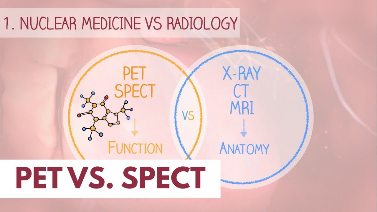


5.0 / 5 (0 votes)
