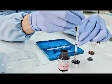
Counting Cells with a Hemocytometer
Summary
TLDRThis video script demonstrates the process of using a hemocytometer to count cells, including the use of Trypan blue to differentiate between living and dead cells. It outlines the steps for preparing the cell suspension, loading the hemocytometer, and counting cells within a grid pattern under a microscope. The script also covers counting rules, handling artifacts, and calculating the percentage of viable cells and the concentration of cells in the culture, providing a clear guide for cell counting and viability assessment.
Takeaways
- 🔬 A hemocytometer is a specialized tool used for counting cells, featuring two identical wells on a microscope slide.
- 🧪 Cells are prepared for counting by diluting a cell suspension with Trypan blue, a dye that differentiates living from dead cells.
- 📏 Each well in the hemocytometer is divided into a grid pattern of 9 large squares, each with a volume of 10^-4 mL.
- 🔍 The counting procedure involves focusing on specific squares: the 4 corner squares and the center square.
- 📐 Cells touching the top and left boundaries of the grid are counted, while those on the right and bottom are ignored.
- 🔵 Dead cells, which have taken up Trypan blue, are identified by their blue color under the microscope.
- 🚫 Artifacts, such as blurry debris, are excluded from the cell count to ensure accuracy.
- 🔄 Proper handling and storage of the hemocytometer are crucial for minimizing artifacts.
- 🧮 The total viable cell count is determined by summing counts from the designated squares and calculating the percentage of viable cells.
- 🧪 The average number of cells per square is calculated by dividing the total viable cell count by the number of squares counted.
- 🔢 The concentration of viable cells is calculated by considering the dilution factor and the average count per square, resulting in cells/mL.
Q & A
What is a hemocytometer used for?
-A hemocytometer is used for counting cells, specifically it is a device that helps in determining the concentration of cells in a suspension.
How is a hemocytometer different from a regular microscope slide?
-A hemocytometer is a modified microscope slide that contains two identical wells or chambers for holding a small volume of cell suspension.
What is the purpose of using Trypan blue when counting cells with a hemocytometer?
-Trypan blue is used to distinguish between living and dead cells. It passes through the membranes of dead cells, staining them blue, while living cells exclude the dye and appear mostly clear.
How should the cell suspension be loaded onto the hemocytometer?
-The cell suspension should be loaded onto both chambers of the hemocytometer by pipetting it under the cover slip.
What is the volume of suspension contained in each square of the grid pattern on the hemocytometer?
-Each square on the hemocytometer contains 10^-4 mL of suspension.
Which squares are counted for cell enumeration in the given lab's procedure?
-In the given lab's procedure, cells are counted in the 4 large corner squares and the center square of the hemocytometer's grid pattern.
How are cells that touch the boundaries of the counting squares handled during counting?
-Cells that touch the top and left boundaries of the counting squares are counted, while cells touching the right and bottom boundaries are ignored.
What is an artifact as mentioned in the script and why is it important to note it?
-An artifact is an object or debris that appears blurry and lacks a well-defined shape, which can interfere with cell counting. It is important to note because artifacts are not included in the cell count to ensure accuracy.
How is the percentage of viable cells calculated from the counted cells?
-The percentage of viable cells is calculated by dividing the number of viable cells by the total number of cells counted, and then multiplying by 100 to get the percentage.
What is the dilution factor and how is it calculated in this context?
-The dilution factor is the ratio of the final volume of the solution to the initial volume of the cells. It is calculated by dividing the final volume by the initial volume of cells.
How is the concentration of viable cells determined from the hemocytometer counts?
-The concentration of viable cells is determined by multiplying the average number of cells per square by the dilution factor and the volume of suspension per square (10^-4 mL).
Outlines
🔬 Hemocytometer Cell Counting Procedure
The paragraph describes the process of using a hemocytometer to count cells. A hemocytometer is a specialized microscope slide with two wells for cell suspension. The cells are prepared by removing 100 µL of suspension and diluting it with 100 µL of Trypan blue, which differentiates living from dead cells by staining only the dead ones blue. The suspension is then loaded under a cover slip, and the hemocytometer is observed under a microscope. The grid pattern on the chamber is used to count cells in specific squares, with rules for counting cells touching boundaries. Artifacts are ignored. The counts of viable and non-viable cells are recorded for each square, and the total counts are used to calculate the percentage of viable cells and the average number of cells per square.
📊 Calculating Cell Concentration and Viability
This paragraph focuses on the calculations following the cell counting process. The dilution factor is determined by dividing the final volume of the cell suspension by the initial volume of cells, resulting in a factor of 2. Using the average count per square and the dilution factor, the concentration of viable cells is calculated to be 216,000 cells/mL, expressed scientifically as 2.16 x 10^5 cells/mL. The paragraph concludes with the final cell culture concentration and the percentage of viable cells, which is approximately 94.7%.
Mindmap
Keywords
💡Hemocytometer
💡Trypan blue
💡Viable cells
💡Non-viable cells
💡Micro-centrifuge tube
💡Cover slip
💡Grid pattern
💡Dilution factor
💡Concentration of viable cells
💡Artifacts
💡Clumps of cells
Highlights
A hemocytometer is used for counting cells and is a modified microscope slide with two identical wells.
100 µL of cell suspension is removed and placed in a micro-centrifuge tube.
Trypan blue is added to the cell suspension to distinguish between living and dead cells.
The hemocytometer's chambers are loaded by pipetting the suspension under the cover slip.
Each chamber is divided into a grid pattern of 9 large squares, each with a volume of 10^-4 mL.
Counting cells involves focusing on the 4 large corner squares and the center square.
Cells touching the top and left boundaries are counted, while those touching the right and bottom are ignored.
Dead cells appear blue under the microscope, while living cells are mostly clear.
Artifacts, such as blurry objects without a well-defined shape, are not included in the cell count.
Proper storage, cleaning, and handling of the hemocytometer minimize artifacts.
The total number of viable cells from all 5 squares is 54, and the total number of non-viable cells is 3.
The percentage of viable cells is calculated as 94.7%.
The average number of cells-per-square is determined to be 10.8 cells.
The dilution factor is calculated as 2, based on the final volume of 200 µL.
The concentration of viable cells is calculated to be 216,000 cells/mL.
The concentration of cells in the culture is expressed in scientific notation as 2.16 x 10^5 cells/mL.
The method of counting clumps of cells varies by lab and should be followed as per local procedure.
Transcripts
A hemocytometer is a device that is used for counting cells.
It's a modified microscope slide, containing two identical wells, or chambers, into which
a small volume of a cell suspension is pipetted.
We have already removed 100 µL of our cell suspension and placed it in a micro-centrifuge tube.
Dilute the suspension by adding 100 µL of Trypan blue. Trypan blue is a dye that helps
us distinguish between living and dead cells. The dye passes through the membranes of dead
cells so they will appear blue under a microscope.
Living cells exclude and will appear mostly clear.
Load both chambers by pipetting the suspension under the cover slip.
Now place the hemocytometer under the microscope.
Each chamber is divided into a grid pattern, consisting of 9 large squares.
Each square has the same dimensions and contains 10 to the negative-fourth power mL of suspension.
The rules for counting cells sometimes differ from lab-to-lab.
In our lab, we count cells in the 4 large corner squares and the center square.
Let's count the cells in the first square.
One, Two, Three,
Four, Five, Six,
Seven, Eight...
So what about the cells that are touching the outside boundaries of the square?
In our lab we count the cells that touch the top and left boundaries, and we ignore the
cells that touch the right and bottom boundaries.
Nine. Ten.
We need to count the number of both living and dead cells. Remember, the dead cells will
appear blue.
Occasionally you will see artifacts - objects or debris that appear blurry and don't have
a well-defined shape.
This is an example of an artifact. We won't include it in our count.
Proper storage, cleaning, and handling of the hemocytometer will minimize the number
of artifacts.
There are 10 viable cells and 1 non-viable cell in the first square.
Now, the top-right square.
There are 9 viable cells and no non-viable cells.
Next let's count the bottom-right square.
There are 11 viable cells and no non-viable cells.
And now the bottom-left square.
There are 10 viable cells and 2 non-viable cells.
And finally, let's count the cells in the center square.
Sometimes cells will appear as clumps or small groups.
It may be difficult to determine exactly how many cells are in a group.
The method of counting clumps of cells differs from lab to lab, so be sure to follow the
procedure in your lab.
We will count this clump as 2 cells.
There are 14 viable cells and no non-viable cells in the center square.
The total number of viable, or living cells from all 5 squares is 54.
The total number of non-viable cells is 3.
Now that we have counted our cells, there are several calculations we need to perform.
First, let's calculate the percentage of viable cells.
Here's the formula. 54 viable cells, divided by 57Éthe total number of cellsâ
gives us 0.947.
Multiply by 100 and the percentage of viable cells is 94.7%.
Next, let's determine the average number of cells-per-square.
We counted 54 viable cells. We divide 54 by 5, because we counted in 5 squares.
The average number of cells-per-square is 10.8 cells.
Now let's calculate the dilution factor.
The dilution equals the final volume divided by the volume of cells.
Our final volume is 200 µL, because we started with 100 µL of cells
and added another 100 µL of trypan blue.
200 divided by 100 is 2. Therefore the dilution factor is 2.
Next we need to calculate the concentration of viable cells - the number of living cells/mL
Our average count-per-square is 10.8.
The dilution factor is 2.
10.8 times 2 times 10-to-the-fourth-power equals 216,000 cells/mL.
We can write the concentration using scientific notation as 2.16 times 10-to-the-fifth power cells/mL.
From our calculations, we now know the concentration of cells in our culture is 216,000 cells-per-milliliter
and approximately 94.7 percent of those cells are viable, living cells.
Посмотреть больше похожих видео
Cell culture techniques 4 - Cryopreservation
Onion Peel Under the Microscope | How to Prepare Stained Temporary Mount of Onion Peel
Microscopy - How to use a microscope - GCSE Science Required Practical
Cell Culture: Cell Culture Basics
Onion Skin Epidermal Cells: How to Prepare a Wet Mount Microscope Slide
Chromosome Numbers During Division: Demystified!
5.0 / 5 (0 votes)