Sirkulasi Darah Fetus aka Janin

Catatan En
6 Oct 202011:54

Summary

TLDRThis video script delves into the fetal circulation system, highlighting the differences between adult and fetal circulation. It explains how a fetus receives oxygen and nutrients through the placenta, as its lungs are not yet functional. The script outlines the anatomical differences in the heart, with the presence of shunts like the ductus venosus and foramen ovale, which facilitate circulation. After birth, these shunts close as the baby's lungs begin to function, and the heart transitions to adult circulation. The video aims to educate viewers on the fascinating changes in the circulatory system from fetal life to infancy.

Takeaways

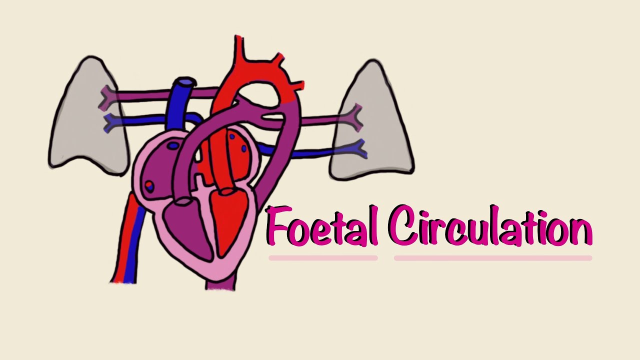
  • 😀 The fetal circulation system differs significantly from that of an adult, as the fetus obtains oxygen and nutrients from the mother through the placenta rather than directly from the environment.
  • 🌟 The fetus receives oxygenated and nutrient-rich blood from the mother via the umbilical vein, which then bypasses the liver and mixes with deoxygenated blood in the heart.
  • 💡 The ductus venosus shunts blood from the umbilical vein directly to the inferior vena cava, bypassing the liver and ensuring the heart receives well-oxygenated blood.
  • 🔍 The foramen ovale, a fetal heart structure, allows blood to flow from the right atrium to the left atrium, ensuring the circulation of oxygenated blood to the body.
  • 🚫 The lungs of the fetus are non-functional in utero, leading to the presence of the ductus arteriosus, which shunts blood away from the lungs and into the systemic circulation.
  • 🌱 After birth, the umbilical cord is clamped and cut, triggering a series of physiological changes that lead to the closure of fetal circulatory pathways.
  • 📉 The increase in oxygen levels post-birth and the decrease in prostaglandins cause the ductus arteriosus to constrict and eventually close.
  • 🛑 The lungs begin to function after birth, increasing blood oxygenation and leading to the closure of the foramen ovale as the left atrial pressure rises.
  • ⏳ The closure of fetal circulatory structures such as the ductus venosus and foramen ovale typically occurs within hours to days after birth, but the exact timing can vary.
  • 🔎 The exact time frame for the complete disappearance of fetal circulatory structures is not well-defined and can range from days to weeks, emphasizing the complexity of the transition from fetal to neonatal circulation.

Q & A

  • What is the main difference between adult circulation and fetal circulation?

    -The main difference is that adults receive oxygen directly from the outside environment through their lungs, while fetuses receive oxygen and nutrients through the placenta from their mother.

  • How does a fetus obtain oxygen and nutrients?

    -A fetus obtains oxygen and nutrients through the placenta from the mother's blood supply.

  • What are the two main blood vessels that connect the fetus to the placenta?

    -The two main blood vessels are the umbilical vein, which carries oxygen-rich blood and nutrients to the fetus, and the umbilical arteries, which carry deoxygenated blood and waste away from the fetus.

  • What is the function of the ductus venosus in fetal circulation?

    -The ductus venosus allows oxygen-rich blood from the umbilical vein to bypass the liver and directly enter the inferior vena cava, which leads to the heart.

  • Why does the foramen ovale open in a fetus?

    -The foramen ovale opens to allow blood to bypass the non-functioning lungs by directing it from the right atrium to the left atrium, ensuring oxygenated blood is circulated to the rest of the body.

  • What is the role of the ductus arteriosus in fetal circulation?

    -The ductus arteriosus allows blood to bypass the lungs by shunting it from the pulmonary artery to the aorta, ensuring that blood is oxygenated through the placenta.

  • How does the circulation change after a baby is born?

    -After birth, the baby's lungs begin to function, and the ductus arteriosus and foramen ovale close, redirecting blood flow to the lungs for oxygenation.

  • What causes the closure of the ductus arteriosus and foramen ovale after birth?

    -The closure is triggered by the increase in oxygen levels in the blood as the baby starts breathing, and the decrease in prostaglandins which were maintaining the patency of these structures in utero.

  • What happens to the umbilical vessels after the baby is born?

    -After the baby is born, the umbilical cord is clamped and cut, and the umbilical vessels undergo vasoconstriction and eventually close off.

  • How long does it take for the fetal blood vessels to close after birth?

    -The timing can vary, but the ductus arteriosus typically constricts within 1-2 hours after birth. The foramen ovale and ductus venosus may take days to weeks to close completely.

  • What is the significance of the changes in fetal circulation after birth?

    -The changes in fetal circulation after birth are significant as they transition the baby from a placental-based oxygen supply to a lung-based respiratory system, which is essential for the baby's survival outside the womb.

Outlines

00:00

🌟 Fetal Circulation and Heart Anatomy

This paragraph discusses the differences in blood circulation between adults and fetuses. In adults, oxygen is directly obtained from the outside environment through the lungs, while fetuses receive oxygen and nutrients through the placenta from the mother. The speaker briefly touches on the anatomy of an adult human, detailing the path of oxygenated blood through the heart's chambers and the pulmonary and systemic circuits. In contrast, fetal circulation involves a shunt or bypass that allows blood to mix between the mother and the fetus. The fetus has a different heart anatomy with structures like the ductus arteriosus and foramen ovale that facilitate this unique circulation pattern. After birth, as the baby's lungs begin to function, the heart transitions to an adult-like circulation.

05:01

🔍 Detailed Explanation of Fetal Blood Circulation

The second paragraph delves deeper into the specifics of fetal blood circulation. It starts with the exchange of blood in the placenta, where the fetus receives oxygen-rich blood and nutrients. The paragraph describes the umbilical arteries and veins, which carry deoxygenated blood from the fetus to the placenta and oxygenated blood back to the fetus, respectively. The ductus venosus is highlighted as a shunt that allows blood to bypass the liver and mix with blood returning from the placenta. The foramen ovale is another critical structure that allows blood to bypass the lungs by directing it from the right atrium to the left atrium. The ductus arteriosus is mentioned as a final shunt that channels blood away from the non-functional lungs to the rest of the body. After birth, these structures close as the baby's lungs begin to function, and the blood circulation pattern shifts to that of an adult.

10:04

🛑 Changes in Circulation Post-Birth

The final paragraph addresses the changes in blood circulation that occur after birth. It explains how the umbilical cord is cut, leading to resistance in the placental blood vessels, which in turn triggers vasoconstriction and the eventual closure of the fetal circulation pathways. The paragraph also discusses the role of oxygen levels in promoting the closure of the ductus arteriosus and the reduction of prostaglandins, which are responsible for keeping the ductus open during fetal life. The closure of the foramen ovale and ductus venosus is also mentioned, with the timing of these closures being variable and dependent on the baby's development and lung function. The paragraph concludes by emphasizing the complexity of the fetal circulation system and the intricacies of its transition to an adult circulation pattern after birth.

Mindmap

Keywords

💡Circulation

Circulation in the context of the video refers to the process of blood flow through the body. It is central to the video's theme as it discusses the differences between fetal and adult circulation. In the script, it is mentioned that an adult's blood circulation is direct from the lungs to the heart and then to the rest of the body, while a fetus receives oxygenated blood and nutrients through the placenta, highlighting the unique circulation patterns within the womb.

💡Placenta

The placenta is a vital organ in the video's discussion of fetal development. It serves as the interface between the mother and the fetus, facilitating the exchange of oxygen, nutrients, and waste products. The script explains that the fetus receives oxygen and nutrients through the placenta, which is essential for its growth and development within the womb.

💡Ductus Venosus

Ductus venosus is a shunt in the fetal circulatory system that allows blood rich in oxygen and nutrients to bypass the liver and travel directly to the heart. The video script describes how this shunt plays a crucial role in fetal circulation, ensuring that the fetus receives the necessary oxygen and nutrients for its development.

💡Foramen Ovale

Foramen ovale is a hole in the heart of a fetus that allows blood to bypass the lungs by directing it from the right atrium to the left atrium. The video script explains that this is essential for fetal circulation since the lungs are not yet used for respiration. After birth, the foramen ovale typically closes as the lungs begin to function, and the circulatory system adjusts to the postnatal state.

💡Ductus Arteriosus

Ductus arteriosus is a blood vessel that connects the pulmonary artery to the aorta in a fetus, allowing blood to bypass the non-functioning lungs. The video script mentions that after birth, the ductus arteriosus closes as the lungs begin to function, and the circulatory system adapts to the new oxygen-rich environment.

💡Umbilical Arteries and Veins

Umbilical arteries and veins are the blood vessels that connect the fetus to the placenta. The video script describes how the umbilical arteries carry deoxygenated blood from the fetus to the placenta, where it picks up oxygen and nutrients. The oxygenated blood then returns to the fetus through the umbilical vein, which is a critical part of the fetal circulation system.

💡Vasconstriction

Vasconstriction refers to the narrowing of blood vessels, which is a physiological response mentioned in the video script. After birth, the placenta is detached, and the umbilical vessels undergo vasconstriction, leading to their closure. This process is part of the transition from fetal to neonatal circulation, as the baby's body adapts to breathing and oxygenation through the lungs.

💡Prostaglandins

Prostaglandins are hormone-like substances that play a role in maintaining the patency of the ductus arteriosus during fetal life. The video script explains that after birth, a decrease in prostaglandin levels contributes to the closure of the ductus arteriosus, as the baby's circulatory system shifts to use the lungs for oxygenation.

💡Fetal Circulation

Fetal circulation refers to the unique blood flow patterns within a developing fetus. The video script details how fetal circulation differs from adult circulation, with special pathways like the ductus venosus, foramen ovale, and ductus arteriosus facilitating the exchange of oxygen and nutrients. This concept is central to understanding the developmental changes that occur as a baby transitions from the womb to the external environment.

💡Transitional Circulation

Transitional circulation is the process by which a newborn's circulatory system adapts from fetal to neonatal patterns. The video script discusses how structures like the ductus venosus, foramen ovale, and ductus arteriosus close after birth, as the baby's lungs begin to function, and the heart assumes its adult-like circulation pattern.

Highlights

Circulation in fetuses differs from adults due to the reliance on the mother for oxygen and nutrients.

Adults receive oxygen directly from the outside air through fully functioning lungs.

Fetuses receive oxygen and nutrients through the placenta from the mother.

The fetus's heart has a different anatomy, including a shunt or bypass, allowing circulation between the mother and the fetus.

The umbilical arteries and veins play a crucial role in fetal circulation.

The umbilical vein carries oxygen-rich blood from the placenta to the fetus.

The umbilical arteries carry deoxygenated blood away from the fetus to the placenta.

The ductus venosus allows oxygen-rich blood to bypass the liver and enter the heart.

The foramen ovale is a channel that allows blood to flow from the right atrium to the left atrium.

The ductus arteriosus is a blood vessel that connects the pulmonary artery to the aorta, bypassing the lungs.

After birth, the umbilical cord is cut, leading to changes in fetal circulation vessels.

The placenta's resistance increases after birth, causing vasconstriction in fetal circulation vessels.

The increase in oxygen levels after birth supports vasconstriction of fetal circulation vessels.

The ductus arteriosus typically constricts within 1-2 hours after birth due to increased oxygen levels.

The foramen ovale closes shortly after birth as the lungs begin to function properly.

The closure of fetal circulation vessels is a complex process that may take days or weeks.

The exact timing of the complete disappearance of fetal circulation vessels is not fully understood.

Transcripts

play00:00

Hai di sini aku akan membahas mengenai

play00:02

Sistem peredaran darah fetus disini

play00:05

kalau kita lihat secara garis besar

play00:07

terdapat perbedaan antara sirkulasi dari

play00:12

orang dewasa dan bayi kalau misalnya

play00:14

dari orang dewasa dia mendapatkan

play00:16

oksigen itu langsung dari luar tubuh

play00:19

karena paru-parunya itu tidak bisa

play00:21

berfungsi secara sempurna Aya bisa

play00:23

memperoleh oksigen dari hidung sementara

play00:25

by disini bayi dalam rahim ibu jadi dia

play00:29

menerima oksigen dan zat-zat nutrisi

play00:31

yang diperlukan lainnya itu itu melalui

play00:34

ibu belum bisa belum bisa secara

play00:36

independen dan itu melalui plasenta

play00:40

plasenta ke baik nah oke langsung saja

play00:46

saya disini sedeku akan bahas sekilas

play00:50

mengenai anatomi dari manusia dari orang

play00:55

yang terang dewasa

play00:57

Hai nah disini darah normalnya itu yang

play01:01

terdoktrin nasi akan masuk melalui

play01:03

penangkapan Superior dan inferior

play01:06

kemudian Devina ventrikel kanan terus ke

play01:10

penting kekiri melalui katup trikuspid

play01:12

terus akan pergi ke paruh melalui katup

play01:15

pulmonal dan Arteri pulmonal terus ke

play01:18

paru untuk di oksigen nasib PTT sudah

play01:21

dioksidasi maka akan kembali ke jantung

play01:24

ke atrium kiri melalui Vena pulmonal

play01:26

trus ke ventrikel kiri terus dialirkan

play01:31

keseluruh tubuh melalui katup aorta

play01:33

terus kuota dan ke Arteri Arteri kecil

play01:36

lainnya nah ini berbeda dengan apa yang

play01:40

ada pada bayi karena Anatomi jantung

play01:45

pada bayi atau pada fetus itu itu

play01:48

terdapat Sunshine atau penyeberangan

play01:50

atau Bypass yang dapat memungkinkan

play01:54

terjadinya sirkulasi antara ibu

play01:57

bye jadi terciptalah Sansan ini yang

play02:00

akan kita bahas selanjutnya dan juga ada

play02:03

beberapa Pembuluh darah yang

play02:04

memungkinkan terjadinya sirkulasi ini

play02:09

jadi terdapat perbedaan antara Anatomi

play02:11

jantung bagi orang dewasa dan juga baik

play02:15

tapi jika bayi tersebut sudah keluar the

play02:20

keluar dari rahim ibu dan sudah

play02:22

berkembang dan sudah menggunakan

play02:24

paru-paru maka jantungnya itu akan

play02:28

berfungsi seperti orang dewasa

play02:31

Hai Oke selanjutnya Mari kita bahas

play02:33

peredaran darah fetus peredaran darah

play02:36

bayi ketika masih dalam rahim oke di

play02:41

sini kita mulai dari pertukaran darah di

play02:46

plasenta sebelum mana yang aku jelaskan

play02:49

sebelumnya bahwa plasenta ini berfungsi

play02:50

sangat penting Dimana tempat bayi atau

play02:54

fetus mendapatkan darah yang kaya akan

play02:57

oksigen dan juga nutrisi jadi plasenta

play03:00

ini terdapat bagiannya ibu atau

play03:06

uteroplasenta yaitu bagian ini dan

play03:09

bagian bayi atau fetoplasenta ini nah

play03:17

nah bagiannya Ibu ini ini akan muncul

play03:21

Arteri arteri dan vena yang berfungsi

play03:25

sebagaimana dengan fungsinya kalau

play03:26

Arteri dialog berfungsi untuk memberikan

play03:29

darah yang kaya akan oksigen

play03:31

nutrisi kalau Vena dia fungsi untuk

play03:34

menangkap cairan-cairan yang sudah tidak

play03:37

dipakai lagi dalam tubuh baik lalu akan

play03:40

ditangkap oleh Venny Bu nah terus kita

play03:42

bisa lihat disini Di sini ada Arteri dan

play03:47

juga Vena dari bayi di sini Mereka

play03:51

berfungsi untuk menangkap O2 nutrisi

play03:54

yang diperlukan terus akan dihantarkan

play03:56

ke bayi dan Selain itu ada-ada Arteri

play04:01

umbilikalis dimana Arteri umbilikalis

play04:03

ini dia berfungsi membuang zat-zat yang

play04:07

tidak diperlukan bayi lagi diperas entah

play04:13

dan ditangkap oleh Vena Ibu Nah dari

play04:16

sini kita

play04:18

Ayo kita bisa lihat ini akan menyatu

play04:20

menjadi tali pusatnya keriput saat ini

play04:22

dia kaya seperti jalinan antara Vena dan

play04:27

Arteri umbilikalis arteri umbilikalis

play04:30

dan vena umbilicalis terbesarnya disini

play04:32

jadi Vena di sini ada satu Vena terus

play04:35

ada dua Arteri umbilikalis nah Venom

play04:40

bilik alis ini karena yang mengandung

play04:41

darah yang kaya akan oksigen dan nutrisi

play04:43

sementara Arteri umbilikalis dia

play04:46

mengandung darah yang terdeoksigenasi

play04:49

Nah selanjutnya Vena ini Vena yang kaya

play04:54

akan oksigen dia akan masuk di akan

play04:58

melewati

play05:00

di Indonesia nah tidur disini masih

play05:03

belum berfungsi tapi oksigen juga dan

play05:07

nutrisi gue baru melewati hati untuk

play05:09

pertumbuhan hati dan selanjutnya akan

play05:12

terjadi Bypass atau penyeberangan

play05:15

melalui pembuluh duktus venosus di sini

play05:19

naik ini Khan yang pertama yaitu duktus

play05:23

venosus jadi darah akan langsung design

play05:26

karena langsung dialihkan ke dokter

play05:28

spinosus akan masuk ke atrium kanan

play05:31

terus darah ini akan bercampur dengan

play05:34

darah darah yang terdoktrin Asy baik itu

play05:38

dari Vena cava Superior dan vena cava

play05:40

inferior jadi akan penuh dengan darah

play05:45

yang kaya akan

play05:47

CO2 dan co 2 termix berhubung paru-paru

play05:52

belum bekerja dengan sempurna reformasi

play05:57

tumbuh dan berkembang maka akan terjadi

play05:59

resistensi dan resistensi ini yang

play06:03

membuat darah yang ada di atrium kanan

play06:06

akan tersusun ke atrium kiri jadi dia

play06:09

akan masuk ke atrium kiri melalui

play06:13

foramen ovale nah foramen ovale ini

play06:16

adalah Chan yang

play06:19

Hai nah Khan ini dan darah dari atrium

play06:23

kanan ke atrium kiri juga ini bisa

play06:25

terjadi karena tekanan di Atrium kiri

play06:28

juga ini masih rendah karena kan darah

play06:31

yang kembali dari paru-paru juga masih

play06:33

sedikit paru-paru belum berfungsi jadi

play06:36

tekannya tekanannya itu cenderung dari

play06:39

atrium kanan ke atrium kiri dan di darah

play06:43

akan masuk dari atrium kanan ke atrium

play06:46

kiri terus dari Atrium kiri ke ventrikel

play06:48

kiri terus akan masuk ke aorta terus

play06:52

akan disebarkan keseluruh tubuh nah

play06:55

sebagian darah yang ada di ventrikel

play06:57

kanan ini akan masuk di ventrikel kiri

play06:59

terus akan masuk ke Arteri pulmonalis

play07:04

nah saat masuk ke Arteri pulmonalis kini

play07:07

sebagaimana yang kita ketahui bahwa

play07:09

paru-paru belum bekerja dan terjadi

play07:12

resistensi dari pembuluh-pembuluh darah

play07:14

paruh karena belum

play07:19

tak bisa bekerja jadi darah ini akan

play07:23

masuk ke Doctors arteriosus nah 21

play07:27

racing adalah San yang ketiga San yang

play07:31

terakhir jadi termasuk kesini dan akan

play07:34

disebarkan keseluruh tubuh nah sadar

play07:38

masuk ke seluruh saudara disebarkan

play07:41

keseluruh tubuh ya kan ke bagian-bagian

play07:44

yang memerlukan oksigen dana ada dua

play07:48

pembuluh darah yaitu Arteri umbilikalis

play07:51

dia akan mengembalikan darah-darah yang

play07:55

terdokumentasi di plasenta jadi langsung

play08:00

dikembalikan haji ke plasenta Nah itulah

play08:06

Hai predaran darah saat bayi dan masih

play08:10

dalam rahim nah bagaimana kalau bayi

play08:14

sudah lahir maka akan terjadi

play08:18

perubahan-perubahan bagi Shining dan

play08:21

juga bagi pembuluh-pembuluh darah yang

play08:23

ada nah saat bayi lahir by keluar maka

play08:28

terpusat ini akan dipotong akan diklaim

play08:32

Nah otomatis akan terjadi resistensi

play08:36

disini resistansi plasenta jadi

play08:41

pembuluh-pembuluh ini sudah tahu kalau

play08:43

mereka ini sudah tidak lagi berfungsi

play08:46

dan mereka akan mengalami vasokonstriksi

play08:50

apalagi Dika misalnya bayi sudah

play08:52

bernapas dan oksigen kadar oksigen dalam

play08:56

pemuda itu sudah tinggi maka ini akan

play08:58

semakin mendukung terjadinya

play09:00

vasokontriksi dari pembuluh darah baik

play09:03

itu 20 venosus trus

play09:06

venomrx alis dan juga Arteri umbilikalis

play09:09

maka ini akan terjadi vasokonstriksi dan

play09:11

akan menghilang Nah untuk duktus

play09:14

arteriosus ini menurut salah satu sumber

play09:19

dia akan mengalami vasokonstriksi

play09:22

sekitar 1-2 jam setelah bayi baru lahir

play09:27

nah ini sama seperti alasan yang lain

play09:30

ini karena terjadi peningkatan kadar

play09:32

oksigen dalam pembuluh darah dan juga

play09:35

ini dikarenakan karena pada saat bayi

play09:38

masih dalam rahim itu terjadi produksi

play09:40

prostaglandin prostat ini yang akan

play09:43

mempertahankan Doctors arteriosus

play09:45

membuka dan jika bayi sudah lahir maka

play09:49

terjadi penurunan prostaglandin naprosta

play09:52

brand ini yang membuat Doktor Satrio sus

play09:55

mengalami vasokonstriksi

play09:58

Hai nah terus sama juga dengan korame

play10:04

Noval dia juga penutupannya itu terjadi

play10:08

secara singkat setelah bayi itu lahir

play10:11

nah penutupan ini dikarenakan karena

play10:14

paruh sudah bisa mengembang tidak

play10:16

terjadi pertukaran gas CO2 dan O2 dalam

play10:19

paru otomatis pengembalian Vena

play10:22

pulmonalis ke dalam ventrikel q-free

play10:25

dalam Atrium kiri juga meningkat Jadi

play10:28

ini akan mendorong oramen oval untuk

play10:31

menutup jadi promo Noval akan menutup

play10:34

setelah terjadi pengembangan paru

play10:37

setelah paru-paru tidak bisa berfungsi

play10:39

dengan baik setelah atrium kanan sudah

play10:43

berfungsi dengan baik tetapi sumber

play10:46

untuk pembuluh-pembuluh darah pemilik

play10:49

alvein Arteri pulmonalis dan duktus

play10:52

venosus untuk sumber pasti Kapan mereka

play10:54

hilang

play10:56

Hai itu masih belum jelas ada yang

play10:58

mengatakan eh secara dalam hitungan hari

play11:03

atau dalam hitungan Minggu dan foramen

play11:07

ovale juga ada yang menetapkan setelah

play11:10

bayi lahir dan duktus venosus ada yang

play11:13

mengatakan 15-24 jam Setelah bayi lahir

play11:19

dan secara sempurna itu bisa sampai 3-4

play11:23

Minggu jadi harus Didik di pelatih harus

play11:26

bicara lagi sumbernya Kapan secara jelas

play11:30

pembuluh-pembuluh darah yang dipakai

play11:33

untuk dalam rangka sirkulasi Fetal ini

play11:36

terjaga ini Menghilang itu Kapan mereka

play11:41

menghilang secara sempurna itu kapan ya

play11:43

sangat rumit oke itu dia sistem

play11:47

peredaran darah fetus Aku harap bisa

play11:50

dimengerti dan semoga video ini

play11:52

bermanfaat

Rate This

5.0 / 5 (0 votes)

Связанные теги
Fetal CirculationAdult AnatomyHeart AnatomyBlood FlowPlacenta FunctionFetal DevelopmentPostnatal ChangesMedical EducationHealthcareAnatomy
Вам нужно краткое изложение на английском?