Inside the Cell Membrane
Summary
TLDRThe Amoeba Sisters explore the concept of osmosis through an egg lab, demonstrating how the egg's membrane mimics a cell's semi-permeability. They delve into the importance of surface area to volume ratios in cells, emphasizing the cell membrane's structure, including the phospholipid bilayer, cholesterol, and proteins. Highlighting the role of glycoproteins in cell signaling and disease resistance, the video educates on the fundamental aspects of cell biology.
Takeaways
- 🔬 The script discusses an educational lab involving eggs soaked in vinegar to model osmosis and cell membrane function.
- 🥚 The eggshell is removed through soaking in vinegar, leaving behind the egg white which acts as a semi-permeable membrane similar to a cell membrane.
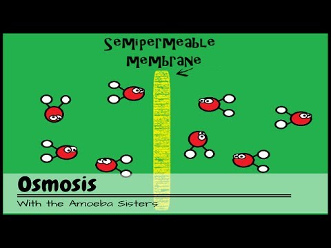
- 🌊 Osmosis is the process where water moves through a semi-permeable membrane, which can be demonstrated using the egg model.
- 📏 The importance of surface area to volume ratio in cells is highlighted, explaining why cells cannot be as large as a chicken egg.
- 📊 A mathematical comparison is made between smaller and larger cube models to illustrate the difference in surface area to volume ratios.
- 🧬 The cell membrane's structure is described as being critical for cell function, with the Fluid Mosaic Model being a key concept.
- 💊 Cholesterol in the cell membrane plays a role in maintaining fluidity and preventing the membrane from becoming too packed or too fluid.
- 🌀 Phospholipids form the phospholipid bilayer, with polar heads and nonpolar tails, contributing to the semi-permeable nature of the cell membrane.
- 🔄 The fluidity of the cell membrane is emphasized, allowing for the movement of its components, including phospholipids and proteins.
- 🛡️ Proteins, both peripheral and integral, serve various functions in the cell membrane, including transport and cell signaling.
- 🦠 Glycoproteins and glycolipids on the cell membrane are important for cell recognition and immune response, with relevance to diseases like HIV.
Q & A
What is the main purpose of the egg lab described in the script?
-The egg lab is designed to model osmosis by using the egg white's membrane to mimic a cell membrane, which is semi-permeable, allowing students to explore different scenarios of water and solute movement across the membrane.
Why are raw eggs used in the egg lab instead of hardboiled eggs?
-Raw eggs are used because the shell can be removed after soaking in vinegar, revealing the egg white's membrane which acts as a model for a semi-permeable cell membrane. Hardboiled eggs have a solidified interior and would not serve this purpose.
What is the significance of the cell membrane's semi-permeability in biological processes?
-The semi-permeability of the cell membrane is crucial for controlling the movement of substances into and out of the cell, including nutrients, waste products, and molecules necessary for metabolic processes.
Why can't a body cell be as large as a chicken egg?
-A body cell can't be as large as a chicken egg because the surface area to volume ratio decreases with increasing size, which would reduce the efficiency of nutrient intake and waste removal, essential for cell function.
What is the ratio of surface area to volume in the smaller cube model mentioned in the script?
-In the smaller cube model, the surface area to volume ratio is 6:1, indicating that the surface area is six times greater than the volume.
How does the cell membrane's structure contribute to its functionality?
-The cell membrane's structure, often described by the Fluid Mosaic Model, allows for flexibility and selective permeability due to the movement of its components, such as phospholipids, cholesterol, and proteins.
What is unique about the structure of a phospholipid in the context of the cell membrane?
-A phospholipid is unique because it has a polar head that is hydrophilic (attracts water) and a nonpolar tail that is hydrophobic (repels water), allowing it to form a bilayer that separates the cell's interior from its exterior.
What role does cholesterol play in the cell membrane?
-Cholesterol in the cell membrane acts as a regulator of fluidity, preventing the phospholipids from packing too closely together in cold temperatures and from becoming too fluid in warm temperatures.
How do integral proteins differ from peripheral proteins in the cell membrane?
-Integral proteins span the entire cell membrane and are often involved in transport, while peripheral proteins are loosely attached to the membrane's periphery and can have various functions, such as enzymatic activity or attachment to the cytoskeleton.
Why are glycoproteins and glycolipids important for cell recognition and signaling?
-Glycoproteins and glycolipids are important for cell recognition and signaling because they can identify the cell as belonging to the organism, aid in self/non-self recognition, and are involved in various cell signaling processes, including immune response.
How does the HIV virus exploit the CD4 glycoprotein?
-The HIV virus exploits the CD4 glycoprotein by using it as a binding site to attach to Helper T cells, which it can then infect, highlighting the importance of cell surface proteins in both immune function and pathogen interaction.
Outlines
🥚 Osmosis and the Egg Lab Experiment
The script begins with the narrator's intention to enhance an osmosis lab activity. Osmosis is the process where water moves across a semi-permeable membrane, such as a cell membrane. The narrator was intrigued by an 'egg lab' where soaking eggs in vinegar for 24-48 hours removes the shell, leaving a semi-permeable membrane that can be used to model cell osmosis. The egg's membrane, once the shell is removed, remains intact and is likened to a cell membrane. The narrator discusses the importance of surface area in relation to volume for cells, explaining why a cell cannot be as large as an egg due to the decreased surface area to volume ratio, which is crucial for efficient nutrient intake and waste removal. The narrator emphasizes the significance of the cell membrane's structure, which is semi-permeable and controls the passage of substances in and out of the cell.
🧬 The Fluid Mosaic Model of the Cell Membrane
The second paragraph delves into the structure of the cell membrane, known as the Fluid Mosaic Model. It describes the phospholipid bilayer, which is composed of phospholipids with a polar head and nonpolar tail, creating a barrier that separates the cell's interior from its exterior. The fluid nature of the membrane allows phospholipids to move around, providing flexibility. Cholesterol is also discussed as a critical component that modulates membrane fluidity depending on temperature. Proteins, integral to the cell membrane, are categorized as peripheral or integral, with the latter playing a vital role in transport across the membrane. The importance of glycoproteins and glycolipids in cell recognition and signaling is highlighted, with an example of the HIV virus exploiting the CD4 glycoprotein for infection. The paragraph concludes by emphasizing the importance of understanding these components in combating diseases.
Mindmap
Keywords
💡Osmosis
💡Semi-permeable membrane
💡Cell membrane
💡Surface area to volume ratio
💡Phospholipid bilayer
💡Cholesterol
💡Proteins
💡Glycoproteins and Glycolipids
💡Fluid Mosaic Model
💡Cell theory
💡Metabolic processes
Highlights
The innovative egg lab technique for teaching osmosis, where the eggshell is removed to model a cell membrane.
The semi-permeable nature of the egg's membrane post-shell removal, simulating a cell membrane's function.
The importance of surface area in cell function and the limitations of using a chicken egg as a cell model due to its size.
The mathematical explanation of surface area to volume ratios in cell models and their biological significance.
The introduction of the Fluid Mosaic Model as a way to describe the structure of the cell membrane.
The amphiphilic nature of phospholipids, with polar heads and nonpolar tails, and their arrangement in a bilayer.
The fluidity of the cell membrane due to the movement of phospholipids within the bilayer.
The role of cholesterol in the cell membrane, acting as a temperature buffer to maintain membrane fluidity.
The presence of proteins in the cell membrane, with peripheral and integral proteins serving different functions.
The significance of glucose transport into cells via integral proteins, essential for cellular metabolism.
The function of peripheral proteins as enzymes or in cell shape maintenance, and their loose attachment to the membrane.
The role of glycoproteins and glycolipids in cell identification and immune system function.
The relevance of the CD4 glycoprotein in immune cell activation and its exploitation by the HIV virus.
The educational value of the egg lab in demonstrating cell transport processes and the importance of the cell membrane.
The universal presence of cell membranes across all living organisms, highlighting the significance of the cell theory.
The practical application of the egg lab in teaching students about osmosis and the structure of cell membranes.
The cautionary note on the limitations of using a chicken egg as a model for a body cell due to size and surface area considerations.
Transcripts
Captions are on! Click CC button at bottom right to turn off.
Updates on Twitter (@AmoebaSisters) and Facebook!
In my second year of teaching, I was looking to improve an osmosis lab I had had done the
year previously.
Osmosis, if you remember from our osmosis video, involves water traveling through a
semi-permeable membrane.
Potentially a cell membrane.
And I wanted a cool way that students could model this in different scenarios.
And one of my colleagues told me about this egg lab.
I won’t get into the whole lab though- it was actually one of the very first steps of
the procedure that got to me.
“To prepare for the lab,” my colleague told me, “you can soak eggs in vinegar for
24-48 hours and the shell comes off…”
“Oh, so I need to make some hardboiled eggs then…”
“No, no, raw eggs.”
“But if the shell comes off…”
“That’s the whole point, what’s underneath the shell is going to mimic a cell membrane.
It’s kind of modeling how a cell membrane would function…you know, if the whole egg
was actually a cell.
So then you can run different scenarios with it for your osmosis lab, because it will be
semi-permeable like a cell membrane.”
I couldn’t visualize this …if the shell comes off…but it’s raw…how does it stay
together?!
So I have this area in my house that is designated for things for me to try out.
Don’t worry, I always clean up afterwards.
I tried this experiment out in advance, just to be sure.
The hard shell is removed, but the membrane that was always there remains.
We often visualize the membrane of a cell this way, like this membrane around the chicken
egg.
The cell membrane is semi-permeable, meaning it lets some materials through but not others.
We have an entire video all about cell transport and how materials can pass through the membrane.
A [*body*] cell could never be as large as a single chicken egg though.
Why?
Well, it turns out surface area is a really important thing.
Remember, that surface area determines the surface measurements of that cell membrane…and
the cell membrane controls what goes in and out of the cells.
That includes food coming in as well as molecules that are essential for metabolic processes---and
then also, waste going out.
If volume, which is all this space inside the cell, increases then you will have more
need surface area as you will more of a need for food to enter, more of a need for waste
to be removed, and more metabolic reactions occurring in this larger volume in the first
place.
If we do a little bit of math here between these two models, and I’m going to use popular
cube models instead of an egg shape model because it’s a little faster for me to do
surface area and volume calculations.
See how, here, there is a big difference in surface area to volume in this smaller model?
6:1 ratio!
That means the surface area in this small model is 6 times more than the volume!
Look at this beautiful ratio with so much surface area!
But if we look at this bigger cube and do some math for this model…that surface area
to volume ratio decreases.
Sure, the surface area is still larger than the volume in this large model, but it’s
only 2 times as large now.
Not 6 times as large.
Cells are way smaller than this small model here to allow for an exceptionally large surface
area to volume ratio.
And a major reason why we’re not going to find a [*body*] cell as big as this chicken egg here.
Surface area is important.
And while we can model a lot of the processes of cell transport from this egg membrane and
how important the membrane is, I don’t want to neglect talking about how amazing the cell
membrane structure is itself.
Because the cell membrane structure---truly---is magnificent.
And since every single living thing is made up of 1 or more cells - which is part of the
cell theory - it’s a big deal because every single cell has a membrane.
So it doesn’t matter whether you’re talking about bacteria or protists or plants or animals
or fungi---even archaea aren’t too cool to have a membrane.
They all have a cell membrane.
The structure can vary some, but we’re going to talk about some major structures of the
membrane that you can actually find in most cells.
We should mention that the Fluid Mosaic Model is often how we describe the cell membrane.
A mosaic, in case you’ve ever created one---we did in some of our art classes over time----arranges
many small pieces together to make some larger piece.
You’ll see what that makes sense when describing the membrane in a minute.
The word “fluid” implies movement, and this is true for the cell membrane, as the
components are floating around, they’re not static.
So let’s take a look at some of these components.
We’re looking first at a phospholipid bilayer.
A phospholipid is a lipid- but an interesting one.
So when you talk about a lipid in general, many lipids are nonpolar.
Think of oil for example.
It’s nonpolar.
It won’t dissolve in water; water is polar.
But a phospholipid is interesting, because one part of it IS polar---the head----and
the other part of it is nonpolar---the tail.
It’s amphiphilic!
Let’s explain what we mean.
We often refer to the polar head of the phospholipid as hydrophilic, which means that part loves
water.
Well, you know if a, lipid could love.
The nonpolar tails are hydrophobic---they do not like water.
These phospholipids arrange themselves into a phospholipid bilayer with the nonpolar areas
here in between, away from any water.
It also allows this area in between to be separated from the outside and inside----
water can be found on the inside and outside areas.
Also, these phospholipids- they don’t just stay put.
They move around---it’s the fluid mosaic model after all.
This gives the cell membrane flexibility.
Phospholipids can even flip-flop around- but that’s far less common.
Remember that this entire phospholipid bilayer borders the whole cell---it would be a sphere
even though we’re just looking at one area of it.
We have an entire video that talks about which molecules can get through this membrane---and
which ones can’t---that you can view, but for now, we’re going to take a look at some
of the other structures more in depth.
Cholesterol.
You know, cholesterol often gets a bad reputation.
And while cholesterol that builds up in arteries can be a problem, cholesterol in your cell
membrane is critical.
If temperatures drop, the cholesterol can actually function kind of like spacers between
these phospholipids---keeping them from becoming too packed.
Or vice versa, the cholesterol can actually function to connect phospholipids to keep
them from being too fluid in warm temperatures.
Proteins.
In protein synthesis, we talk about why it’s so important for cells to make proteins.
Many proteins are found on or in the cell membrane, and they play major roles.
Peripheral proteins, like the name suggests, tend to be on the peripheral area of the membrane.
So while they tend to be on exterior areas of the membrane, they generally are not going
to go through the membrane…that’s for integral proteins.
Integral proteins go through the membrane.
Oh, and, peripheral proteins can sit on them.
Sometimes.
Because of location, these proteins tend to have different functions.
Integral proteins, with their potential to go through the membrane, are frequently involved
in all kinds of transporting methods for all kinds of materials.
Some relevance?
Consider the breakfast you ate this morning.
Your body digests what you ate for breakfast to obtain glucose.
Once in the bloodstream, those glucose molecules can’t just squeeze through the phospholipid
bilayer to enter all of your cells.
The glucose molecules are too big and polar.
But your cells need glucose to survive to make ATP, and they rely on integral proteins
to get it.
Peripheral proteins tend to be more loosely attached since they’re generally not stuck
in the membrane---they can have an assortment of functions such as acting as enzymes to
speed up reactions or attaching to the cytoskeleton structures to help with cell shape.
Both protein types can have carbohydrates bound to them---which can then make them considered
a glycoprotein.
If the carbohydrates attach to the phospholipid, you have what is called a glycolipid.
Glycoproteins and glycolipids can identify the cell as belonging to the organism---self/non-self
recognition---which is very important when you are fighting pathogens.
They can also be involved in many kinds of cell signaling.
In fact, here’s some relevance: a glycoprotein known as CD4 is found on the surface of some
of your immune cells.
The CD4 glycoprotein is essential for some of these immune systems cells to interact
with each other and activate.
However, it is also exploited by the HIV virus.
The HIV virus uses that CD4 glycoprotein as a way to bind to Helper T cells, which it
then can infect.
Understanding the components of the cell membrane and how those components are involved in recognition
and cell signaling is critical to understanding how to fight back against many viral and bacterial
diseases.
Well that’s it for the Amoeba Sisters, and we remind you to stay curious!
関連動画をさらに表示
Cell Membrane Structure And Function - Function Of Plasma Membrane - What Is The Plasma Membrane
Cell Membrane Structure & Functions || Membrane Lipids, Membrane Proteins and Carbohydrates
Cell Membrane Structure and Function
Fluid mosaic model | Cells | Biology | FuseSchool
5 1Membrane111
GENERAL BIOLOGY I - STRUCTURE AND COMPONENTS OF PLASMA MEMBRANE
5.0 / 5 (0 votes)