SDS-PAGE, Sodium Dodecyl Sulfate–PolyAcrylamide Gel Electrophoresis–Animation
Summary
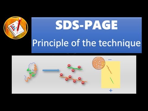
TLDRSDS-PAGE is a powerful technique for protein analysis, separating proteins based on molecular weight using polyacrylamide gel electrophoresis. Proteins are denatured and bound with SDS, then loaded into a gel system with a stacking and separating gel. An electric field drives negatively charged proteins through the gel, allowing size-based separation. The process involves sample preparation, gel polymerization, and electrophoresis, culminating in protein bands that can be visualized and analyzed using staining and immunological methods.
Takeaways
- 🧪 SDS-PAGE is a powerful technique for studying proteins, separating them based on molecular weight using polyacrylamide gel electrophoresis.
- 🔬 Sample preparation involves adding a loading buffer with SDS, beta-mercaptoethanol, bromophenol blue, and glycerol to protein samples, which may come from various biological or environmental sources.
- 🔥 Heating samples at 95°C for five minutes helps denature proteins, disrupting their natural structure and interactions.
- 🌊 SDS is an anionic surfactant that binds to proteins, neutralizing their intrinsic charges and allowing separation based on molecular weight alone.
- 🔗 Amino acids, the building blocks of proteins, are linked by peptide bonds and fold into specific spatial conformations influenced by various interactions.
- 🌡 The gel setup includes a separating gel with pH 8.8 and a stacking gel with pH 6.8, both made of acrylamide and other components to create a porous structure.
- 🚀 The gel polymerization process is initiated by ammonium persulfate and accelerated by TEMED, resulting in a gel with specific porosity for protein separation.
- 🏎️ The stacking gel ensures that proteins enter the separating gel simultaneously, preventing smearing and improving separation clarity.
- 🌀 The running buffer with glycine and chloride ions, along with SDS, maintains the protein charge state during electrophoresis.
- 📊 The electrophoresis process sorts proteins by size, with smaller proteins migrating more easily through the gel matrix than larger ones.
- 🖼️ After separation, proteins can be visualized and analyzed using staining methods like Coomassie blue, which binds to proteins and creates visible bands.
Q & A
What is SDS-PAGE and how is it used in protein studies?
-SDS-PAGE, or Sodium Dodecyl Sulfate Polyacrylamide Gel Electrophoresis, is a method used to separate proteins based on their molecular weight. It involves the use of a polyacrylamide gel and is a powerful technique in the study of proteins.
What is the purpose of adding a loading buffer to protein samples in SDS-PAGE?
-The loading buffer, containing SDS, beta-mercaptoethanol, bromophenol blue, and glycerol, is added to denature proteins, break disulfide bonds, visualize the sample, and increase sample density to ensure it falls to the bottom of the well during electrophoresis.
Why are proteins heated at 95 degrees Celsius before being loaded onto the gel?
-Heating the proteins at 95 degrees Celsius helps to denature them, ensuring that they are in a linear form and that their intrinsic charges are negligible compared to the negative charges from SDS, which is necessary for uniform separation based on molecular weight.
What is the role of SDS in the SDS-PAGE process?
-SDS, an anionic surfactant, denatures the native proteins by disrupting hydrogen, hydrophobic, and ionic interactions. It binds uniformly to the proteins, giving them a net negative charge, which allows for separation based on molecular weight.
How are the separating and stacking gels prepared for SDS-PAGE?
-The separating gel solution has a pH of 8.8, and the stacking gel solution has a pH of 6.8. Both are prepared with acrylamide, ammonium persulfate, and TEMED. The polymerization process involves a free radical initiator and accelerator, resulting in a gel with characteristic porosity.
What is the significance of the pH difference between the separating and stacking gels?
-The pH difference between the separating gel (pH 8.8) and the stacking gel (pH 6.8) is crucial for the migration of proteins. The lower pH in the stacking gel slows down the migration of charged molecules, allowing proteins to concentrate into a narrow zone before entering the separating gel.
Why is a molecular weight size marker loaded onto the gel along with the samples?
-A molecular weight size marker, consisting of proteins of known sizes, is loaded to provide a reference for estimating the sizes of the proteins in the actual samples. This allows for the determination of the molecular mass of unknown proteins.
How does the electric field affect the migration of proteins in the gel?
-The electric field causes the negatively charged proteins to migrate towards the positive electrode. Smaller proteins can move more easily through the gel matrix, while larger proteins are retained and migrate more slowly, leading to separation based on size.
What is the function of the running buffer in SDS-PAGE?
-The running buffer, containing glycine and chloride ions, is used to maintain the pH and ionic strength during electrophoresis. It also ensures that the proteins remain denatured and that the pH conditions are suitable for the migration of proteins and ions.
What happens to the proteins when they enter the separating gel from the stacking gel?
-Upon entering the separating gel, the pH changes to 8.8, causing glycine to become negatively charged and migrate faster than the proteins. This results in the separation of proteins based on their molecular weight, with higher molecular weight proteins moving more slowly.
How can the separated proteins be analyzed after electrophoresis?
-After electrophoresis, proteins can be analyzed using staining methods, such as Coomassie blue, to visualize the protein bands. Additionally, techniques like Western blot can be used for immunological detection of specific proteins.
Outlines
🔬 SDS-PAGE Technique and Sample Preparation
The paragraph introduces the SDS-PAGE (Sodium Dodecyl Sulfate Polyacrylamide Gel Electrophoresis) method, a crucial technique in protein analysis. It explains how proteins, once denatured and combined with SDS, are separated based on their molecular weight in a polyacrylamide gel. The process involves sample preparation with a loading buffer containing SDS, beta-mercaptoethanol, bromophenol blue, and glycerol, followed by heating at 95°C. The paragraph details the denaturation of proteins by SDS, which disrupts the protein's structure and allows separation based on size alone. It also describes the creation of the gel matrix, involving a separating gel with pH 8.8 and a stacking gel with pH 6.8, both composed of acrylamide, ammonium persulfate, and TEMED. The gel's porosity is determined by the ratio of acrylamide to Biss, with different concentrations suitable for various molecular weights. The application of an electric field initiates the migration of negatively charged proteins towards the positive electrode, with smaller proteins moving more easily through the gel matrix.
🚀 Mechanism of Protein Separation and Staining in SDS-PAGE
This paragraph delves into the mechanism of protein separation in the SDS-PAGE process. It explains the role of the stacking gel in ensuring proteins enter the separating gel simultaneously, which is crucial for obtaining sharp bands. The running buffer's composition, consisting of glycine and chloride ions, is highlighted, with glycine's charge state varying based on pH. At the running buffer's pH of 8.3, glycine is negatively charged, while at the stacking gel's pH of 6.8, it becomes predominantly neutral, slowing down the migration of proteins and chloride ions. This concentration of proteins into a narrow zone facilitates their separation in the separating gel, where pH 8.8 allows glycine to migrate faster than proteins, leading to their size-based separation. The paragraph concludes with the description of protein detection post-electrophoresis, mentioning the use of Coomassie blue as a staining agent to visualize proteins as blue bands against a clear background. Molecular weight markers are used to estimate the size of unknown proteins by comparing their migration distance relative to the markers.
Mindmap
Keywords
💡SDS-PAGE
💡Proteins
💡Molecular Weight
💡Acrylamide
💡Beta-Mercaptoethanol
💡Peptide Bonds
💡Denaturation
💡Polymerization
💡Stacking Gel
💡Running Buffer
💡Coomassie Blue
Highlights
SDS-PAGE is a powerful technique for studying proteins, separating them based on molecular weight using polyacrylamide gel electrophoresis.
Sample preparation involves adding a loading buffer containing SDS, beta-mercaptoethanol, bromophenol blue, and glycerol to protein samples.
Proteins are heated at 95 degrees Celsius to denature them, facilitating uniform SDS binding and charge distribution.
Proteins are large biomolecules composed of amino acid residues linked by peptide bonds, folding into specific spatial conformations influenced by various interactions.
SDS is an anionic surfactant that denatures proteins by disrupting hydrogen bonds, hydrophobic interactions, and ionic bonds.
Beta-mercaptoethanol acts as a reducing agent to cleave disulfide bonds in proteins.
The polyacrylamide gel's pore size is determined by the ratio of acrylamide to bis-acrylamide, with low concentrations preferred for high molecular weight samples.
The gel is created through a polymerization process initiated by ammonium persulfate and accelerated by TEMED.
A separating gel with pH 8.8 and a stacking gel with pH 6.8 are used, both contributing to the gel's characteristic porosity.
The stacking gel ensures that all proteins enter the separating gel simultaneously, preventing smeared bands.
The running buffer, containing glycine and chloride ions, maintains the proteins' denatured state throughout the electrophoresis process.
Molecular weight markers are used alongside samples to estimate the sizes of proteins in the samples.
Bromophenol blue in the samples helps visualize the sample in the well, while glycerol increases sample density for proper well placement.
The application of an electric field causes negatively charged proteins to migrate towards the positive electrode.
Smaller proteins migrate more easily through the gel matrix, while larger proteins are retained and migrate slower.
The pH of the running buffer and the gel affects the charge state of glycine, influencing protein migration speed.
Proteins are separated by size in the separating gel, with Coomassie blue being a popular staining method for visualization.
After electrophoretic separation, proteins can be analyzed using other methods such as Western blot for immunological detection.
The separating gel can be stained and the molecular weight of unknown proteins determined by comparing to size markers.
Transcripts
sds-page your sodium dodecyl sulfate
polyacrylamide gel electrophoresis is an
electrophoresis method and very powerful
technique in the study of proteins an
sds-page proteins are separated in a
poly acrylamide gel based on their
molecular weight for sample preparation
a loading buffer containing SDS beta
mercaptoethanol bromophenol blue and
glycerol is added to the protein samples
protein samples maybe biologically
derived for example from prokaryotic or
you create excels tissues viruses
environmental samples or purified
proteins when the sample buffer has been
added the samples are then heated at 95
degrees Celsius for five minutes
proteins are large biomolecules
consisting of one or more long chains of
amino acid residues
Meno acids are organic compounds that
contain a mined and carboxyl functional
groups along with the sidechain proteins
are formed by linking amino acids with
peptide bonds and fold into one or more
specific spatial confirmations driven by
a number of interactions such as
hydrogen bonds hydrophobic interaction
disulfide bonds and ionic bonds SDS is
an anionic surfactant and contain a
polar head group with a net negative
charge at the end of a long hydrophobic
carbon chain SDS denatures the native
proteins by disturbing the hydrogen
bonds hydrophobic and ionic interactions
while the reducing agent beta
mercaptoethanol is used to cleave the
disulfide bonds
also when a protein mixture is heated
SDS binds uniformly to the protein and
the intrinsic charges of this protein
become negligible when compared to the
negative charges contributed by SDS this
treatment brings the folded proteins
down to linear molecules with net
negative charge therefore these proteins
can be separated in a poly acrylamide
gel based on their molecular weight
Vergil preparation a separating gel
solution with ph 8.8 and a stacking gel
solution with ph 6.8 are used and both
consist of acrylamide this acrylamide
ammonium persulfate and tea med the gel
is produced by polymerization between
two glass plates anchored vertically in
a cassette the separating gel is poured
first
the polymerization reaction is a vinyl
addition polymerization initiated by a
free radical generating system ammonium
persulfate is used as the free radical
initiator while tea med accelerates the
rate of formation of free radicals from
persulfate these free radicals convert
acrylamide monomers to free radicals
which react with unactivated monomers to
begin the polymerization chain reaction
the elongating polymer chains are
randomly cross-linked by Biss acrylamide
resulting in a gel with a characteristic
porosity
the pore size of the gel is related to
the ratio of acrylamide to Biss
acrylamide low concentration is
preferred for high molecular weight
samples the stacking gel solution is
poured on top of the solid separating
gel
and a plastic comb placed on the top of
the stacking gel during polymerization
enables the formation of small wells in
the gel once the gel completely
polymerizes the comb is removed and the
gel cassette is placed vertically
between two electrodes positive
electrode located at the bottom of the
gel whereas negative electrode
positioned at the top of the gel
the gel is inserted into a chamber
and at risk lysine chloride buffer
system with ph 8.3 is poured to allow
the conduction of current through the
gel SDS is also present in the gel and
in the running buffer to make sure that
once the proteins are denatured they
stay that way throughout the run
for sample application in addition to
the samples a molecular weight size
marker is usually loaded onto the gel
this consists of proteins of known sizes
and thereby allows the estimation of the
sizes of the proteins in the actual
samples which migrate in parallel in
different tracks of the gel each sample
is pivoted into its own well in the gel
and as we have seen previously Roma
phenol blue and glycerol are present in
the samples bromophenol blue is a dye
that is useful for visualizing the
sample in the well and glycerol
increases the density of a sample and
used to make the sample fall to the
bottom of the sample well rather than
just flow out and mix with the running
buffer
after the sample application procedure
an electric field is applied across the
gel
causing the negatively charged proteins
to migrate across the gel away from the
negative electrode and towards the
positive electrode
also small proteins migrate relatively
easily through the mesh of the gel while
larger proteins are more likely to be
retained and thereby migrate more slowly
through the gel the stacking gel is used
to ensure that all of the proteins
arrive at the separating gel at the same
time because gel wells are around one
centimeter deep so in the absence of a
stacking gel proteins would all enter
the separating gel at different times
resulting in very smeared bands
as we have seen previously the running
buffer consists of glycine and chloride
ions so in the sample wells we will have
proteins chloride ions and lysine
proteins and chloride ions are
negatively charged
well glycine can exist in three
different charged States depending on
the pH the ice electric pointer pH I of
glycine is equal 5.97 at this pH glycine
carries neutral charge due the gain and
loss of protons at a pH below its pH I
glycine carries a net positive charge
and at a pH above its pH I it becomes
negatively charged
the running buffer pH is equal 8.3 which
above the pH I of glycine so at this pH
glycine is negatively charged once the
electric current is applied proteins
chloride ions and lysine are forced to
enter the stacking gel where the pH is
equal six point eight at this pH glycine
switches predominantly to this material
ik neutrally charged State this loss of
charge causes them to move very slowly
in the electric field the chloride ions
on the other hand move much more quickly
chloride ions migrate in front of the
proteins as leading ions and lysine
molecules migrate behind the proteins as
initial trailing ions consequently
proteins are trapped and concentrated
into a narrow zone between the chloride
ions and glycine this procession carries
on until it hits the separating gel
where the pH switches 28.8 at this pH
the glycine molecules are mostly
negatively charged and can migrate much
faster than the proteins proteins are
separated so higher molecular weight
proteins move more slowly through the
porous acrylamide gel than lower
molecular weight proteins
due to the relatively small molecule
size of Brahma phenyl blue it migrates
faster than proteins and by optical
control of the migrating colored band
the electrophoresis can be stopped
before the dye and before the samples
have completely migrated through the gel
and leave it at the end of the electro
phoretic separation all proteins are
sorted by size and can then be analyzed
by other methods for example protein
staining methods and immunological
detection such as the Western blot
technique after removing the glass
plates the stacking gel is discarded and
the separating gel can be stained
Kumasi blue is the most popular protein
stain it is an anionic dye which nan
specifically binds to proteins the
structure of Kumasi blue is
predominantly nonpolar and it is usually
used in methanol excision acidified with
acetic acid proteins in the gel are
fixed by acetic acid and simultaneously
stained after staining the proteins are
detected as blue bands on a clear
background the molecular weight size
markers in a separate lane in the gel
can be used to determine the approximate
molecular mass of unknown biomolecules
by comparing the distance traveled
relative to the marker
you
5.0 / 5 (0 votes)