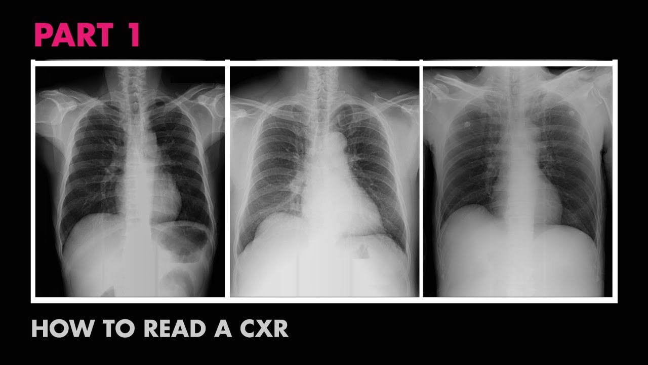
How To Read A Chest X-ray
Summary
TLDRThis video provides an insightful guide on how to read a chest X-ray, focusing on essential steps such as understanding opacity and lucency, ensuring correct patient positioning, and performing quality checks like rotation, exposure, and inspiration. The guide highlights key areas to observe, such as the trachea, heart size, mediastinum, hilum, diaphragm, and angles, while explaining how to detect abnormalities like pleural effusion. The video emphasizes a systematic approach for interpreting chest X-rays, helping medical professionals become proficient in this crucial diagnostic skill.
Takeaways
- 😀 X-rays show areas of opacity (white) and lucency (black), with opacity representing dense structures like bones and lucency indicating less dense areas like lungs filled with air.
- 😀 When reading a chest X-ray, always assume the patient is facing you, meaning the right side of the X-ray corresponds to the patient's left side, and vice versa.
- 😀 Before interpreting the X-ray, confirm key details such as the patient's name, the reason for the X-ray, and the X-ray view (PA or AP).
- 😀 Three key quality checks for chest X-rays: rotation (check the distance between clavicles and the spinous process), exposure (check visibility of the lower thoracic vertebrae), and inspiration (check the number of visible ribs).
- 😀 A well-centered X-ray shows equal distances between the lateral borders of the clavicles and the spinous process. Rotation causes lung field asymmetry and incorrect X-ray interpretation.
- 😀 Adequate exposure on an X-ray means the lower thoracic vertebrae are barely visible. If they are clearly visible, the X-ray is overexposed; if not visible, it’s underexposed.
- 😀 Proper inspiration is confirmed by seeing 5-6 anterior ribs and 8-10 posterior ribs above the diaphragm in the X-ray.
- 😀 The trachea should be positioned midline or slightly tilted due to the aortic arch, and you should check for foreign bodies or masses in the trachea.
- 😀 The cardiothoracic ratio (cardiac diameter divided by thoracic diameter) is used to assess heart size, with a normal value of <0.5 in PA view and >0.5 indicating cardiomegaly.
- 😀 The mediastinum contains vital structures like the right atrium, superior vena cava, left ventricle, and aorta. These should be carefully examined for abnormalities.
- 😀 The diaphragm's left dome is typically lower than the right dome due to the position of the heart. The stomach bubble is a normal finding, indicating gas in the stomach.
- 😀 Blunting of the costophrenic and cardiophrenic angles can indicate pleural effusion. Regularly compare both lung fields, chest walls, and bones for abnormalities.
Q & A
What are the white and black areas seen in a chest X-ray?
-In a chest X-ray, the white areas are known as areas of opacity, which are caused by denser structures like bones. The black areas are areas of lucency, which represent less dense structures like the lungs filled with air.
Why does the X-ray beam produce different colors for different body structures?
-The X-ray beam passes through various structures in the body, such as bones, lungs, and the heart, all of which have different densities. Denser structures, like bones, block the X-ray more and appear white, while less dense structures, like the lungs, allow the X-ray to pass through and appear black.
How should you visualize a chest X-ray in terms of patient positioning?
-When reading a chest X-ray, always assume the patient is facing you. The right side of the X-ray corresponds to the patient's left side, and the left side of the X-ray corresponds to the patient's right side.
What basic details should you confirm before interpreting a chest X-ray?
-Before interpreting a chest X-ray, confirm the patient's name, the date and time the X-ray was taken, the reason for the X-ray request, and the type of view (e.g., PA or AP).
What are the three parameters to check the quality of an X-ray?
-The three parameters to check the quality of an X-ray are rotation, exposure, and inspiration.
How can you assess rotation in a chest X-ray?
-To assess rotation, look at the distance between the lateral border of the clavicle and the spinous process on both sides. If these distances are equal, the X-ray is well-centered. If one side has a larger gap, it indicates rotation.
What does exposure mean in terms of chest X-ray quality?
-Exposure refers to the adequacy of the X-ray beam. To assess it, check the lower thoracic vertebrae margins. If they are barely visible, the exposure is adequate. If they are clearly visible, the X-ray is overexposed, and if not visible, it is underexposed.
How can you determine if a chest X-ray has adequate inspiration?
-To assess adequate inspiration, you should be able to see 5-6 anterior ends and 8-10 posterior ends of the ribs above the diaphragm. This indicates that the patient properly inflated their lungs during the X-ray.
What is the cardiothoracic ratio, and how is it used to assess heart size?
-The cardiothoracic ratio is the ratio of the cardiac diameter to the thoracic diameter. A ratio of less than 0.5 in a PA view indicates normal heart size, between 0.5 and 0.55 is borderline, and above 0.55 is abnormal. In an AP view, a ratio above 0.6 indicates false cardiomegaly.
What are the main components of the mediastinum that should be identified in a chest X-ray?
-The mediastinum consists of the heart (right atrium, right ventricle, left atrium, and left ventricle), major blood vessels (superior vena cava, inferior vena cava, aorta), and other structures like the trachea. Each side of the mediastinum is formed by different parts of the heart and major vessels.
Outlines

このセクションは有料ユーザー限定です。 アクセスするには、アップグレードをお願いします。
今すぐアップグレードMindmap

このセクションは有料ユーザー限定です。 アクセスするには、アップグレードをお願いします。
今すぐアップグレードKeywords

このセクションは有料ユーザー限定です。 アクセスするには、アップグレードをお願いします。
今すぐアップグレードHighlights

このセクションは有料ユーザー限定です。 アクセスするには、アップグレードをお願いします。
今すぐアップグレードTranscripts

このセクションは有料ユーザー限定です。 アクセスするには、アップグレードをお願いします。
今すぐアップグレード関連動画をさらに表示

CARA MEMBACA RONTGEN PARU / THORAX ‼️ HAPAL SAMPAI KAPANPUN 😱😱

Wheelchair Chest

Chest X-ray Interpretation | How to Read a CXR | OSCE Guide | UKMLA | CPSA | PLAB 2

How to Interpret a Chest X-Ray (Lesson 1 - An Introduction)
Anatomy of a Chest X-Ray - How to Read a Chest X-Ray (Part 1)

Assessment of CXR Quality - How to Read a Chest X-Ray (Part 5) - MEDZCOOL
5.0 / 5 (0 votes)
