UQx Bioimg101x 3.2.4 CT Reconstruction & Back Projection
Summary
TLDRThis video offers an insightful overview of Computed Tomography (CT), explaining how X-ray radiation is used to create detailed 3D images of the body's internal structures. It delves into the principles of image reconstruction and back projection, highlighting the role of the computer in processing signals from detectors to form voxels, the 3D equivalent of pixels. The video also touches on the significance of Hounsfield units and the enhancement of image clarity through mathematical filtering, providing a comprehensive introduction to the technology behind CT scans.
Takeaways
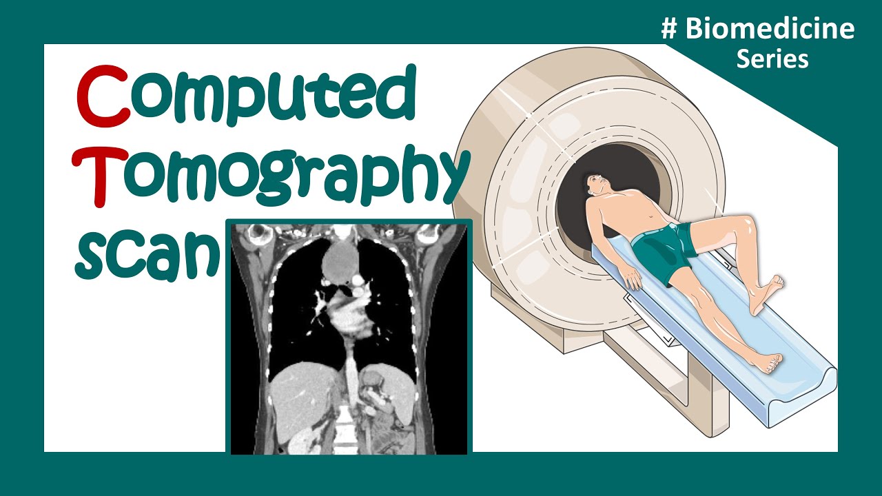
- 🩻 Computed Tomography (CT) uses X-ray radiation to create 3D images of the body by taking measurements from multiple angles.
- 📡 X-ray tubes and detectors rotate around the patient, where detectors capture the varying X-ray attenuation across different tissues.
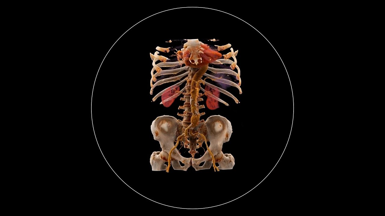
- 🧠 A CT image is constructed by splitting the body into tiny volumes called voxels, which are the 3D equivalent of pixels.
- 🦴 Dense tissues like bones have higher attenuation values, meaning they block more X-rays, while less dense tissues like skin have lower attenuation.
- 📏 Each voxel is assigned an attenuation value, called a Hounsfield unit, which represents the tissue’s ability to block X-ray radiation.
- 🎯 The process of image reconstruction involves summing the attenuation values from different angles to generate a full image, known as back projection.
- ⚙️ Back projection can result in blurry images, which can be sharpened using mathematical filtering techniques.
- 🔍 Filtering helps refine CT images, making them clearer and easier to interpret for medical purposes.
- 💻 The computer plays a key role in processing the X-ray measurements and assembling them into detailed 3D images of the body.
- 📚 For more advanced information, users are encouraged to explore additional resources like Dr. Andrew Janke's presentation on CT topics.
Q & A
What is the primary function of the X-ray tube and detectors in CT scanning?
-The X-ray tube and detectors rotate around the patient, with the tube emitting X-ray radiation that passes through the body. The detectors measure the radiation intensity that passes through different tissues, which is then used to create CT images.
How does a CT scanner create three-dimensional images compared to a conventional X-ray?
-While conventional X-rays can only produce two-dimensional images, CT scanners capture X-ray radiation from multiple angles, which allows the computer to reconstruct three-dimensional images by dividing the body into small volumes called voxels.
What role do voxels play in CT imaging?
-Voxels are the three-dimensional equivalents of pixels and represent small volumes of tissue. Each voxel is assigned an attenuation coefficient, or 'mu' value, based on how much it attenuates X-ray radiation.
What is an attenuation coefficient and how is it used in CT imaging?
-The attenuation coefficient, represented as 'mu,' reflects how much X-ray radiation a particular tissue absorbs. Higher mu values indicate higher attenuation, such as in bones, while lower values reflect tissues like skin, which attenuates less radiation.
How do Hounsfield units (HU) relate to attenuation values in CT imaging?
-Hounsfield units (HU) are a standardized way of expressing attenuation values in CT imaging. They provide a scale where water has a value of 0, air has negative values, and bones have positive values, corresponding to the tissue’s X-ray attenuation.
What is back projection in the context of CT imaging?
-Back projection is a method used to reconstruct images from multiple X-ray views taken at different angles. The data from each view are summed along the X-ray paths, which gradually forms an image of the scanned object.
Why does the initial CT image appear blurry, and how is it improved?
-The initial CT image appears blurry because back projection alone doesn’t perfectly reconstruct the image. Filtering techniques are applied to the data to improve image sharpness and clarity by removing noise and enhancing edges.
What is the significance of filtering in CT image reconstruction?
-Filtering is crucial in CT image reconstruction as it enhances image quality. Without filtering, the images would remain blurry, but after applying mathematical filters, the image becomes sharper and clearer.
Can you explain the process of CT image reconstruction in simple terms?
-CT image reconstruction involves measuring X-ray radiation passing through the body from different angles, converting the data into electrical signals, and using algorithms to combine the data. Back projection and filtering techniques are then applied to generate a clear, three-dimensional image.
What is the importance of the different angles of X-ray radiation during a CT scan?
-Different angles of X-ray radiation provide a complete view of the internal structures from all sides, allowing the computer to accurately reconstruct a 3D image of the body. Each angle contributes additional data that is crucial for precise image creation.
Outlines

このセクションは有料ユーザー限定です。 アクセスするには、アップグレードをお願いします。
今すぐアップグレードMindmap

このセクションは有料ユーザー限定です。 アクセスするには、アップグレードをお願いします。
今すぐアップグレードKeywords

このセクションは有料ユーザー限定です。 アクセスするには、アップグレードをお願いします。
今すぐアップグレードHighlights

このセクションは有料ユーザー限定です。 アクセスするには、アップグレードをお願いします。
今すぐアップグレードTranscripts

このセクションは有料ユーザー限定です。 アクセスするには、アップグレードをお願いします。
今すぐアップグレード関連動画をさらに表示

CT (Computed Tomography) Scans - A Level Physics
CT scan | computerized tomography (CT) scan |What is a CT scan used for? | Clinical application

How X-rays see through your skin - Ge Wang
What is Computed Tomography (CT) and how does it work?

Diagnostic Imaging Explained (X-Ray / CT Scan / Ultrasound / MRI)

Biomedical instrumentation- CT scan (Computed Tomography)
5.0 / 5 (0 votes)
