Radiation Exposure ,Radiation safety- Everything You Need To Know - Dr. Nabil Ebraheim

nabil ebraheim
19 Jun 201507:46

Summary

TLDRThis script discusses the risks of radiation from medical imaging like X-rays and CT scans, emphasizing the potential for cancer induction. It highlights the importance of minimizing exposure, especially in children, and stresses the need for justified use of scans. The script also covers radiation protection measures, such as distance, shielding, and monitoring with a datometer badge, and the sensitivity of rapidly dividing cells to radiation. It concludes with a call for patient education and universal access to medical imaging records to avoid unnecessary exposure.

Takeaways

  • 🔬 **X-rays and Radiation Risk**: X-rays can ionize human tissue, depositing energy that may lead to harmful changes and an increased cancer risk.
  • ⚠️ **Cumulative Dose**: The radiation dose from x-rays is cumulative, and unnecessary exposure should be avoided, especially in sensitive groups like children.
  • 🏥 **Medical Use Controversy**: While there's documented cancer risk from radiation, the medical community often downplays the risk associated with routine procedures like x-rays.
  • 📉 **Low-Level Radiation Effects**: The effects of low-level radiation, such as that from medical x-rays, are not fully understood, and the safe level is unknown.
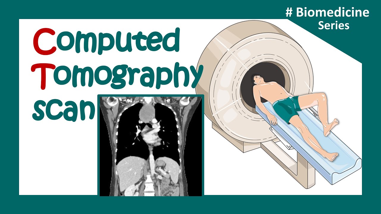
  • 🧐 **Radiation in Medical Imaging**: Certain medical procedures like CT scans, fluoroscopy, and mammography expose patients to higher levels of radiation, particularly concerning in young females.
  • 🛡️ **Optimizing Radiation Doses**: It's crucial to balance the medical benefits with the risks, ensuring patients receive only the necessary radiation for a clear image.
  • 🚫 **Growing Concerns**: There's a growing concern about the potential long-term effects of radiation from medical imaging, especially with the increasing use of CT scans.
  • 👶 **Children's Sensitivity**: Children are significantly more sensitive to radiation than adults, with a higher risk of developing cancer from CT scans.
  • 📏 **Inverse Square Law**: The intensity of radiation decreases with distance from the source, emphasizing the importance of positioning in radiation protection.
  • 🥼 **Protection Measures**: Using lead gowns and aprons can significantly reduce radiation exposure, and it's essential to wear them during procedures involving radiation.
  • 📚 **Patient Education**: Patients should be educated about the necessity of x-ray procedures and advocate for the lowest possible dose that maintains image quality.

Q & A

  • What is the primary risk associated with X-ray exposure?

    -The primary risk associated with X-ray exposure is the potential for harmful changes within the body due to ionization of human tissue, which may lead to cancer.

  • Why is radiation dose considered cumulative?

    -Radiation dose is cumulative because each exposure adds to the total amount of radiation received over time, increasing the potential risk of developing radiation-induced health issues, such as cancer.

  • How does the government try to mitigate unnecessary radiation exposure from medical imaging?

    -The government attempts to avoid unnecessary CT scans and X-rays by promoting careful consideration before ordering such imaging tests to minimize radiation exposure and reduce cancer risk.

  • How does the radiation dose from a CT scan compare to that of a regular X-ray?

    -A CT scan exposes a patient to a significantly larger dose of radiation compared to a regular X-ray. For example, radiation from a CT scan of the pelvis equals the same amount as 100 chest X-rays.

  • Why are children more sensitive to radiation than adults?

    -Children are more sensitive to radiation because their cells are still rapidly dividing, and their tissues are more vulnerable to the effects of radiation. Additionally, children have a longer life expectancy, giving more time for radiation-induced cancer to develop.

  • What are some tissues in the body that are highly susceptible to radiation-induced tumors?

    -Tissues highly susceptible to radiation-induced tumors include bone marrow, breast tissue, gonads, and lymphatic tissue.

  • What protective measures are suggested for minimizing radiation exposure during medical imaging?

    -Protective measures include using lead aprons and shields, monitoring radiation exposure with a dosimeter badge, optimizing radiation doses to only necessary levels, and maintaining distance from the radiation source to reduce intensity.

  • What is the inverse square law in relation to radiation exposure?

    -The inverse square law states that radiation intensity decreases as the square of the distance from the radiation source. For example, doubling the distance from the source reduces the radiation intensity to one-quarter of its original value.

  • What long-term effects can result from exposure to low levels of radiation, and why is it difficult to determine a 'safe' radiation level?

    -The long-term effects of low-level radiation are not fully understood, making it difficult to establish a 'safe' radiation level. While high-level radiation is known to be carcinogenic, the effects of low-level exposure are controversial and under-researched.

  • Why is there growing concern about the use of CT scans in children?

    -There is growing concern about CT scan use in children because they are more sensitive to radiation, and studies show that large doses of radiation from CT scans can increase the risk of developing cancer later in life. Additionally, about 1,500 out of 3 to 4 million children receiving CT scans may develop cancer two decades later.

Outlines

00:00

⚠️ Radiation Exposure Risks from X-Rays and CT Scans

This paragraph discusses the potential risks of radiation exposure from X-rays and CT scans, which can ionize human tissue and cause harmful changes in the body. The risk of cancer is highlighted, especially since the radiation dose is cumulative. The government aims to reduce unnecessary use of CT scans and X-rays to minimize exposure. While high levels of radiation are known to be carcinogenic, the effects of low-level exposure remain uncertain. Medical procedures such as CT scans, fluoroscopy, mammography, and X-rays expose patients to high radiation doses, particularly young females. The risk of radiation exposure should be carefully balanced with the medical benefits, and doctors are urged to minimize the dose required for clear imaging.

05:00

👶 Children’s Sensitivity and Risks from CT Scans

This paragraph emphasizes that children are significantly more sensitive to radiation than adults, with millions receiving CT scans annually. A considerable number of these children may develop cancer later in life due to the high doses of radiation they receive. It is advised that children should not be exposed to adult doses of radiation. Specific risks associated with CT scans, particularly to sensitive tissues like the skin, bone marrow, and gonads, are addressed. Radiation protection measures, such as the use of minimal fluoroscopy or CR arms, are recommended to reduce exposure.

📏 Distance and Radiation Exposure in Fluoroscopy

This section explains how radiation exposure from fluoroscopy follows the inverse square law, meaning that increasing the distance from the radiation source significantly reduces exposure. For instance, at 2 meters from the source, the radiation intensity drops to one-quarter of what it would be at 1 meter. Healthcare professionals are advised to monitor exposure, avoid being in the direct path of the radiation beam, and be aware of increased exposure when imaging larger body parts.

🛡️ Radiation Monitoring and Protection

This paragraph focuses on radiation protection measures such as using datometer badges to monitor accumulated exposure, though these devices do not offer protection themselves. Lead aprons and gowns help shield against radiation, blocking about 95% of scattered radiation. Certain rapidly dividing cells in the body, like those in the sperm and lymphocytes, are particularly vulnerable to radiation damage. External signs of damage, such as skin burns, and internal effects, like a drop in white blood cell count, are outlined. Studies have also shown that frequent fluoroscopy users may be at higher risk of developing cataracts.

📝 Justifying and Limiting CT Scans

This section discusses the overuse of CT scans, with a large portion considered unnecessary. These excess scans increase cancer risk without providing significant medical benefits. The lack of strict oversight in the administration of CT scans is a concern, and better patient education is needed. Patients are encouraged to ask whether the scan is necessary and if a lower radiation dose can be used without compromising the study’s effectiveness. The paragraph also advocates for a universal X-ray bank, where patients’ X-rays can be accessed by any hospital to avoid unnecessary repeats.

🔬 Susceptible Tissues and Radiation-Induced Tumors

This paragraph summarizes the specific tissues most susceptible to radiation-induced tumors, including bone marrow, breast tissue, gonads, and lymphatic tissue. It reinforces the importance of shielding vulnerable areas, such as the gonads, during exposure. Additionally, it stresses the need for wearing protective lead aprons and continuously monitoring radiation exposure with datometer badges. The paragraph closes with a reminder that the video is educational and urges viewers to consult with their doctors before making medical decisions.

Mindmap

Keywords

💡radiation

Radiation refers to the emission of energy as electromagnetic waves or as moving subatomic particles. In the context of the video, it specifically addresses ionizing radiation from X-rays, which has enough energy to remove tightly bound electrons from atoms, creating ions. This ionization can lead to harmful changes within the body, such as DNA damage, which may result in cancer.

💡carcinogen

A carcinogen is any substance, radionuclide, or radiation that is an agent directly involved in the promotion of cancer. The video mentions that X-rays are considered carcinogens due to their ability to cause cellular changes that can lead to cancer, which is why there is an effort to limit unnecessary exposure.

💡cumulative

Cumulative refers to the total amount of something that has been accumulated over time. In relation to the video, it discusses how the dose of radiation from X-rays is cumulative, meaning that the body's exposure to radiation adds up with each X-ray or CT scan, increasing the potential risk over time.

💡CT scans

CT scans, or computed tomography scans, are a type of medical imaging that uses X-rays to create detailed cross-sectional images of the body. The video points out that CT scans expose patients to a higher level of radiation compared to standard X-rays, which is a concern due to the potential increased cancer risk.

💡fluoroscopy

Fluoroscopy is a medical imaging technique that uses X-rays to obtain real-time moving images of the body's structures. The video script notes that fluoroscopy emits a significant amount of radiation, and the dose increases with proximity to the radiation source, highlighting the need for careful patient and physician protection.

💡inverse square law

The inverse square law states that the intensity of radiation is inversely proportional to the square of the distance from the source. This principle is mentioned in the video to explain how increasing the distance from the radiation source can significantly reduce the radiation dose received by the patient and the physician.

💡datometer badge

A datometer badge is a radiation monitoring device worn by individuals to measure their exposure to radiation. The video emphasizes the importance of using such badges to monitor radiation exposure, as they help in understanding the total dose received and ensuring safety limits are not exceeded.

💡shielding

Shielding refers to the use of materials or barriers to protect against harmful radiation. The video discusses the use of lead gowns and aprons as shielding protection to stop exposure to radiation, particularly during fluoroscopy, by blocking the radiation and reducing scattered radiation by about 95%.

💡susceptible

Susceptible means being more likely to be harmed or affected by something, such as radiation. The video identifies certain tissues like bone marrow, breast tissue, gonads, and lymphatic tissue as being more susceptible to radiation-induced tumors, emphasizing the need for special care in shielding these areas during X-ray procedures.

💡justification

Justification in the context of medical imaging means having a valid medical reason for performing a procedure, such as a CT scan. The video argues that many CT scans are performed without proper justification, potentially exposing patients to unnecessary radiation and increasing their cancer risk.

💡patient education

Patient education involves informing patients about the procedures they are undergoing, including the benefits and risks. The video script suggests that patient education is crucial in the context of radiation exposure, encouraging patients to ask questions about the necessity of X-ray studies and the lowest possible dose that can be used.

Highlights

X-rays ionize human tissue and deposit energy that can cause harmful changes within the body.

There is a cancer risk from x-rays, and the dose of radiation is cumulative.

X-rays are considered a carcinogen by the government, and efforts are being made to avoid unnecessary CT scans and x-rays.

Doctors must pay close attention to the cancer risk involved with the use of x-rays.

The cancer risk from radiation exposure has been documented in cases of atomic bomb survivors, but the risk for medical uses is controversial.

Radiation at high levels is carcinogenic, though the effects of low-level radiation are not fully understood.

CT scans, fluoroscopy, mammography, and x-rays expose the public to high levels of radiation, especially young females.

Children are 10 times more sensitive to radiation than adults, and about 1500 children who receive CT scans may develop cancer decades later.

The use of CT scans has increased in adults and children, potentially exposing patients to unnecessary high doses of radiation.

Radiation dose from a CT scan of the pelvis equals the amount of 100 chest x-rays.

Radiation intensity follows the inverse square law: doubling the distance from the source reduces radiation intensity by four times.

Monitoring and shielding, such as using lead aprons, are essential to reduce exposure to fluoroscopy radiation.

Rapidly dividing cells, such as sperm, lymphocytes, and stomach lining cells, are most sensitive to radiation exposure.

The first external signs of radiation damage, such as skin burns, are usually seen after a drop in white blood cell count.

There should be more oversight and standardization to avoid unnecessary radiation exposure from CT scans, and patients should be educated to ask if the study is necessary.

Transcripts

play00:07

radiation

play00:09

exposure x-rays ionize human tissue and

play00:12

deposit energy that can cause harmful

play00:14

changes within the

play00:17

body there is a cancer risk from

play00:22

x-rays the dose of radiation is

play00:25

cumulative x-rays are considered for

play00:27

carcinogen list

play00:30

the government is attempting to avoid

play00:32

the use of unnecessary CT scans and

play00:34

x-rays to avoid unnecessary exposure to

play00:37

radiation this highlights the cancer

play00:39

risk doctors need to pay close attention

play00:42

to the risk involved with the use of

play00:45

x-rays the cancer risk associated with

play00:47

radiation exposure is documented in

play00:49

cases of atomic bomb

play00:54

survivors the risk for medical uses is

play00:56

controversial and usually played down by

play00:58

physicians

play01:03

radiation at a high level is

play01:07

carcinogenic the level of radiation from

play01:09

x-ray exposure is low the effects of

play01:12

low-level radiation is not known so what

play01:15

is the safe radiation

play01:18

level the safe level is not

play01:22

known it is known that CT scans

play01:25

fluoroscopy mamography and x-rays expose

play01:28

the public to a high level of radiation

play01:30

especially in young

play01:33

females the risk of exposure should

play01:36

balance the medical

play01:41

benefits optimize radiation doses by

play01:44

exposing the patient only to enough

play01:46

radiation to get a clear

play01:52

image there is a growing concern about

play01:55

the risk associated with giving a

play01:56

patient large doses of

play01:58

radiation the use of CT scans has

play02:01

increased recently in adults and

play02:02

children possibly exposing the patient

play02:04

to an unnecessary high dose of

play02:06

radiation CT scan is the method most

play02:09

often used to diagnose cancer diseases

play02:12

fractures and it exposes the patient to

play02:14

a much larger dose of radiation than

play02:16

x-rays radiation from CT scan of the

play02:19

pelvis equals the same amount as 100

play02:21

chest x-rays children are 10 times more

play02:24

sensitive to radiation than adults

play02:30

3 to four million children receive CT

play02:32

scans and about 1500 of them will

play02:34

develop cancer two decades

play02:38

later children should not be given an

play02:40

adult dose of

play02:44

radiation radiation dose limits CT scan

play02:48

of the pelvis has the highest level of

play02:49

exposure to the skin marrow and gonads

play03:05

use a Min fluroscopy CR arm whenever

play03:21

possible radiation exposure with

play03:23

fluoroscopy fluoroscopy admits a lot of

play03:26

radiation

play03:30

the closer the extremity is to the

play03:32

radiation Source the higher the dose of

play03:34

radiation the patient

play03:37

receives when the distance from the beam

play03:39

increases the dose of radiation is

play03:43

less radiation intensity follows the

play03:45

inverse Square law it is all about

play03:48

distance if the intensity of radiation

play03:50

at 1 M from the source is 100 m per hour

play03:53

then the intensity of radiation at 2 m

play03:55

from the source is 1/4 or 25 M per hour

play03:59

in same unit area at 3 m from The Source

play04:03

the intensity of radiation is 1 nth the

play04:05

original or 11.1 M Rankin per

play04:26

hour there is less exposure to the

play04:29

physician when Imaging a smaller body

play04:34

part larger body parts create an

play04:36

increased exposure to The Physician when

play04:38

Imaging a patient with the

play04:47

CR do not be in the direct path of the

play04:50

radiation

play04:53

beam radiation protection is of

play04:55

importance with monitoring shielding and

play04:58

position

play05:00

monitoring protection should be used

play05:01

with a datometer badge a datometer badge

play05:04

only records how much radiation you have

play05:06

received it does not protect you from

play05:08

exposure to

play05:10

radiation shielding protection of

play05:12

fluoroscopy radiation lead gowns and

play05:14

aprons work to stop exposure let aprons

play05:17

accentuate scattered radiation by about

play05:20

95% rapidly dividing cells are most

play05:23

sensitive to radiation exposure such as

play05:25

sperm lymphocytes the small intestines

play05:28

and stomach

play05:30

radiation damage seldom appears at the

play05:32

time of a radiation the first effects of

play05:34

radiation damage is usually seen as a

play05:37

drop in the white blood cell count the

play05:39

first external sign of damage is seen as

play05:41

a skin

play05:43

burn studies suggests that people who

play05:45

use fosc opy extensively have a higher

play05:47

rate of

play05:50

cataracts early effects of radiation

play05:52

exposure could include death hematologic

play05:55

depression chromosome aberation skin

play05:59

uretha

play06:00

or

play06:01

epilation here are some facts about team

play06:03

exposure to

play06:17

radiation CT scan examination is usually

play06:20

done without justification by most

play06:22

insurances it is opinion that 1/3 of CT

play06:25

scan studies that are given could be

play06:27

avoided giving unnecessary study

play06:30

causes an added cancer risk with no

play06:32

benefit the CT scan study should be

play06:36

justified there is no close oversight or

play06:38

uniform standard in order to eliminate

play06:40

radiation exposure this must be

play06:44

done patient education is important the

play06:47

patient should ask if the study is

play06:48

necessary and what is the lowest dose

play06:50

possible that can be given without

play06:52

compromising the

play06:53

study there should be a universal x-ray

play06:56

Bank where patients x-rays can be

play06:58

accessible anywhere from from any

play06:59

hospital this would avoid the

play07:01

unnecessary repeating of

play07:04

x-rays in summary the bone marrow breast

play07:07

tissue gonads and lymphatic tissue are

play07:10

susceptible to radiation induced tumors

play07:12

it's important to Shield the gonads from

play07:16

exposure always wear lead aprons and

play07:18

monitor the radiation exposure with a

play07:20

datometer

play07:23

badge this video is for educational

play07:26

purposes only please consult your doctor

play07:30

before you make any decision about your

play07:32

medical care

Rate This

5.0 / 5 (0 votes)

関連タグ
Radiation RiskX-ray SafetyCancer LinkMedical ImagingCT Scan ConcernsRadiation ProtectionFluoroscopyChild ExposureRadiation ShieldingPatient Education
英語で要約が必要ですか?