Types of joints in the human body - Anatomy & Examples | Kenhub

Kenhub - Learn Human Anatomy
28 Aug 201911:37

Summary

TLDRThis tutorial delves into the anatomy of human joints, distinguishing them by structure, mobility, and range of motion. It covers synovial, fibrous, and cartilaginous joints, highlighting six synovial subtypes like ball-and-socket and hinge joints. The video explains how joint stability is influenced by factors like articular surface contact, ligaments, and muscle tone, emphasizing the trade-off between mobility and stability.

Takeaways

  • 🦴 A joint is a connection between two bones in the skeleton and can be classified by structure, mobility, and range of motion.
  • 🔍 There are three main types of joints based on structure: synovial, fibrous, and cartilaginous joints.
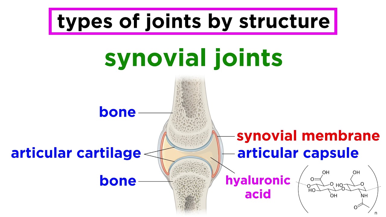
  • 🌀 Synovial joints are the most common and are surrounded by an articular capsule with an outer fibrous layer and an inner synovial layer.
  • 🧽 The articular surfaces of synovial joints are covered in hyaline cartilage which reduces friction and assists in shock absorption.
  • 🔄 Synovial joints can be further subclassified into six types based on the shape of their articular surfaces and range of motion: ball-and-socket, hinge, pivot, condylar, saddle, and plane joints.
  • 💺 Fibrous joints have little to no mobility and include sutures, gomphoses, and syndesmoses.
  • 🦷 Gomphoses are a type of fibrous joint found in the mouth where the roots of teeth articulate with the dental alveoli.
  • 🔗 Cartilaginous joints are connected by fibrocartilage or hyaline cartilage and include synchondroses and symphyses.
  • 🤸‍♂️ Diarthroses are freely mobile joints like the knee joint, while amphiarthroses and synarthroses are less mobile or immobile, respectively.
  • 🔄 Joints can be classified by their range of motion into uniaxial (one axis), biaxial (two axes), and polyaxial/multiaxial (three axes).
  • 🏋️‍♀️ Factors contributing to joint stability include the degree of contact between articulating surfaces, presence of ligaments, and tone of surrounding muscles.

Q & A

  • What is the definition of a joint in the human skeleton?

    -A joint is defined as a connection between two bones in the skeleton.

  • How can joints be classified according to their structure?

    -Joints can be classified into synovial joints, fibrous joints, and cartilaginous joints based on their structure.

  • What are the characteristic features of synovial joints?

    -Synovial joints are surrounded by an articular capsule with an outer fibrous layer and an inner synovial layer, have articular surfaces covered in hyaline cartilage, and may contain additional structures like articular discs and bursae.

  • What are the different types of fibrous joints mentioned in the script?

    -The script mentions sutures, gomphoses, and syndesmoses as the types of fibrous joints.

  • How are the bones connected in a cartilaginous joint?

    -In a cartilaginous joint, the bones are connected by either fibrocartilage or hyaline cartilage.

  • What are the key terms for classifying joints based on their mobility?

    -The key terms are diarthrosis for freely mobile joints, amphiarthrosis for slightly mobile joints, and synarthrosis for immobile joints.

  • How are joints classified according to their range of motion?

    -Joints are classified as uniaxial, biaxial, or polyaxial based on the number of axes they can move along.

  • What is the only polyaxial joint in the human body?

    -The ball-and-socket joint is the only polyaxial joint in the human body.

  • What are the factors that contribute to joint stability?

    -The degree of contact between articulating surfaces, the presence of ligaments, and the tone of the surrounding muscles are the factors that contribute to joint stability.

  • Why are some joints more susceptible to dislocation or injury?

    -Some joints are more susceptible to dislocation or injury because there is a trade-off between mobility and stability; the more mobile a joint is, the less stable it will be.

  • What are the movements allowed by the different types of synovial joints?

    -Ball-and-socket joints allow flexion/extension, adduction/abduction, and internal/external rotation. Hinge joints allow flexion/extension. Pivot joints allow rotation. Condylar joints allow radial/ulnar deviation and flexion/extension. Saddle joints allow abduction/adduction, flexion/extension, and circumduction. Plane joints allow sliding or gliding movements.

Outlines

00:00

🦴 Introduction to Joints

This paragraph introduces the tutorial on the types of joints found in the human body. It explains that joints are connections between bones and can be classified by structure, mobility, and range of motion. The paragraph focuses on structural classification, identifying three types: synovial, fibrous, and cartilaginous joints. Synovial joints are highlighted as the most common, with features like an articular capsule, hyaline cartilage, and possible additional structures like menisci and bursae. Subclassifications of synovial joints such as ball-and-socket, hinge, pivot, condylar, saddle, and plane joints are mentioned, setting the stage for further detailed discussion.

05:03

🔍 Classification of Joints by Mobility and Range of Motion

The second paragraph delves into classifying joints by their mobility and range of motion. It starts by differentiating between diarthroses (freely mobile), amphiarthroses (slightly mobile), and synarthroses (immobile) joints. The paragraph then explains the concept of movement axes and how joints can be uniaxial, biaxial, or polyaxial based on the number of axes they can move along. Examples of each type of joint are provided, such as the ball-and-socket joint being the most mobile (polyaxial), hinge joints being uniaxial, and condylar and saddle joints being biaxial. The paragraph also covers the plane joint, which does not move around an axis but allows gliding movements.

10:05

🏥 Clinical Significance of Joint Stability

The final paragraph addresses the clinical importance of joint stability, explaining the trade-off between mobility and stability. Factors contributing to joint stability are discussed, including the degree of contact between articulating surfaces, the presence of ligaments, and the tone of surrounding muscles. The paragraph uses the glenohumeral joint as an example to illustrate how less contact can lead to greater mobility but less stability. It also mentions the role of ligaments in increasing stability and restricting movement, using the knee joint as an example. The tone of muscles, exemplified by the rotator cuff muscles, is highlighted as a key factor in joint stability, noting how a loss of tone can make joints more susceptible to dislocation. The paragraph concludes by encouraging further learning on the topic through resources like articles, quizzes, and anatomical images.

Mindmap

Keywords

💡Joint

A joint is defined as a connection between two bones in the skeleton, which is central to the video's theme of explaining the human body's anatomical structure. Joints are essential for movement and stability. The video discusses various types of joints, such as synovial, fibrous, and cartilaginous, each serving different functions and found in different parts of the body.

💡Synovial Joint

Synovial joints are the most common type of joint in the human body and are characterized by their high degree of mobility. They are surrounded by an articular capsule and contain synovial fluid for lubrication. The video uses the knee joint as an example of a synovial joint, highlighting its structural features and role in movement.

💡Fibrous Joint

Fibrous joints are bound by tough fibrous connective tissue and exhibit little to no mobility. They are further classified into sutures, gomphoses, and syndesmoses. The video explains that these joints provide stability with minimal movement, such as in the skull's sutures or the connection between teeth and jawbone.

💡Cartilaginous Joint

Cartilaginous joints are connected by either fibrocartilage or hyaline cartilage. They are divided into synchondroses, found in growing bones, and symphyses, found along the body's midline. The video mentions that these joints offer some flexibility while maintaining structural integrity, like the pubic symphysis.

💡Diarthrosis

Diarthrosis refers to freely mobile joints, all of which are synovial. The video uses the knee joint as an example, explaining that diarthroses allow for a wide range of motion, which is crucial for functions like walking and running.

💡Amphiarthrosis

Amphiarthrosis is a term used for slightly mobile joints, such as the pubic symphysis. The video explains that these joints provide some movement but are more stable than diarthroses, serving as a balance between mobility and stability.

💡Synarthrosis

Synarthrosis describes immobile joints, like the coronal suture of the skull. The video mentions that these joints do not allow for movement but provide significant stability and protection to the structures they connect.

💡Ball-and-Socket Joint

A ball-and-socket joint is a type of synovial joint that allows for movement in multiple directions, making it polyaxial. The video uses the shoulder and hip joints as examples, explaining how this joint type enables a wide range of motion necessary for activities like lifting and rotating the arms.

💡Hinge Joint

Hinge joints allow for movement around a single axis, similar to the hinge of a door. The video mentions the elbow and knee joints as examples, explaining that these joints facilitate flexion and extension, crucial for bending and straightening limbs.

💡Pivot Joint

Pivot joints, also known as rotary joints, allow for rotation around a single axis. The video uses the atlantoaxial joint as an example, explaining how this joint type enables the head's rotational movements, such as shaking one's head.

💡Condylar Joint

Condylar joints, also known as ellipsoid joints, permit movement around two axes at right angles to each other, making them biaxial. The video mentions the wrist joint as an example, explaining how this joint type allows for various wrist movements, including flexion, extension, and deviation.

💡Saddle Joint

Saddle joints are biaxial joints where the bones meet with one bone's concave surface articulating with the other's convex surface. The video uses the carpometacarpal joint of the thumb as an example, explaining how this joint type allows for the thumb's opposition, essential for grasping objects.

💡Plane Joint

Plane joints, also known as gliding joints, permit sliding or gliding movements along the plane of the articular surface. The video mentions the acromioclavicular joint as an example, explaining how this joint type increases the shoulder's flexibility without moving around an axis.

Highlights

A joint is defined as a connection between two bones in the skeleton.

Joints can be classified by structure, mobility, and range of motion.

Synovial joints are the most common type in the human body.

Synovial joints are surrounded by an articular capsule.

Articular surfaces of synovial joints are covered in hyaline cartilage.

Synovial joints may contain articular discs like the menisci in the knee.

Bursae in synovial joints reduce friction caused by muscles and tendons.

Synovial joints are further classified by articular surface shape and range of motion.

Examples of synovial joints include ball-and-socket, hinge, pivot, condylar, saddle, and plane joints.

Fibrous joints have little to no mobility, such as sutures in the skull.

Gomphoses are fibrous joints where tooth roots articulate with the dental alveoli.

Syndesmoses are fibrous joints held by ligaments and membranes, like between the radius and ulna.

Cartilaginous joints connect bones with fibrocartilage or hyaline cartilage.

Synchondroses are primary cartilaginous joints found between the diaphysis and epiphysis of growing bones.

Symphyses are secondary cartilaginous joints found along the body's midline, like the pubic symphysis.

Diarthroses are freely mobile joints, like the knee joint.

Amphiarthroses are slightly mobile joints, such as the pubic symphysis.

Synarthroses are immobile joints, like the coronal suture of the skull.

Joints are classified by the axes of movement: sagittal, frontal, and vertical.

Uniaxial joints move along a single axis, like hinge joints.

Biaxial joints move about two distinct axes, like condylar and saddle joints.

Polyaxial joints, like the ball-and-socket joint, can move through all three axes.

Ball-and-socket joints permit a wide range of movements, including flexion, extension, adduction, abduction, internal rotation, and external rotation.

Hinge joints allow flexion and extension movements.

Pivot joints allow rotation around a single axis.

Condylar joints are biaxial, permitting radial and ulnar deviation, and flexion and extension.

Saddle joints are biaxial, allowing a range of movements including abduction, adduction, flexion, extension, and circumduction.

Plane joints permit sliding or gliding movements.

Joint stability is influenced by the degree of contact between articulating surfaces, presence of ligaments, and muscle tone.

More mobile joints are less stable, highlighting a trade-off between mobility and stability.

Transcripts

play00:01

Do you feel like all of your anatomy knowledge hinges on this tutorial?

play00:05

Maybe you're worried that it won't be plain sailing.

play00:08

But don't be anxious, because we're going to have a ball – and socket.

play00:15

Saddle up for our tutorial on the types of joints found in the human body.

play00:24

A joint is defined as a connection between two bones in the skeleton and there are many

play00:28

ways in which joints can be classified.

play00:30

For example, they can be classified according to their structure, by their mobility, and

play00:36

according to their range of motion.

play00:38

Okay, let's begin with the structural classification of the joints.

play00:43

There are three distinct types – the synovial joint, the fibrous joint, and the cartilaginous

play00:53

joint.

play00:54

Let’s start with the synovial joint as it's the most common joint found in the human body.

play01:00

Synovial joints have several characteristic features which we can see using this sagittal

play01:04

illustration of the knee joint.

play01:06

First of all, synovial joints are surrounded by an articular capsule.

play01:10

This capsule consists of an outer fibrous layer which helps us to stabilize the joint

play01:14

and an inner synovial layer which absorbs and secretes synovial fluid lubricating the

play01:19

joint.

play01:20

Next, it's important to know that the articular surfaces of a synovial joint are covered in

play01:25

hyaline cartilage.

play01:26

This cartilage is also known as articular cartilage and acts to reduce friction and

play01:31

assists in shock absorption.

play01:33

Additional structures may also be found within synovial joints such as articular discs.

play01:38

We can see examples of these in the knee joint – the medial and lateral menisci.

play01:43

We may also see bursae, which are small sacs lined by synovial membrane and filled with

play01:48

synovial fluid.

play01:50

The bursae act to reduce friction caused by muscles and tendons which are located over

play01:54

bony joints.

play01:55

Before we move on to the next type of joint, it's worth mentioning that synovial joints

play01:59

can be further subclassified according to the shape of their articular surfaces and

play02:03

their range of motion.

play02:05

These include the ball-and-socket joint, the hinge joint, the pivot joint, the condylar

play02:13

joint, the saddle joint, and the plane joint.

play02:17

We’ll talk about these joints in more detail later on in our tutorial.

play02:20

Ok, let's move on to our next joint type, which is the fibrous joint.

play02:25

In a fibrous joint, the bones are bound by a tough fibrous connective tissue.

play02:30

These joints exhibit little to no mobility.

play02:32

The types of fibrous joints we’re going to talk about today are sutures, gomphoses,

play02:38

and syndesmoses.

play02:40

Firstly, we have sutures, which are fibrous joints found exclusively between the bones

play02:46

of the skull.

play02:47

We can see a nice example in our illustration here, which is the coronal suture.

play02:51

This suture connects the frontal bone to the parietal bones via suture ligaments.

play02:57

Next, we have a gomphosis, which is found in the mouth where the roots of the teeth

play03:02

articulate with the dental alveoli at the dentoalveolar joints.

play03:06

The tooth is bound into its socket by the strong periodontal ligament, which we can

play03:10

see here highlighted in green.

play03:12

Lastly, we have a syndesmosis, which is formed by ligaments and a strong membrane that holds

play03:17

two bones in place.

play03:19

We can see an example of this here where the interosseous membrane runs between the radius

play03:24

and the ulna.

play03:25

The last type of joint we're going to talk about is the cartilaginous joint.

play03:29

In a cartilaginous joint, the bones are connected by a fibrocartilage or hyaline cartilage.

play03:34

These joints can be subclassified into two types – synchondroses and symphyses.

play03:40

First, we'll talk about a synchondroses or primary cartilaginous joint where the bones

play03:45

are connected by hyaline cartilage.

play03:48

In our histological image, we can see that this type of joint is found between the diaphysis

play03:53

and the epiphysis of a growing long bone and will eventually become completely ossified

play03:58

in adulthood.

play04:00

The second type of cartilaginous joint is known as a symphyses or secondary cartilaginous

play04:04

joint where the bones are connected by fibrocartilage.

play04:08

This type of joint is found primarily along the midline of the body; for example, the

play04:12

pubic symphysis which we can see here highlighted in green.

play04:16

Alright, time to move on to our next mode of classification, which is mobility.

play04:21

What are the key terms?

play04:23

A diarthrosis is a freely mobile joint, and an example of this is the knee joint which

play04:29

is a synovial hinge joint.

play04:30

It’s worth noting that every synovial joint is a diarthrosis.

play04:35

An amphiarthrosis is a slightly mobile joint, and an example of this is the pubic symphysis

play04:41

which is a secondary cartilaginous joint.

play04:44

The syndesmosis, which is a fibrous joint, is also an example of an amphiarthrosis.

play04:50

Lastly, we have a synarthrosis, which is an immobile joint and an example of this is the

play04:56

coronal suture of the skull.

play04:58

Other examples of this type of joint include the gomphosis, which is a fibrous joint, and

play05:03

the synchondrosis, which is a primary cartilaginous joint.

play05:07

Keep in mind that although a joint may be slightly mobile, it does not perform functional

play05:11

movements; therefore, when we talk about range of motion, we'll be talking about our freely

play05:16

mobile diarthroses or our synovial joints.

play05:19

The final way in which joints can be classified is according to their range of motion.

play05:26

When classifying a joint based on its range of motion, it's important to first understand

play05:30

the various axes of movement that the movements are occurring along.

play05:34

In joint movement, there are three main axes – the sagittal axis which passes horizontally

play05:39

from anterior to posterior, the frontal axis which passes horizontally from left to right,

play05:45

and the vertical axis which passes vertically from superior to inferior.

play05:50

We can then describe joints by the number of axes they can move along.

play05:54

Joints that can move back and forth along a single axis are called uniaxial.

play05:58

Examples of these are the hinge joint and the pivot joint.

play06:02

Joints that move about two distinct axes are called biaxial; for example, the condylar

play06:06

joint and the saddle joint.

play06:09

Finally, joints that can move through all three axes are called polyaxial or multiaxial.

play06:15

The only example of this is the ball-and-socket joint.

play06:19

Okay, time to bring together what we've learned so far and take a closer look again at the

play06:23

most common type of joint in the human body – the synovial joint.

play06:28

Do you remember the six types?

play06:29

Let’s start off with the ball-and-socket joint, which is also known as the spheroid

play06:33

joint.

play06:34

This is the only polyaxial joint and, therefore, the most mobile of all joint types.

play06:39

The movements that occur at these joints are flexion and extension, which occur around

play06:44

a frontal axis; adduction and abduction, which occur around a sagittal axis; and internal

play06:49

rotation and external rotation, which occur around a vertical axis.

play06:54

For example, ball-and-socket joints are pretty useful when you want to play in the snow and

play06:59

make a snow angel.

play07:01

Two examples of this joint are the acetabulofemoral joint, otherwise, known as the hip joint and

play07:06

the glenohumeral joint or the shoulder joint, which is shown here.

play07:11

Using the glenohumeral joint as a reference, we can see that this ball-and-socket joint

play07:14

has one ball-shaped articular surface – the head of the humerus – and one socket-like

play07:19

articular surface - the glenoid cavity.

play07:22

Next, we have the hinge joint which is only one axis of motion making it uniaxial.

play07:28

Hinge joints allow movement around the frontal axis, which passes transversely through the

play07:33

joint.

play07:34

Therefore, the movements that occur at this joint are flexion and extension.

play07:38

Two examples of these joints are the tibiofemoral joint or the knee joint and the elbow joint.

play07:44

So we use our hinge joints when we show off – I mean flex – our biceps!

play07:49

Like the hinge joint, the pivot joint is also uniaxial.

play07:53

So pivot joints, also known as rotary joints, allow movement around a single axis – this

play07:57

movement being rotation.

play07:59

The best example of this is the atlantoaxial joint, which occurs between the anterior arch

play08:04

of the atlas and the front of the odontoid process of the axis.

play08:09

The atlantoaxial joint allows you to shake your head when you've had enough.

play08:13

Let’s move on to talk about the condylar joint, which is also known as the ellipsoid

play08:17

joint.

play08:18

Condylar joints allow movement around two axes that are at right angles to each other;

play08:22

therefore, they are described as biaxial joints.

play08:26

An example of this joint is the radiocarpal joint or the wrist joint.

play08:30

The movements that take place at this joint are radial deviation and ulnar deviation which

play08:35

occur around the sagittal axis and flexion and extension which occur around the frontal

play08:39

axis.

play08:40

These movements combine to produce circumduction of the wrist joint.

play08:44

So this is the joint that comes in handy when you want to wave hello.

play08:48

Similarly to the condylar joint, the saddle joint is a biaxial joint.

play08:53

In this particular joint, we can see that the bones involved meet at the concave articular

play08:57

surface of one bone and the convex articular surface of the other and this is the connection

play09:02

that forms the saddle-shaped articulation.

play09:05

Again, the movements that take place at this joint are abduction, adduction, flexion, extension,

play09:13

circumduction.

play09:14

An example of such a joint is the carpometacarpal joint of the thumb, which is the joint necessary

play09:19

for the opposition of the thumb.

play09:21

So, this is the joint that allows you to text your bestie about how bad your day was.

play09:27

The final type of joint we're going to have a look at in the body is the plane joint,

play09:31

which is also known as the gliding joint.

play09:33

This type of joint is unlike the other joints we've discussed in that it doesn't move around

play09:37

an axis and only permits movement along the plane of the articular surface.

play09:41

Therefore, it cannot be classified as a uniaxial, biaxial, or polyaxial joint.

play09:46

Instead, the plane joint performs a sliding or a gliding movement where one bone moves

play09:51

across the surface of another.

play09:54

An example of this joint is the acromioclavicular joint, which increases the flexibility of

play09:58

the shoulder.

play09:59

Before we conclude this tutorial, I'd like to take a clinical detour and talk about joint

play10:04

stability.

play10:08

Joint stability is of great clinical significance and helps explain why some joints are more

play10:12

susceptible to dislocation or injury than others.

play10:16

Ultimately, it's a trade-off.

play10:17

The more mobile a joint is the less stable it will be.

play10:20

However, there are specific factors that contribute towards joint stability.

play10:25

The first factor to consider is the degree of contact between the two articulating surfaces.

play10:30

With less contact, the joint is more mobile but less stable.

play10:33

A good example of this is the glenohumeral joint where the humeral head is much larger

play10:38

than the glenoid fossa resulting in less contact between the two bones.

play10:43

The second factor to consider is the presence of ligaments.

play10:46

Ligaments increase the stability of the joint, but they also restrict movement.

play10:51

In our illustration, we can see some of the ligaments associated with the knee joint.

play10:56

The third and final factor to consider is the tone of the surrounding muscles.

play11:00

The best example of this is the rotator cuff muscles, which stabilize the glenohumeral

play11:04

joint.

play11:05

If there is a loss of tone such as seen in old age, the shoulder will be more susceptible

play11:10

to dislocation.

play11:11

So that brings us to the end of our tutorial on the types of joints in the human body.

play11:15

But don't let your learning stop there, visit kenhub.com where you can read interesting

play11:19

articles, test your knowledge with challenging quizzes, explore our atlas with beautiful

play11:23

anatomical images, or watch more video tutorials like this one.

play11:27

Yes, you'll find everything you need to master anatomy in no time.

play11:31

Go on, click the button.

play11:32

You know you want to.

Rate This

5.0 / 5 (0 votes)

関連タグ
AnatomyJointsSynovialFibrousCartilaginousDiarthrosisAmphiarthrosisSynarthrosisJoint StabilityMedical Education
英語で要約が必要ですか?