Internal Spinal Cord (Gray Matter, White Matter, Funiculus) - Anatomy
Summary
TLDRIn this educational video, the host delves into the anatomy of the Central Nervous System, focusing on the internal structure of the Spinal Cord. The discussion covers the distribution of grey and white matter, detailing the functions of key nuclei within the grey matter and the tracts within the white matter. The video explains the roles of these components in processing sensory information and initiating motor responses, distinguishing between conscious and unconscious pathways. It also touches on the significance of the spinal cord's sympathetic and parasympathetic segments. A quiz is provided at the end to test viewers' understanding.
Takeaways
- 🧠 The Central Nervous System (CNS) is composed of the encephalon and the spinal cord.
- 🔎 The internal surface of the spinal cord is divided into grey and white matter, with the grey matter containing cell bodies and dendrites, and the white matter containing myelinated axons and glial cells.
- 🏓 The grey matter is structured into three horns: anterior, posterior, and lateral horns, each with specific functions related to motor and sensory activities.
- 🧪 The central canal within the spinal cord is filled with cerebrospinal fluid and lined by ependymal cells.
- 🌐 The anterior horn primarily contains motor nuclei, whose axons form the motor root of the spinal nerve.
- 🔍 The posterior horn is associated with sensory information, housing several nuclei that process different types of sensory data.
- 🔄 The intermediate zone contains nuclei that are involved in proprioception and are situated between the anterior and posterior horns.
- 📏 The lateral horn, present in specific spinal cord segments, contains nuclei related to the sympathetic and parasympathetic nervous systems.
- 🧵 The white matter is divided into three funiculi (posterior, lateral, and anterior), each containing different tracts responsible for ascending and descending signals.
- 🔺 Ascending tracts in the spinal cord convey sensory information to the brain, while descending tracts transmit motor commands from the brain to the body.
Q & A
What are the two main parts of the Central Nervous System?
-The Central Nervous System consists of the encephalon and the spinal cord.
What is the difference between grey matter and white matter in the spinal cord?
-Grey matter consists of nerve tissue rich in nerve cell bodies and dendrites, while white matter consists of nerve tissue rich in myelinated axons and glial cells.
Why are myelinated axons in the white matter considered white?
-Myelinated axons are white because they are rich in lipids, which are white in color.
What are the three horns of the grey matter in the spinal cord?
-The grey matter consists of an anterior horn, a posterior horn, and a lateral horn.
What is the function of the anterior horn in the spinal cord?
-The anterior horn primarily consists of motor nuclei whose axons leave the spinal cord as the motor root of the spinal nerve.
What type of sensory information is processed in the posterior horn of the spinal cord?
-The posterior horn is associated with receiving sensory information, including pain, touch, and temperature.
What is the function of the central canal in the spinal cord?
-The central canal is filled with cerebrospinal fluid and is lined by ependymal cells, serving as a pathway for the fluid within the spinal cord.
How are the tracts within the white matter of the spinal cord organized?
-The tracts are organized into ascending (afferent) and descending (efferent) tracts, which further divide into conscious and unconscious sensory information tracts, and involuntary and voluntary movement tracts.
What are the two types of sensory information carried by the ascending tracts in the spinal cord?
-The ascending tracts carry both conscious sensory information, which goes to the cortex of the cerebrum, and unconscious sensory information, which goes to the cerebellum.
What is the role of the corticospinal tracts in the spinal cord?
-The corticospinal tracts, which include the anterior and lateral corticospinal tracts, are responsible for voluntary movements and originate from the pyramidal cells of the primary motor cortex.
Outlines

このセクションは有料ユーザー限定です。 アクセスするには、アップグレードをお願いします。
今すぐアップグレードMindmap

このセクションは有料ユーザー限定です。 アクセスするには、アップグレードをお願いします。
今すぐアップグレードKeywords

このセクションは有料ユーザー限定です。 アクセスするには、アップグレードをお願いします。
今すぐアップグレードHighlights

このセクションは有料ユーザー限定です。 アクセスするには、アップグレードをお願いします。
今すぐアップグレードTranscripts

このセクションは有料ユーザー限定です。 アクセスするには、アップグレードをお願いします。
今すぐアップグレード関連動画をさらに表示

External Spinal Cord (Surface, Segments, Spinal Nerve, Enlargements, Reflex Arch) - Anatomy
Sistem Saraf (Overview) | Ilmu Biomedik Dasar | Brainy Panda

Overview of the CNS (Pars, Neurons, Neuroglia, White & Grey Matter, Development) - Anatomy
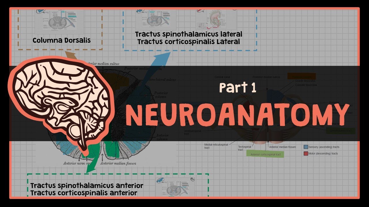
Overview Dasar Sistem Neurologi : #1 NEUROANATOMY

BAB 2 Sistem Koordinasi Manusia || Sistem Saraf Manusia || IPA SMP/MTs Kelas 9 Kurikulum Merdeka
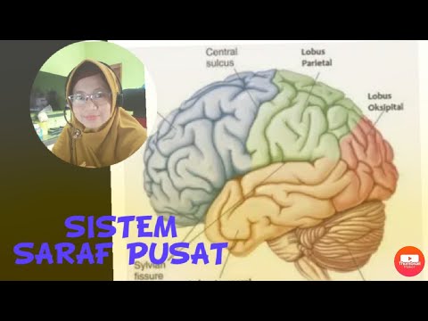
Sistem Saraf Pusat
5.0 / 5 (0 votes)
