Cranium - Part 2
Summary
TLDRThis educational video explores the intricacies of the human skull, focusing on the neurocranium, sphenoid bone, and their topography. It delves into the sphenoid bone's components, including the lesser and greater wings and the pterygoid process, highlighting the sinuses and foramina. The video also covers the ethmoid bone's structure, featuring the cribriform plate, perpendicular plate, and ethmoidal cells. It touches on the maxilla, nasal bones, and mandible, providing a comprehensive overview of the skull's key features.
Takeaways
- 💡 The video discusses the anatomy of the skull, focusing on four main topics: neurocranium, viscerocranium, topography, and important structures.
- 🔍 The sphenoid bone is located in the posterior part of the skull and consists of several parts including the body, lesser wing, greater wing, and the pterigoideus process.
- 🧠 The sphenoid sinus is observed within the body of the sphenoid, with the right and left sphenoid sinuses separated by the septum sphenoidalis.
- 👁️🗨️ On the superior surface of the sphenoid body, the sella turcica is found, which will be occupied by the pituitary gland, and laterally, there are the carotid sulci.
- 🕳️ The superior orbital fissure is a narrow gap between the greater and lesser wings of the sphenoid bone.
- 🦴 The greater wing of the sphenoid has five foramina: the cerebral, temporal, infratemporal, maxillary, and orbital.
- 🗝️ The pterygoid process has two laminae: the lateral and medial, with the medial lamina housing the fossa scaphoidea and the canalis pterygoideus.
- 🦴 The ethmoid bone is composed of the cribriform plate, perpendicular plate, and the ethmoidal labyrinth.
- 👃 The nasal bone articulates with the frontal bone to form the frontonasal structure, and with the maxilla to form the nasomaxillary structure.
- 🦷 The maxilla is the bone that forms the upper jaw and a large part of the human face, articulating with the frontal bone, sphenoid, ethmoid, and palatine bones.
Q & A
What are the four main topics discussed in the video about the cranial bones?
-The four main topics discussed in the video are neurocranium, viscerocranium, topography, and important structures.
Where is the sphenoid bone located?
-The sphenoid bone is located in the skull, specifically in the splanchnocranium.
What are the parts of the sphenoid bone?
-The sphenoid bone consists of several parts including the body (corpus), lesser wing (ala minor), greater wing (ala major), and the pterygoid process (processus pterygoideus).
What are the sinuses found within the sphenoid bone?
-Within the body of the sphenoid bone, there are sinuses such as the sphenoid sinus (sinus sphenoidalis), the right (dexter) and left (sinister) sphenoid sinuses, which are separated by a septum called the septum sphenoidalis.
What is the function of the sella turcica on the sphenoid bone?
-The sella turcica is a saddle-shaped depression on the body of the sphenoid bone that houses the pituitary gland (glandula pituitaria).
What are the five fossae found in the greater wing of the sphenoid bone?
-The five fossae in the greater wing of the sphenoid bone are the cerebral fossa (facies cerebralis), temporal fossa (facies temporalis), infratemporal fossa, maxillary fossa (facies maxillaris), and orbital fossa (facies orbitalis).
What are the four margins of the greater wing of the sphenoid bone?
-The four margins of the greater wing of the sphenoid bone are the zygomatic margin (margo zygomaticus), frontal margin (margo frontalis), parietal margin (margo parietalis), and the squamous margin (margo squamous).
What is the function of the pterygoid process of the sphenoid bone?
-The pterygoid process (processus pterygoideus) of the sphenoid bone has two laminae, the lateral lamina and the medial lamina. It is involved in the formation of the pterygopalatine fossa and provides attachment for various muscles and ligaments.
What are the components of the ethmoid bone as mentioned in the video?
-The ethmoid bone consists of the cribriform plate (lamina cribrosa), the perpendicular plate (lamina perpendicularis), and the ethmoidal labyrinth (labyrinthus ethmoidalis).
What are the three parts of the palatine bone?
-The palatine bone consists of two horizontal plates (lamina horizontalis) and a perpendicular plate (lamina perpendicularis), as well as three processes: the palatine process (processus palatinus), the orbital process (processus orbitalis), and the pyramidal process (processus pyramidalis).
What is the maxilla's role in the facial structure?
-The maxilla is a bone that forms the upper jaw and a major part of the facial skeleton. It articulates with the opposite maxilla, the frontal bone, the sphenoid, the ethmoid, the zygomatic, the palatine, and the nasal bones.
Outlines

このセクションは有料ユーザー限定です。 アクセスするには、アップグレードをお願いします。
今すぐアップグレードMindmap

このセクションは有料ユーザー限定です。 アクセスするには、アップグレードをお願いします。
今すぐアップグレードKeywords

このセクションは有料ユーザー限定です。 アクセスするには、アップグレードをお願いします。
今すぐアップグレードHighlights

このセクションは有料ユーザー限定です。 アクセスするには、アップグレードをお願いします。
今すぐアップグレードTranscripts

このセクションは有料ユーザー限定です。 アクセスするには、アップグレードをお願いします。
今すぐアップグレード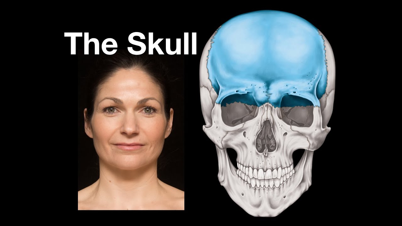


5.0 / 5 (0 votes)
