Humerus bone osteology Animation : Bony mandmarks, Development and Clinical anatomy 🦴🦴🦴
Summary
TLDRThe video script offers an in-depth look at the anatomy of the humerus, the largest long bone in the upper limb. It details its structure from the head to the lower end, highlighting key features such as the anatomical and surgical neck, greater and lesser tubercles, intertubercular sulcus, and the shaft's borders and surfaces. The script also discusses ossification centers, clinical correlations including nerve associations and common fracture sites, and touches on shoulder joint dislocation, particularly the inferior type.
Takeaways
- 📐 The humerus is the largest and longest bone in the upper limb, with its upper end comprising the head, neck, greater and lesser tubercles, and the intertubercular sulcus.
- 🔺 The head of the humerus is smooth,球形 (spherical) in shape, and covered with articular cartilage, articulating with the glenoid cavity of the scapula to form the shoulder joint.
- 💡 The anatomical and surgical necks of the humerus are important for understanding the bone's structure and potential sites of injury or fractures.
- 🏥 The surgical neck is a common site for fractures due to its location at the junction between the upper end and the shaft of the humerus.
- 💪 The greater and lesser tubercles are prominent features of the humerus, contributing to the shoulder's shape and providing attachment points for various ligaments and muscles.
- 🏋️♂️ The intertubercular sulcus, also known as the bicipital groove, houses the long head of the biceps brachii and the ascending branch of the anterior circumflex humeral artery.
- 📐 The shaft of the humerus is cylindrical above and triangular below, with three borders and three surfaces that are significant for muscle attachment and bone structure.
- 🔗 The lower end of the humerus is broad and has both an articular part (capitulum) and a non-articular part, with the radial fossa being a notable feature.
- 🦴 The determination of the side of the humerus can be made by observing the position of the head, lesser tubercle, intertubercular sulcus, and medial epicondyle.
- 🧬 The humerus develops from multiple ossification centers, with the upper end fusing by age seven and the lower end by age 14, while the full fusion with the shaft occurs in early adulthood.
- 🚑 Common fractures of the humerus include the surgical neck, shaft, and supracondylar region, which can result in injury to nerves and blood vessels in the area.
Q & A
What is the humerus and where is it located?
-The humerus is the largest and strongest long bone in the upper limb, located in the arm.
Describe the upper end of the humerus.
-The upper end of the humerus consists of the head, neck, greater and lesser tubercles, and the intertubercular sulcus. The head is smooth, medially backwards and upwards directed, and articulates with the glenoid cavity of the scapula to form the shoulder joint.
What are the two necks of the humerus and their functions?
-The anatomical neck is the constricted portion surrounding the head and gives attachment to the shoulder joint capsule, except medially. The surgical neck is the junction between the upper end and the shaft and is a common site for fractures.
What is the significance of the greater and lesser tubercles on the humerus?
-The greater tubercle forms the prominence of the shoulder and has the lateral margin that gives attachment to the transverse ligament. The lesser tubercle is smaller, located in the anterior aspect of the upper end, and its lateral margin also gives attachment to the transverse ligament.
What does the intertubercular sulcus contain?
-The intertubercular sulcus, also known as the bicipital groove, contains the long head of the biceps brachii and the ascending branch of the anterior circumflex humeral artery.
Describe the shaft of the humerus and its features.
-The shaft of the humerus is cylindrical above and flat, triangular below, consisting of three borders (anterior, lateral, and medial) and three surfaces (anteromedial, anterolateral, and posterior). The anterior border extends from the lower end of the greater tubercle to above the radial fossa, while the medial border extends from the lesser tubercle to the medial epicondylar sulcus.
What are the two fossae located at the lower end of the humerus and their functions?
-The radial fossa is a hollow depression on the posterior surface of the lower end, lodging the radial head of the radius during elbow extension. The coronoid fossa is a small depression situated just above the trochlea, lodging the anterior margin of the coronoid process of the ulna during elbow flexion.
How many centers of ossification are involved in the development of the humerus and when do they typically appear?
-There is one primary center for the shaft, three secondary centers for the upper end, and four secondary centers for the lower end of the humerus. The upper end centers appear in the first year for the head, third year for the greater tubercle, and fifth year for the lesser tubercle. The lower end centers appear in the second year for the capitulum and lateral lip of the trochlea, in the 10th year for the medial part of the trochlea, and in the 16th year for the lateral epicondyle.
What are the clinical implications of humerus fractures and common sites?
-Common sites for humerus fractures include the surgical neck, shaft, and the supracondylar region. These fractures can injure nerves such as the auxiliary nerve around the surgical neck, radial nerve in the radial groove, and the ulnar nerve behind the medial epicondyle. Supracondylar fractures often occur in young individuals due to falls on outstretched hands.
What is the most common dislocation involving the humerus and why does it occur?
-The most common dislocation involving the humerus is the inferior dislocation of the shoulder joint. This occurs more frequently due to the loose nature of the capsule and the large size of the head compared to the glenoid cavity.
Outlines

このセクションは有料ユーザー限定です。 アクセスするには、アップグレードをお願いします。
今すぐアップグレードMindmap

このセクションは有料ユーザー限定です。 アクセスするには、アップグレードをお願いします。
今すぐアップグレードKeywords

このセクションは有料ユーザー限定です。 アクセスするには、アップグレードをお願いします。
今すぐアップグレードHighlights

このセクションは有料ユーザー限定です。 アクセスするには、アップグレードをお願いします。
今すぐアップグレードTranscripts

このセクションは有料ユーザー限定です。 アクセスするには、アップグレードをお願いします。
今すぐアップグレード関連動画をさらに表示

Ossos dos Membros Superiores: Sistema Esquelético 5/5
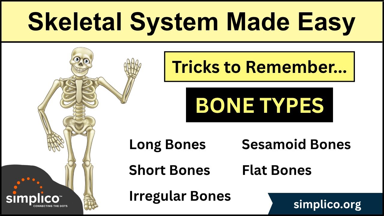
Skeletal System: Types of Bones in Under 10 Minutes [Anatomy Physiology Human Body]

Muscles of the Upper Arm (glenohumeral and elbow joints)
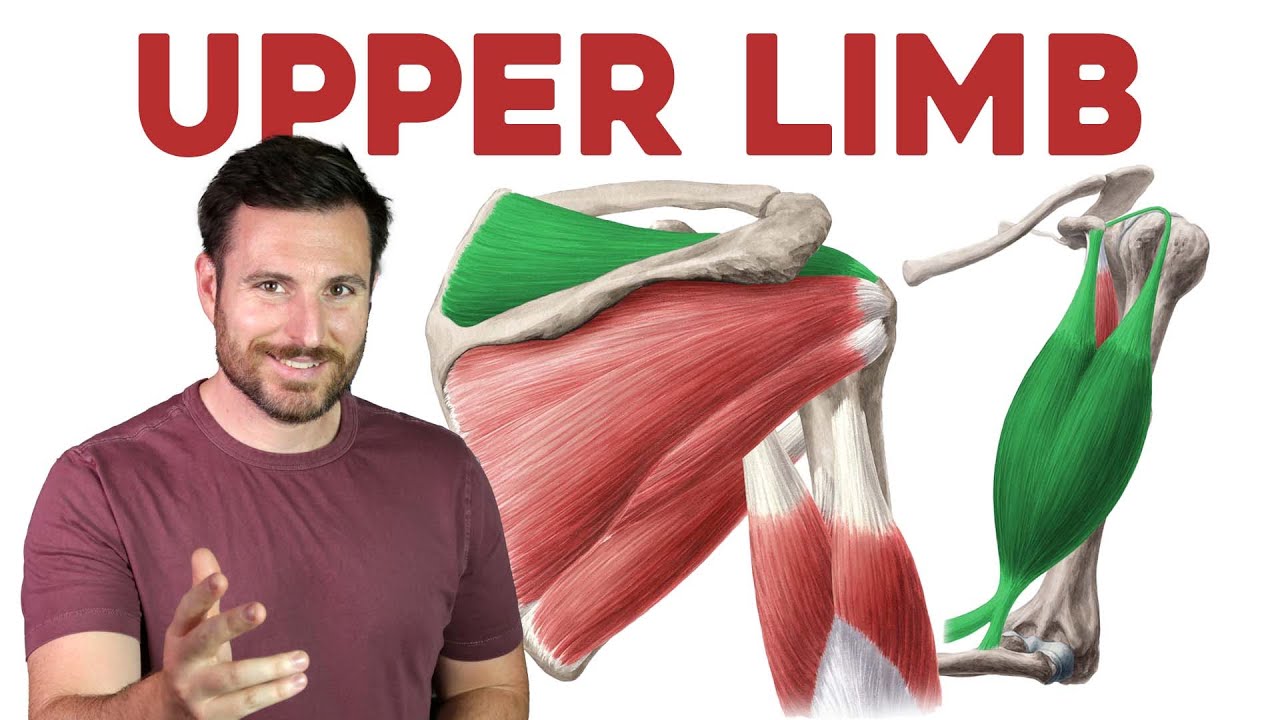
How To Remember Every Muscle in the Upper Limb and Arm | Corporis
Musculi Regio Brachii (video 16)
Clavicle Bone Anatomy Animation : Bony landmarks and Development
5.0 / 5 (0 votes)
