Avaliação Semiológica e Diagnóstico em Pequenos Animais - Aula 10 a 10.1
Summary
TLDRThis veterinary lecture delves into the anatomy of the female urinary and reproductive systems in small animals, focusing on structures such as the ovaries, uterus, and vagina. Key points include the importance of understanding anatomical locations, such as the ovaries’ proximity to the duodenum and spleen, for surgical procedures like spaying. The speaker emphasizes the need for knowledge of vascularization and fixation ligaments, like the suspensory ligament and uterine ligaments, to prevent complications during surgery. Additionally, the role of various anatomical features in clinical diagnosis and surgical intervention, particularly in cases like pyometra, is highlighted.
Takeaways
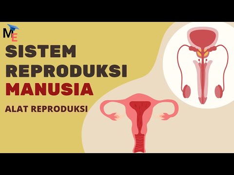
- 😀 The female reproductive tract includes the ovaries, uterine tubes, uterus, cervix, vestibule, vagina, and vulva.
- 😀 Knowing the anatomy and location of reproductive structures is crucial, especially for procedures like castration.
- 😀 The ovaries are located near the duodenum and the spleen, and their proximity to major blood vessels makes castration surgeries risky due to potential hemorrhage.
- 😀 The ovarian blood supply comes from the ovarian arteries and veins, which are high-pressure vessels requiring careful management during surgery.
- 😀 The fallopian tubes (uterine horns) are located laterally to the ovaries and are long and narrow in shape.
- 😀 The uterus in female dogs is typically shaped with small body and long uterine horns, whereas in cats, the uterus is shaped like a 'Y'.
- 😀 The cervix acts as a barrier to prevent the ascent of bacteria into the uterus, especially when not in estrus, and is critical during pregnancy and parturition.
- 😀 The uterus is supported by ligaments, including the broad ligament and round ligament, which are important in both anatomical and surgical considerations.
- 😀 The vagina is relatively long in female dogs and cats, and has structures like the vestibule, clitoris, and vulva for examination.
- 😀 A good understanding of the anatomical structure and blood supply is essential for successful clinical examination, diagnostics, and surgical procedures.
Q & A
What are the key components of the female reproductive system discussed in the transcript?
-The key components of the female reproductive system discussed are the ovaries, uterine tubes (or cornual parts), the body of the uterus, cervix, vestibule, vagina, and vulva.
Why is the anatomical knowledge of the female reproductive system important in small animal clinical practice?
-Anatomical knowledge is important for performing semiological exams, diagnosing issues, and conducting surgeries such as castration to avoid future reproductive problems, especially in small animals.
Where are the ovaries located in the female animal body?
-The right ovary is located near the duodenum and the right lateral abdominal wall, while the left ovary is located near the spleen.
What is the significance of the suspensory ligament in the female reproductive system?
-The suspensory ligament is crucial for the support of the ovaries and the uterus, especially during pregnancy. It is visible to the naked eye and aids in the ventral traction of the ovaries during surgical procedures.
How are the ovaries vascularized and why is this important during surgery?
-The ovaries are vascularized by the ovarian artery and vein, which are branches of the abdominal aorta. This is important because they carry high-pressure blood, making it critical to prevent hemorrhage during surgeries like castration.
What is the structure and function of the uterine tubes (cornu uterinum)?
-The uterine tubes, or cornua uterinum, are small, measuring 5-8 cm long, and are located lateral and medial to the ovarian bursa. They are important for transporting eggs from the ovaries to the uterus.
What are some common pathologies associated with the uterus in small animals?
-Common uterine pathologies in small animals include cystic endometrial hyperplasia, which is excessive secretion from the endometrium, and pyometra, which is an infection leading to uterine inflammation.
What is the role of the cervix in the female reproductive system?
-The cervix acts as a barrier, especially during pregnancy, to prevent the ascension of bacteria and other microorganisms into the uterus. It also produces a mucus plug before and during pregnancy to seal the uterus.
How is the uterine body positioned in relation to the rest of the reproductive organs?
-The uterine body is located in the pelvic region, and it is much smaller in size compared to the uterine horns, which are longer and located in the abdominal area.
What role does the large ligament play in the female reproductive system?
-The large ligament (ligamentum latum) helps fixate the uterus to the dorsal and lateral walls of the pelvic cavity, ensuring its stability during pregnancy and other conditions.
Outlines

Cette section est réservée aux utilisateurs payants. Améliorez votre compte pour accéder à cette section.
Améliorer maintenantMindmap

Cette section est réservée aux utilisateurs payants. Améliorez votre compte pour accéder à cette section.
Améliorer maintenantKeywords

Cette section est réservée aux utilisateurs payants. Améliorez votre compte pour accéder à cette section.
Améliorer maintenantHighlights

Cette section est réservée aux utilisateurs payants. Améliorez votre compte pour accéder à cette section.
Améliorer maintenantTranscripts

Cette section est réservée aux utilisateurs payants. Améliorez votre compte pour accéder à cette section.
Améliorer maintenantVoir Plus de Vidéos Connexes


5.0 / 5 (0 votes)
