Circulatory System and Pathway of Blood Through the Heart
Summary
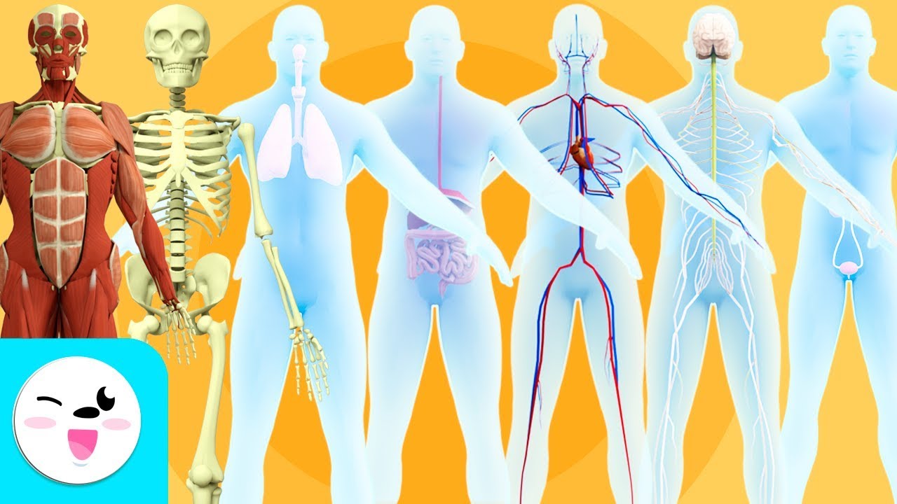
TLDREl script del video ofrece una introducción al sistema circulatorio humano, destacando su importancia en el transporte de glucosa, oxígeno y dióxido de carbono. Se menciona la sangre como el medio de transporte, y se describen sus componentes: plasma, glóbulos rojos, glóbulos blancos y plaquetas. La anatomía del corazón se divide en dos partes: una para sangre desoxigenada y otra para sangre oxigenada, con cuatro cámaras y válvulas que evitan la refluxo sanguíneo. Se sigue la trayectoria de la sangre desde la yema del dedo hasta su oxigenación en los pulmones y distribución a través del cuerpo. Además, se destaca la importancia de la coordinación en los latidos cardíacos y se mencionan algunas afecciones cardíacas, como el defecto septal auricular, que pueden alterar la flujo sanguíneo. El video concluye animando a la curiosidad y la exploración del campo de la cardiología.
Takeaways
- 🍳 La digestión del desayuno y el transporte del glucógeno en el cuerpo es un proceso fascinante que involucra al sistema circulatorio.
- 💨 El oxígeno se transporta en la sangre y el dióxido de carbono se elimina a través del proceso respiratorio.
- 🔍 El sistema circulatorio es complejo y este video ofrece solo una introducción a su funcionamiento.
- 🩸 La sangre es un medio de transporte para glucógeno y gases, y su color rojo varía según la cantidad de oxígeno presente.
- 🌡️ La sangre tiene múltiples funciones, incluyendo mantener el pH, la temperatura y la presión osmótica, lo que es crucial para el homeostasis.
- 🛑 Las venas y arterias a menudo se representan en diagramas de color azul o rojo para indicar sus concentraciones de oxígeno, pero la sangre no es de esos colores.
- 🧫 La sangre está compuesta por plasma, glóbulos rojos, glóbulos blancos y plaquetas, cada uno con funciones específicas.
- 🚫 Las arterias generalmente llevan sangre desde el corazón, mientras que las venas la llevan hacia él.
- 🫀 El corazón humano está dividido en dos compartimentos separados: uno desoxigenado y otro oxigenado.
- ❤️ Los valvulas del corazón son estructuras de un solo sentido que ayudan a separar las cámaras y evitan la refluxión de la sangre.
- 🔄 El recorrido de la sangre a través del corazón comienza en la vena cava inferior, continúa hasta los pulmones y luego se distribuye por todo el cuerpo a través de la aorta.
- 🚑 El suministro de sangre al corazón ocurre a través de las arterias coronarias, y en caso de afecciones cardíacas, pueden requerirse medicamentos o cirugía.
Q & A
¿Cómo se transporta la glucosa después de la digestión de la comida?
-La glucosa se transporta a través del sistema circulatorio, principalmente en las hemoglobinas de los glóbulos rojos sanguíneos.
¿Cómo se transporta el oxígeno en el cuerpo después de inhalarlo?
-El oxígeno se une a la hemoglobina en los glóbulos rojos y es transportado a través de las arterias hasta los tejidos y órganos del cuerpo.
¿Cómo se elimina el dióxido de carbono del cuerpo?
-El dióxido de carbono se transporta desde los tejidos y órganos a través de la sangre de regreso a los pulmones, donde es exhalado al ambiente.
¿Cuáles son las funciones principales del sistema circulatorio?
-El sistema circulatorio tiene como funciones mantener la homeostasis, transportar hormonas, nutrientes y gases, y ayudar en la coagulación de la sangre.
¿Por qué la sangre humana es roja?
-La sangre es roja debido a la presencia de la hemoglobina, una proteína que contiene hierro en los glóbulos rojos sanguíneos.
¿Cómo se divide la sangre en el corazón humano?
-La sangre en el corazón humano se divide en una parte desoxigenada y otra oxigenada, con dos compartimentos separados y distintos dentro del corazón.
¿Cuáles son las estructuras que ayudan a separar las cámaras del corazón y a prevenir el reflujo de sangre?
-Las válvulas cardíacas son estructuras de un solo sentido que ayudan a separar las cámaras del corazón y a prevenir el reflujo de sangre.
¿Cómo se llama la vía por la que la sangre desoxigenada llega al corazón?
-La sangre desoxigenada llega al corazón a través de la vena cava, específicamente a través de la vena cava inferior.
¿Qué sucede con la sangre en los pulmones?
-En los pulmones, los glóbulos rojos sanguíneos absorben oxígeno y liberan dióxido de carbono, convirtiendo la sangre en oxigenada.
¿Cómo se llama la arteria que lleva la sangre oxigenada a todo el cuerpo?
-La aorta es la arteria principal que transporta sangre oxigenada a todo el cuerpo.
¿Cómo recibe el corazón su propia fuente de sangre para oxígeno y glucosa?
-El corazón recibe su propia fuente de sangre a través de las arterias coronarias, que se ramifican de la aorta y finalmente entregan sangre a los capilares del corazón.
¿Qué es un defecto en la septum interauricular y cómo afecta el flujo sanguíneo?
-Un defecto en la septum interauricular es una abertura en la pared muscular que separa el lado derecho y el lado izquierdo del corazón, lo que puede causar una mezcla de sangre rica en oxígeno con sangre pobre en oxígeno, pudiendo generar problemas a largo plazo como taquicardia, accidente cerebrovascular o fallo cardíaco en casos severos.
Outlines
😀 Introducción al Sistema Circulatorio
Este primer párrafo introduce el tema del sistema circulatorio, destacando su importancia en el transporte de glucosa y gases en el cuerpo humano. Se menciona que la sangre es roja y su color puede variar según la concentración de oxígeno presente. Además, se aclara un malentendido común sobre las venas y arterias, que a menudo se representan de color azul o rojo en diagramas, pero en realidad no son de esos colores. La sangre tiene múltiples funciones, incluyendo el mantenimiento de la homeostasis, y está compuesta por plasma y componentes celulares como glóbulos rojos, glóbulos blancos y plaquetas. La hemoglobina, una proteína conteniendo hierro en los glóbulos rojos, es la fuente del color rojo de la sangre. Finalmente, se describe la anatomía del corazón, dividido en dos compartimentos: uno desoxigenado y otro oxigenado, y se mencionan las arterias y venas, así como los capilares donde ocurre la entrega de oxígeno y la recolección de dióxido de carbono.
💓 El Recorrido de la Sangre a Través del Corazón
El segundo párrafo sigue el recorrido de la sangre desoxigenada desde una yema de un dedo del pie hasta su oxigenación en los pulmones y posterior distribución a través del cuerpo. Se describe con detalle la secuencia a través de las diferentes cámaras del corazón: la aurícula derecha y ventriculo derecho, y luego la aurícula izquierda y ventriculo izquierdo, pasando por las válvulas tricúspide, pulmonar, mitral (bícuspide) y aórtica. Se destaca la importancia de las válvulas como estructuras de un solo sentido que evitan la refluxo sanguíneo y separan las cámaras cardíacas. Además, se menciona el suministro de sangre al propio corazón a través de las arterias coronarias y las venas coronarias. Se invita al espectador a trazar de nuevo el recorrido sanguíneo y se destaca la complejidad y coordinación del ciclo cardíaco, que no se aborda en este video introductorio. Finalmente, se mencionan algunas afecciones cardíacas, como el defecto septal auricular, que pueden cambiar el flujo sanguíneo y causar problemas futuros, y se hace un llamado a la curiosidad y al interés por la cardiología.
Mindmap
Keywords
💡Circulatory system
💡Glucose
💡Oxygen
💡Carbon dioxide
💡Blood
💡Hemoglobin
💡Heart
💡Arteries
💡Veins
💡Capillaries
💡Valves
💡Cardiac cycle
Highlights
El sistema circulatorio es fascinante y altamente involucrado en el transporte de glucosa y gases en el cuerpo.
La sangre es el medio de transporte de glucosa y gases, y su color rojo varía según la cantidad de oxígeno presente.
Las venas y arterias a menudo se representan en diagramas de color azul o rojo para indicar sus concentraciones de oxígeno, pero esto es solo una convención gráfica.
La sangre humana tiene múltiples funciones, incluyendo la mantención de pH, temperatura y presión osmótica, que son cruciales para la homeostasis.
El plasma es la parte líquida de la sangre que contiene agua, proteínas, sales y lípidos.
Las células sanguíneas incluyen glóbulos rojos, que transportan gases, glóbulos blancos, que combaten infecciones, y plaquetas, que ayudan a coagular la sangre.
Los glóbulos rojos contienen una proteína con hierro llamada hemoglobina, responsable de la coloración roja de la sangre.
El corazón humano está dividido en dos compartimentos separados: uno desoxigenado y otro oxigenado.
Las arterias generalmente transportan sangre lejana del corazón y son ricas en oxígeno, aunque hay excepciones.
Las venas generalmente transportan sangre hacia el corazón y son pobres en oxígeno, aunque hay excepciones.
Las capilarias son vasos sanguíneos pequeños donde se entrega oxígeno a los órganos y tejidos y se recoge dióxido de carbono.
El derecha del corazón (del punto de vista de la persona) bombea sangre desoxigenada, mientras que el lado izquierdo bombea sangre oxigenada.
El corazón contiene válvulas que son estructuras de un sentido para separar las cámaras y prevenir la refluxo de sangre.
La sangre desoxigenada en una yunta humana viaja a través de la vena cava inferior hasta llegar al corazón.
La sangre oxigenada viaja a través de una vena pulmonar al átrio izquierdo y luego se bombea a lo largo del cuerpo a través de la válvula aórtica y la aorta.
El corazón recibe su propia fuente de sangre a través de las arterias coronarias, que se ramifican desde la aorta.
Las venas coronarias llevan la sangre desoxigenada de vuelta al átrio derecho, donde eventualmente seguirá el camino para volver a oxigenarse.
El corazón humano late más de 100,000 veces al día, lo que hace que la coordinación de cada latido y la dirección de la sangre sean significativas.
Las afecciones cardíacas, como el defecto septal auricular, pueden cambiar el flujo de la sangre y causar problemas futuros como taquicardia, accidente cerebrovascular o insuficiencia cardíaca.
Hay avances continuos en el tratamiento de condiciones cardiovasculares, y se anima a los interesados en la cardiología a explorar los vínculos de lectura adicional en los detalles del video.
Transcripts
Captions are on! Click CC at bottom right to turn off.
Did you have breakfast this morning?
Did you ever wonder after you eat breakfast and your food is digested, how is that glucose
getting transported around your body?
Or as you take a breath in, how does that oxygen get transported around?
Or when you exhale, how does that carbon dioxide get transferred out?
Are these only questions I ponder?
Maybe.
But your circulatory system is absolutely fascinating and highly involved in this.
In this short intro video of the circulatory system, we will mention some basics about
its functions and trace the pathway of how blood travels through your heart, but please
know before we get started, there are gigantic textbooks on the circulatory system itself.
So, obviously, this video is just an intro.
We’re going to first talk about blood: the medium of how we transport glucose and gases.
As we mention in our body systems intro video from many years ago- there are some misconceptions.
Human blood is red and always red although the shade of red can vary based on how much
oxygen is in the blood.
Veins and arteries are often drawn in diagrams as blue or red to show whether they have lower
or higher concentrations of oxygen, but that’s just how it is used in most diagrams.
It doesn’t mean the blood, or the veins, or arteries are actually that color.
Veins that you see under your skin may look blue or green by the way, but that involves
the way they appear under the skin and the reason for this would make a great physics
topic ----but I digress.
Human blood has a lot of functions.
It maintains a certain pH, temperature, osmotic pressure – all of this is very important
for homeostasis.
It transports things like hormones, nutrients, and gases.
And it’s made up of different components.
One component includes plasma, the liquid portion.
Water, proteins, salts, lipids- you’ll find them in this liquid portion of blood known
as plasma.
Another component includes cellular components.
This means red blood cells, which do the transporting of gases.
White blood cells which can fight infections.
And platelets, which are actually cellular fragments, and they’re involved with helping
your blood clot.
Very important when there is damage to the body.
Red blood cells have an iron-containing protein called hemoglobin, and that is where that
red coloring of blood comes from.
So, when we’re talking about blood, and we’re just introing the circulatory system,
we’re going to focus on how this blood moves around in the human body.
Human heart anatomy observes the heart divided into two distinct and separated partitions;
a deoxygenated, or low-oxygen partition, and an oxygenated partition.
There are some human congenital heart conditions that can result in this oxygenated and deoxygenated
blood mixing, however.
More on that at the end.
Arteries generally carry blood “away” from the heart.
Think “A” for away.
Arteries are typically oxygen-rich but there are exceptions.
Veins generally carry blood “to” the heart.
Veins typically are oxygen-poor but there are exceptions.
Capillaries are small blood vessels and it is at the capillary level where oxygen is
delivered to organs and tissues and where carbon dioxide will also be picked up to travel
back to the lungs.
So, looking at this heart, the right side (and that’s the person’s right, so for
you it will look opposite) pumps deoxygenated blood and the left side pumps oxygenated blood.
We can also see 4 chambers: the right atrium and right ventricle and the left atrium and
left ventricle.
I like to remember that A comes before V in the alphabet so that helps me remember the
A’s – for atria- are at the top of the heart.
V for ventricles- are at the bottom of the heart.
Atria also have thinner walls than the thicker walled ventricles.
The heart also contains valves which we’ll see when we get to tracing the pathway of
blood.
The valves are one-way structures that help separate the chambers and also prevent backflow
of blood.
Ready to take the adventure of a lifetime?
An adventure tracing the pathway of blood through the heart?
We’re going to start with blood that is in a human toe.
This blood is deoxygenated.
It needs to get to the heart so that it can be pumped to the lungs to pick up oxygen and
then be spread throughout the body.
It’s going to get there through the vena cava.
Inferior vena cava to be specific as superior vena cava is above the heart.
The blood enters the right atrium.
The right atrium contracts, pushing the blood through the tricuspid valve into the right
ventricle.
The right ventricle contracts, pumping the blood through the pulmonary valve to the pulmonary
artery.
By the way, when you see the word “pulmonary,” it likely involves the lungs.
The pulmonary artery takes blood to the lungs where the red blood cells in the blood will
take on oxygen and release carbon dioxide.
Now this blood is oxygenated!
It needs to return to the heart so that the heart can pump it throughout the body.
The oxygenated blood travels through a pulmonary vein to the left atrium.
The left atrium contracts and the blood travels through the mitral valve, also known as the
bicuspid valve, into the left ventricle.
The left ventricle contracts and pumps the blood through the aortic valve and out a major
artery known as the aorta.
The aorta is a major artery that carries oxygenated blood throughout the body.
Now I don’t want to neglect the fact that the heart needs its own blood supply to deliver
oxygen and glucose.
The heart can receive this blood supply through coronary arteries.
Coronary arteries branch off the aorta and eventually deliver blood into capillaries.
These capillaries deliver oxygen and glucose to the heart.
Coronary veins will take the deoxygenated blood to the right atrium where the blood
will eventually travel the pathway to become oxygenated.
In fact to quiz yourself, can you pause the video and trace the pathway of blood again?
Ok, all together.
Right atrium, tricuspid valve, right ventricle, pulmonary valve, pulmonary artery, lungs,
back through the pulmonary vein, left atrium, mitral valve (bicuspid valve), left ventricle,
aortic valve, aorta…takes it to the body and then it will eventually return through
the vena cava back to the right atrium again.
*phew* It almost makes you want to turn it into a song!
But we won’t.
The significance of the pathways, how they interact, the coordination of contraction,
and many more elements are part of every beat of your heart.
A human heart beats over 100,000 times per day so it’s significant that every beat
is coordinated and blood is directed where it should go.
The complexity of the cardiac cycle – which is the coordinated sequence of the heart’s
contractions and relaxations – isn’t something this short video can go into; hopefully a
separate video on that soon.
One last thing: there are many conditions in which the heart doesn’t function correctly.
Anatomically, some heart conditions change the pathway flow of blood.
One example that we had mentioned before is an atrial septal defect.
The septum is the muscular wall that separates the right and left side of the heart.
So, a septal defect could mean an opening and oxygen-rich blood could mix with oxygen-poor
blood.
Depending on the size, this can cause future problems such as an abnormal heartbeat, stroke,
or potentially heart failure in severe cases.
Some medications may help the symptoms or surgery can be an option.
There continues to be more advancements for treating cardiovascular conditions.
If you have interest in the amazing field of cardiology, take a look at the suggested
further reading links in the video details!
Well, that’s it for the Amoeba Sisters, and we remind you to stay curious.
5.0 / 5 (0 votes)