Nervous System
Summary
TLDRThe video script delves into the diversity and specialization of cells in the human body, with a focus on the nervous system. It outlines the structure of the nervous system, including the central nervous system (CNS) and peripheral nervous system (PNS), and highlights the roles of the brain's regions. The script emphasizes the importance of neurons and glial cells in the nervous system and explains the concept of action potentials and neurotransmitters, which facilitate communication between neurons. The video aims to dispel common myths about the brain and explores the intricacies of the nervous system's function and structure.
Takeaways
- 🧬 Cell diversity is vast, with specialized cells performing unique functions in different body systems.
- 🧠 The nervous system is divided into the central nervous system (CNS) and the peripheral nervous system (PNS).
- 🌟 The CNS consists of the brain and spinal cord, while the PNS includes all other components like nerves throughout the body.
- 💡 The brain is divided into the hindbrain, midbrain, and forebrain, each with distinct functions such as regulation, alertness, and higher cognitive processes.
- 🏃♂️ The somatic nervous system (SNS) is involved with voluntary motor functions and somatic reflexes.
- 🔄 The autonomic nervous system (ANS) regulates the body's internal environment and can be further divided into the sympathetic and parasympathetic systems.
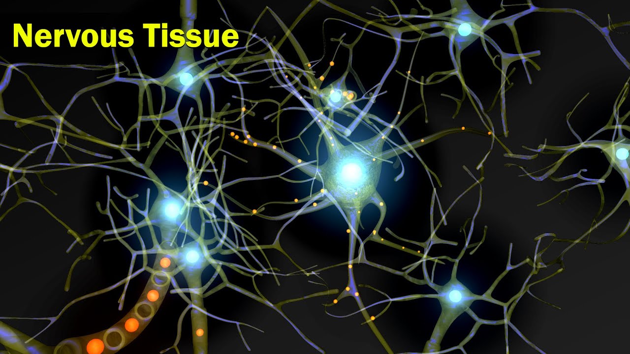
- 🌐 Neurons and glial cells are the two major types of cells in the nervous system, with neurons being the primary communicators and glial cells providing essential support.
- 🚀 Action potentials allow neurons to communicate rapidly by changing the electrical charge along the axon in a process that is 'all or none'.
- 🔌 Synapses are the junction points where neurons communicate, and neurotransmitters are released from synaptic vesicles to bind specific receptors on neighboring neurons.
- 🧠 The brain's structure and function are complex, with myths like 'we only use 10% of our brain' being debunked.
- 🔍 Ongoing research in neurology aims to better understand and treat diseases and conditions of the nervous system.
Q & A
What is the main topic of the video?
-The main topic of the video is the nervous system, its structure, function, and the cells that comprise it.
What are the two general regions the nervous system can be divided into?
-The nervous system can be divided into the central nervous system (CNS), which includes the brain and spinal cord, and the peripheral nervous system (PNS), which includes all other components of the nervous system such as nerves throughout the body.
What are the three general regions the human brain is divided into?
-The human brain is divided into the hindbrain, midbrain, and forebrain.
What are the primary functions of the medulla in the hindbrain?
-The medulla has many regulatory functions such as the regulation of breathing, blood pressure, and heart rate.
How is the peripheral nervous system (PNS) functionally divided?
-Functionally, the PNS is divided into the somatic nervous system (SNS), which is involved with motor functions of skeletal muscle, and the autonomic nervous system (ANS), which deals with the internal environment and involuntary functions.
What are the two major types of cells that make up nervous tissue?
-The two major types of cells that make up nervous tissue are neurons and glial cells.
What is the role of glial cells in the nervous system?
-Glial cells, or glia, play critical roles in the nervous system, including structural support, maintaining the blood-brain barrier, producing myelin to insulate axons, and participating in immune functions within the nervous system.
What is an action potential and why is it important for neurons?
-An action potential is a rapid change in the electrical charge of a neuron that allows it to communicate with other cells. It is important because it enables the fast transmission of signals along the neuron's axon, which is essential for the neuron's function in communication and processing information.
What happens when an action potential reaches the end of an axon?
-When an action potential reaches the end of an axon, it signals synaptic vesicles to release neurotransmitters into the synapse, the space between two neurons. These neurotransmitters then bind to specific receptors on the next neuron, potentially initiating a new action potential in that neuron.
What is the significance of the 'fight or flight' response in the sympathetic nervous system?
-The 'fight or flight' response in the sympathetic nervous system prepares the body for rapid action in response to a perceived threat or stressor. It increases heart rate, blood flow to muscles, and alertness, while temporarily reducing functions like digestion that are not immediately necessary for survival in the short term.
How does the parasympathetic nervous system contrast with the sympathetic nervous system?
-The parasympathetic nervous system is often referred to as the 'rest and digest' system. It works to conserve resources, reduce heart rate, promote digestion, and generally maintain homeostasis in the body, in contrast to the activating effects of the sympathetic nervous system.
What is the myth about the brain that the video aims to dispel?
-The video aims to dispel the myth that 'humans only use 10% of their brain.' This is incorrect, as we use virtually every part of our brain, though not all areas are active at the same time.
Outlines
🌟 Diversity of Body Cells and Overview of the Nervous System
This paragraph introduces the concept of cell diversity within the human body, emphasizing that cells are not all uniform but have specialized functions. It highlights examples such as parietal cells in the stomach that produce stomach acid, mast cells in the immune system that release histamine, and skeletal muscle cells that facilitate muscle contraction. The paragraph then transitions to the main focus of the video, the nervous system, and provides a general tour of its structure, including the central nervous system (CNS) and peripheral nervous system (PNS), and briefly touches on the functions of the brain's regions like the hindbrain, midbrain, and forebrain.
🧠 Neurons and Glial Cells: The Building Blocks of the Nervous System
The second paragraph delves into the two primary cell types of the nervous system: neurons and glial cells. It describes the structure of a neuron, including the cell body, dendrites, and axon, and explains the concept of synapses where neurons communicate. The paragraph also reiterates the importance of glial cells, which were once thought of as merely supportive but are now understood to have critical roles in maintaining homeostasis, producing myelin, and participating in immune responses within the nervous system. Additionally, the paragraph introduces the concept of action potentials, which are essential for rapid communication between neurons, and explains the resting potential of a neuron and the process of depolarization during an action potential.
🚀 The Nervous System's Functioning and Communication
The final paragraph wraps up the discussion on the nervous system by recapping the main points covered in the previous sections. It reiterates the division of the nervous system into the PNS and CNS, the latter including the brain and spinal cord, and the functional differences between the somatic and autonomic nervous systems. The paragraph also summarizes the roles of glial cells and neurons, the process of action potentials, and the release of neurotransmitters in synaptic communication. It concludes by acknowledging the complexity of the nervous system and the ongoing research in neurology, encouraging viewers to stay curious and explore careers in this field.
Mindmap
Keywords
💡Body Cell Diversity
💡Nervous System
💡Neurons
💡Glial Cells
💡Action Potential
💡Synapse
💡Central Nervous System (CNS)
💡Peripheral Nervous System (PNS)
💡Somatic Nervous System (SNS)
💡Autonomic Nervous System (ANS)
💡Myth of Brain Usage
Highlights
Body cell diversity and the specialized functions of different cell types.
Parietal cells in the stomach produce stomach acid, showcasing cell specialization within the digestive system.
Mast cells in the immune system contain histamine, critical for the inflammatory response.
Skeletal muscle cells, or muscle fibers, have a unique cylindrical structure with multiple nuclei and thin and thick filaments for muscle contraction.
The nervous system is composed of the central nervous system (CNS) and the peripheral nervous system (PNS).
The CNS includes the brain and spinal cord, serving as the command center of the body.
The PNS consists of all other components, providing sensory information to the CNS and executing motor responses.
The human brain is divided into the hindbrain, midbrain, and forebrain, each with distinct functions.
The hindbrain includes the medulla, pons, and cerebellum, regulating essential functions like breathing and balance.
The midbrain is involved in alertness, sleep/wake cycles, and motor activity.
The forebrain, including the cerebrum, is responsible for speech, thinking, reasoning, sensing, and emotions.
The PNS can be further divided into the somatic nervous system (SNS) and autonomic nervous system (ANS), based on their functions.
The SNS is involved with motor functions of skeletal muscle, including voluntary actions and somatic reflexes.
The ANS regulates the internal environment, including the gastrointestinal, excretory, endocrine, and smooth and cardiac muscle functions.
The ANS is divided into the sympathetic and parasympathetic systems, responsible for the fight or flight response and rest and digest functions, respectively.
Neurons are the primary cells of the nervous system, with structures including the cell body, dendrites, and axon.
Glial cells, or glia, are essential supporting cells in the nervous system, involved in maintaining homeostasis, producing myelin, and immune functions.
Action potentials are rapid electrical signals that allow neurons to communicate, involving the movement of ions across the neuron's membrane.
The resting potential of a neuron is characterized by a negative charge inside the cell, maintained by the sodium-potassium pump.
Depolarization occurs during an action potential as sodium ions flood into the neuron, changing the electrical charge along the axon.
The action potential is an 'all or none' event, meaning it either occurs fully or not at all, similar to a light switch.
Neurotransmitters are released from synaptic vesicles at the end of an axon, crossing the synaptic cleft to bind to receptors on the next neuron.
Transcripts
You know – sometimes we forget how different the cells in the body can
be – we kind of imagine them as all as little circle blobs when in reality,
there is so much body cell diversity. Parietal cells in the stomach as part
of the digestive system – they can make stomach acid! Thankfully, cells in other systems do not.
Mast cells as part of the immune system – they contain substances like histamine
that they can release which is critical for the inflammatory response. Skeletal
muscle cells -which are also called muscle fibers – as part of the muscular system,
they’re shaped like cylinders with multiple nuclei – and their structure
includes thin and thick filaments which are essential for muscle contraction.
We could on with all the specialized cells in all the body systems and
the cells themselves structurally sure are different – specialized for their
function. And if I had to pick my favorite specialized body cell – it’d be a neuron
–a cell that is part of the nervous system. The system that is the topic of this video.
But before we talk about neurons or other cells in the nervous
system because it’s not just neurons – let’s give a little general tour of the nervous system. Then
we’ll get to cells of the nervous system and briefly mention the action potential!
First, structure wise, you can divide the nervous system into 2 very general regions: the central
nervous system (CNS) – which consists of the brain and spinal cord - and peripheral nervous system
(PNS) –which consists of all other components of the nervous system -such as nerves throughout the
body. The PNS can provide sensory information for the CNS while the CNS can process that information
and act as a command center – the CNS can execute motor responses or regulate body mechanisms.
So, we said the CNS consisted of the spinal cord and brain. Let’s talk a bit about the
amazing human brain – although realize we are being very general here – as we are
going to divide it into 3 general regions: the hindbrain, midbrain, and forebrain.
Let’s look at the hindbrain first. It includes the medulla, pons,
and cerebellum. The medulla has many regulation functions such as the regulation of breathing,
blood pressure, and heart rate. The pons is involved with some of these type of functions
as well and also coordinating signals with this area to the rest of the brain. And the
cerebellum? Balance and movement coordination are some functions of the cerebellum.
The midbrain: deep in the brain, this area is involved in alertness and the sleep/wake cycle,
motor activity, and more. If you’ve heard the term “brainstem,” this includes some
of the structures we just mentioned: the medulla, pons, and midbrain specifically.
Finally, the forebrain. Most notably, this includes the cerebrum, which itself is
divided into two hemispheres: right and left. So many functions are done by our amazing
cerebrum depending on specific location whether it’s our speech, our thinking and reasoning,
our sensing, our emotions – check out the further reading to explore this!
The forebrain also technically includes some structures in it like the thalamus – which is
involved with sensory and motor information- and hypothalamus – which if you remember
from our endocrine system video, has major control of the endocrine system.
There are a lot of myths about the brain. One quick myth I heard all the time as a kid that
I’d like to put to rest. It’s the myth that “humans only use 10% of their brain” – it’s
not correct. We have a great reading suggestion on that as well as some others that circulate around.
Now, that was all the central nervous system (CNS). What about the peripheral nervous
system or PNS? Functionally speaking, we can further divide the PNS based on what it does.
The somatic nervous system (SNS) and autonomic nervous system (ANS). The SNS is involved with
motor functions of skeletal muscle. This will include voluntary actions under conscious
control but also somatic reflexes that involve skeletal muscle. The ANS is all about what’s
going on in the internal environment in regard to gastrointestinal or excretory
or endocrine or smooth and cardiac muscle and it also includes autonomic reflexes.
And the ANS itself can be further divided – I know, I know, there’s a lot of dividing but
stay with me – the ANS can be divided into the sympathetic and parasympathetic systems. The
sympathetic system – the shorter word of the two– helps me remember it’s part of the quick fight
or flight response. I know the whole running from a bear is a very popular example. For me,
it’d be more realistic if I was face to face with my personal nemesis: the copy machine which I may
or may not have had some very bad experiences with before – and the warning bell just rang
so you now know you have 60 seconds to get your copies – but it’s making crazy machine noises
and giving you vague warnings– this also could activate your fight or flight response. A response
that can cause your heart to race and breathing rate to increase and some things to not be active:
like the digestive system. Because if you’re desperately trying to run from a bear or take
on the copy machine, you don’t really need to be digesting your food at that very moment…right?
The parasympathetic system – longer word – this is often called rest and digest.
Heart rate will decrease, digestion will occur – again, rest and digest. Many times,
these two systems can therefore have opposite effects on the same organ.
So let’s talk about two major types of cells in the nervous system that
makes up nervous tissue. That means these are cells that you’ll find in
the central nervous system and the peripheral nervous system.
Most of the time, neurons are what come to mind. There are different types of neurons but to focus
on general neuron structure: you have the cell body – the nucleus and most other organelles
are here. There are dendrites, generally these branched structures are where signals
are received. And you have an axon – I like to think away axon! – because axons are the
fiber where normally a signal will be carried away to some other cell. The junction area
where the neuron will be communicating with another cell is called a synapse.
And the other major cell type? Glial cells. Or you can call them glia. When I was a student
and read that they were supporting cells – I don’t think the word “supporting” emphasized
to me at the time how essential they really are. Structurally, there was a lot of emphasis
on how they actually help the neurons connect in place – the word “glia” comes from a Greek
word that means glue. But glia have huge roles and they are SO much more than that. Some glial
cells keep a balance of certain chemicals in the space between cells – essential for
signaling – and maintain the blood-brain barrier which keeps a lot of substances in the body from
getting into the nervous system. Some glial cells make myelin – which goes around the
axons of neurons as something called a myelin sheath - insulates the axon and transferring
of the signal. Some glial cells produce cerebrospinal fluid which is protective
to the brain and essential for homeostasis - as well as many other critical functions. Some
glial cells have important immune function in the nervous system. These are all just a few examples.
As amazing as glial cells are, it’s time to move on to the action potential. Generally,
action potentials are recognized as something neurons do – but we did link some interesting
reads about certain glial cell types and action potentials. We’re just going to touch
on what an action potential is but we may have a future video to go into more detailed steps.
The main idea is that neurons need to be able to communicate with each other. And to do that
they’ve got to be able to receive a signal in the dendrite and carry it down the axon. And they need
to do that fast – like less than 2 milliseconds fast. The action potential makes that possible.
We can’t talk about an action potential without talking about when the neuron is at rest – meaning
when there is no signal being carried – at rest, a neuron has something called a resting
potential. The resting potential of a neuron is more negative than its surroundings – in fact
it can be measured – it generally is around -70 mv. Yes, mv, which is millivolts- it has
an electrical charge. That’s because there are ions involved inside and outside of the cell:
ions like chloride (Cl-), sodium (Na+), potassium (K+), certain anions (A-). Specifically,
sodium (Na+) and potassium (K+) play huge roles in keeping the resting potential – they should
sound familiar because we talk about the sodium potassium pump in another video and that is a pump
that helps maintain a neuron at resting potential. At rest, generally the sodium (Na+) concentration
is higher outside of the cell and the potassium (K+) concentration is higher inside the cell.
How can we remember that? How about it’s Kool to be K+ resting in the cell. But overall,
at rest, the neuron is more negative inside compared to its surroundings.
So let’s say the dendrite of the neuron receives a signal. This can generate an action potential
along the axon. An action potential is going to rapidly change the charge in the neuron along
the axon - the signal carries from one area of the axon to the next. Ion channels open
allowing Na+ to flood inside the first region of the axon. Recall Na+ is a positive ion. This
event is called depolarization – as the electric charge is becoming more positive in the axon as
Na+ floods in and most K+ channels at that moment stay closed. This spreads to the next region of
the axon and carries along. But as the action potential spreads to a new region of the axon,
the old region where the action potential already occurred will start to be restored
back - to learn more about the different channels that open and close to achieve this amazing
feat – or specific events like the undershoot or refractory period- check out the further reading
links in the video description. Eventually we hope to have an entire video on this process.
Two things to point out about this action potential. 1. If neurons are
myelinated – meaning they have myelin sheaths that insulate the axon and
assist with the transfer of the signal – the action potential can actually jump
from node to node – the nodes being areas of where it’s not myelinated.
2. Important to realize, the action potential is considered an “all or
none” thing. What we mean by that is that it either happens or it doesn’t – like a light
switch it’s either on or off – there isn’t a dimmer switch, there aren’t different levels,
it’s either off or it reaches a threshold of when it’s on and if it’s on, it’s going.
So that’s all good but what happens next? Let’s say you have an action potential and it’s going
to signal another neuron – how? Well that’s one way to introduce neurotransmitters. So the action
potential goes down the axon and gets to the axon terminals – the ends of the axon. We had mentioned
there is this space called a synapse which consists of the area between the two neurons.
The action potential can signal synaptic vesicles in that neuron to release something called
neurotransmitters. There are different types of neurotransmitters and they can be derived
from different substances: for example, amino acids or amino acid precursors. Or even a gas
such as nitric oxide although the release is different than other neurotransmitters.
Generally, when neurotransmitters are released from the synaptic vesicles,
the neurotransmitters only need to travel a small space between the neurons specified
as the synaptic cleft. Then they can bind specific receptors of the next
neuron – specific receptors to the type of neurotransmitter that binds it. The dendrite
area of the other neuron receives the signal and can start an action potential across its axon.
When we cover a lot of things, we think it’s important to recap: so,
we’ve talked about the peripheral nervous system (PNS) and the central
nervous system (CNS). Since the CNS includes the spinal cord and brain,
we also talked some about major areas of the brain. Then we focused on the PNS- how it can
be divided into the somatic nervous system (SNS) and autonomic nervous system (ANS) and then how
the autonomic nervous system (ANS) can be divided into the sympathetic and parasympathetic system.
We then explored major cell types in the nervous system: glial cells and neurons.
And since neurons can communicate with each other using an action potential,
we gave a brief overview of the action potential. We then mentioned that once
the action potential occurs, this can signal the release of neurotransmitters in the synapse
between neurons. Those neurotransmitters bind specific receptors of a neighboring neuron. Phew!
So, with such a complex system that could be so many videos long –there continues to be a
lot of research done to help diseases and conditions of the nervous system. If you
have an interest in this field– there are many careers involved in neurology
to explore. Well that’s it for the Amoeba Sisters, and we remind you to stay curious.
5.0 / 5 (0 votes)