ATM - Articulação temporomandibular - Sistema articular - Anatomia direto ao ponto!
Summary
TLDRThis video provides a concise yet detailed overview of the temporomandibular joint (TMJ), highlighting its complex biaxial structure and unique articulation. It explains the roles of the mandibular fossa, articular tubercle, and mandible head, and how these surfaces interact during mouth opening and chewing. The video covers the articular disc, synovial membrane, and joint capsule, emphasizing their function in stress absorption and joint stability. Key ligaments, including the temporomandibular (lateral) ligament, mandibular styloid, and sphenomandibular ligaments, are described, along with the role of the mandibular branch of the trigeminal nerve in joint sensitivity. The session concludes with practical insights on TMJ movement and muscle involvement.
Takeaways
- 😀 The temporomandibular joint (TMJ) is a complex biaxial joint, functioning like two joints in one.
- 😀 The TMJ allows both sliding and hinge-like movements due to its upper and lower articular surfaces.
- 😀 Key articular surfaces include the mandibular fossa, articular tubercle, and the head of the mandible.
- 😀 When the mouth opens slightly (around 2 cm), the head of the mandible stays in the mandibular fossa; when opened widely, it moves under the articular tubercle.
- 😀 The articular surfaces are covered with cartilage, and the joint contains an articular disc to absorb stress.
- 😀 The TMJ has a joint capsule enclosing the joint, lined with a synovial membrane that produces synovial fluid.
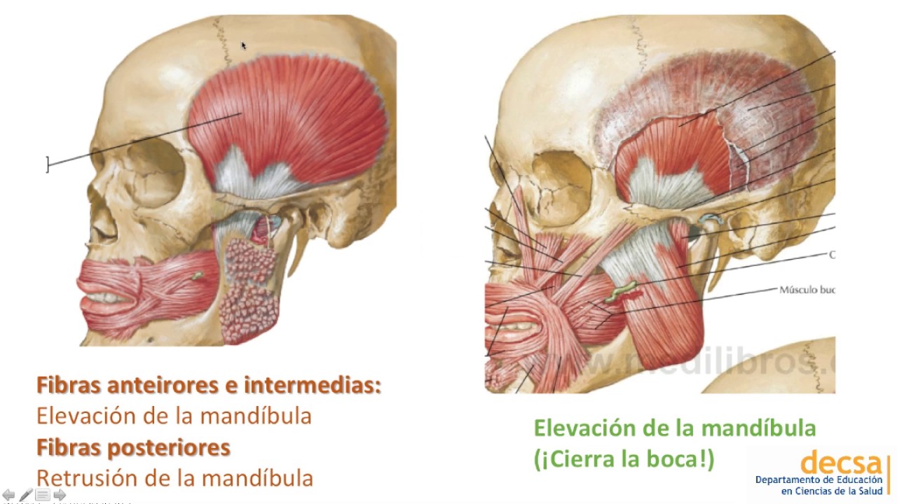
- 😀 Muscles of mastication and ligaments strengthen the TMJ and limit excessive movement.
- 😀 The temporomandibular (lateral) ligament mainly limits jaw depression, while the mandibular styloid and sphenomandibular ligaments provide secondary stabilization.
- 😀 The TMJ is highly sensitive, with sensory innervation provided by a branch of the mandibular division of the trigeminal nerve.
- 😀 Specific movements of the TMJ, such as chewing and wide mouth opening, involve coordinated actions of muscles, ligaments, and articular surfaces.
Q & A
What type of joint is the temporomandibular joint (TMJ)?
-The TMJ is a complex biaxial synovial joint that allows both hinge-like and sliding movements, functioning almost as if it were two joints in one.
Which structures form the articular surfaces of the TMJ?
-The articular surfaces are the mandibular fossa, the articular tubercle (or articular eminence) of the temporal bone, and the head of the mandible.
What is the role of the articular disc in the TMJ?
-The articular disc is a fibrocartilaginous structure that sits between the mandibular head and the temporal bone, absorbing stress and allowing smooth sliding movements.
How does the TMJ move when the mouth opens slightly versus widely?
-With slight opening (~2 cm), the head of the mandible remains in contact with the mandibular fossa. With wide opening, the mandibular head slides forward below the articular tubercle to allow greater jaw depression.
What covers the articular surfaces of the TMJ?
-All articular surfaces are covered with articular cartilage, which reduces friction and protects the bone during movement.
Which ligament primarily limits jaw depression at the TMJ?
-The temporomandibular ligament, also known as the lateral ligament of the TMJ, primarily restricts excessive lowering of the jaw.
What are the other ligaments associated with the TMJ and their roles?
-The sphenomandibular ligament supports the joint medially and runs from the sphenoid spine to the mandible. The stylomandibular ligament limits jaw protrusion but is relatively weak.
How is the TMJ stabilized besides ligaments?
-The muscles of mastication contribute to TMJ stability by controlling movements and maintaining joint alignment during function.
Which nerve provides sensory innervation to the TMJ?
-A branch of the mandibular division of the trigeminal nerve (CN V3) provides sensory innervation, making the TMJ highly sensitive.
What is the function of the synovial membrane in the TMJ?
-The synovial membrane lines the joint capsule, produces synovial fluid, and lubricates the joint to facilitate smooth movement.
Why is the TMJ considered a biaxial joint?
-It is considered biaxial because it allows movement along two axes: hinge-like rotation for opening/closing and sliding (translation) for protrusion and lateral movements.
What happens to the mandibular head during chewing with wide mouth opening?
-During wide mouth opening, the mandibular head moves forward below the articular tubercle, allowing sufficient jaw depression and facilitating chewing.
Outlines

Dieser Bereich ist nur für Premium-Benutzer verfügbar. Bitte führen Sie ein Upgrade durch, um auf diesen Abschnitt zuzugreifen.
Upgrade durchführenMindmap

Dieser Bereich ist nur für Premium-Benutzer verfügbar. Bitte führen Sie ein Upgrade durch, um auf diesen Abschnitt zuzugreifen.
Upgrade durchführenKeywords

Dieser Bereich ist nur für Premium-Benutzer verfügbar. Bitte führen Sie ein Upgrade durch, um auf diesen Abschnitt zuzugreifen.
Upgrade durchführenHighlights

Dieser Bereich ist nur für Premium-Benutzer verfügbar. Bitte führen Sie ein Upgrade durch, um auf diesen Abschnitt zuzugreifen.
Upgrade durchführenTranscripts

Dieser Bereich ist nur für Premium-Benutzer verfügbar. Bitte führen Sie ein Upgrade durch, um auf diesen Abschnitt zuzugreifen.
Upgrade durchführenWeitere ähnliche Videos ansehen
ATM y Músculos de la Masticación - Anatomía

MÚSCULOS DA ATM #medicina #anatomia #anatomy #medico #fisioterapia #anato #odontologia #odonto

Anatomia da Articulação Temporomandibular (ATM) PARTE 1
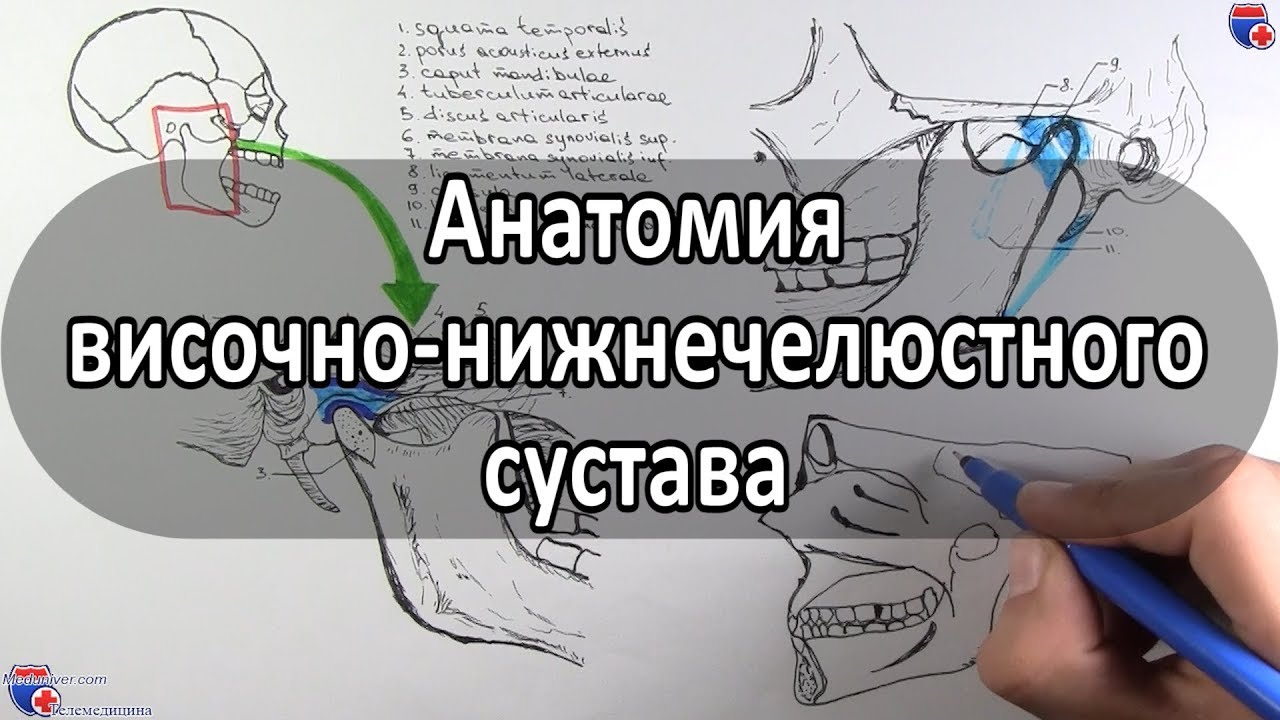
Анатомия височно-нижнечелюстного сустава - meduniver.com

Dentacademy - Anatomi Umum - Sistem Mastikasi - Part 1

Temporomandibular Joint 😲 | Biomechanics Part 2/2
5.0 / 5 (0 votes)
