All of MEDICAL PHYSICS in 7 mins - A-level Physics
Summary
TLDRThis video provides a detailed exploration of different scanning techniques used in medical diagnostics, from traditional X-rays to advanced methods like CT scans, PET scans, and ultrasounds. It explains the science behind each technique, such as how X-rays interact with materials, the process of attenuation, and the role of contrast agents in scans. The video also covers how ultrasound uses sound waves to visualize soft tissues and blood flow, offering insights into the benefits and limitations of each method. The goal is to explain how these technologies work and why they are crucial for medical imaging.
Takeaways
- 😀 Scanning a body is non-invasive, meaning doctors can identify issues without surgery.
- 😀 X-ray scans involve a cathode and anode setup where high potential difference causes electrons to emit X-ray photons.
- 😀 Four key ways X-rays are attenuated: scattering, the photoelectric effect, the Compton effect, and pair production.
- 😀 The attenuation of X-rays depends on the material being scanned, such as bone having a higher attenuation than soft tissue.
- 😀 The equation for X-ray attenuation is I/I0 = e^(-μx), where μ is the material's absorption coefficient.
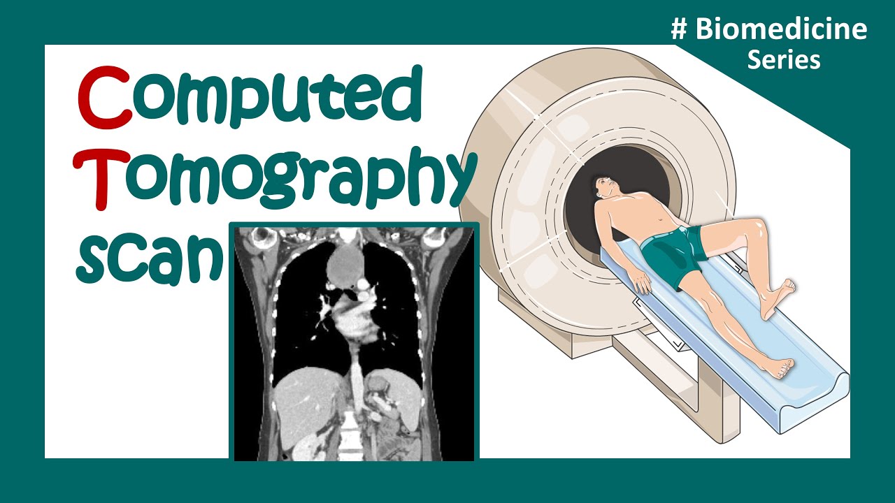
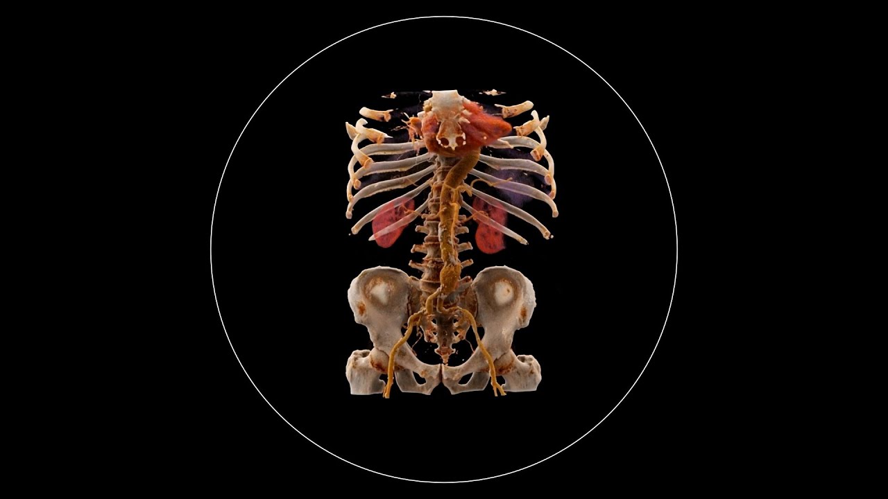
- 😀 CAT scans (CT scans) use rotating X-ray tubes and detectors to create 3D images, offering more detailed insights than regular X-rays.
- 😀 A CT scan may require contrast agents (like iodine or barium) to improve image clarity for soft tissue.
- 😀 PET scans use radioactive tracers to detect gamma emissions, helping visualize blood flow and blockages.
- 😀 Ultrasound scans use high-frequency sound waves to visualize soft tissue, with no harmful radiation involved.
- 😀 Doppler ultrasound measures blood flow by analyzing changes in the sound wave frequency reflecting off moving red blood cells.
- 😀 Ultrasound has the advantages of being quick, inexpensive, and non-invasive, with uses ranging from pregnancy monitoring to blood flow measurement.
Q & A
What is the purpose of scanning a body using X-rays?
-The purpose of scanning a body using X-rays is to provide a non-invasive method for detecting issues inside the body without the need for surgery. X-rays allow for visualizing bones, tissues, and abnormalities.
How do X-ray scans generate images?
-X-ray scans generate images by using a cathode and anode with a high potential difference. Electrons from the cathode are accelerated toward a heavy metal target like tungsten, which emits X-ray photons when the electrons collide with it. These photons are then attenuated by the material being scanned, producing an image on a film or sensor.
What are the four ways that X-ray photons can be attenuated?
-The four ways X-ray photons can be attenuated are: 1) Simple scattering (Thomson scattering), 2) The photoelectric effect (photon absorbed by an electron), 3) The Compton effect (photon scattered with lower energy), and 4) Pair production (photon converts into a particle-antiparticle pair).
What is the equation for the exponential decay of X-ray intensity?
-The equation for the exponential decay of X-ray intensity is: I/I0 = e^(-μx), where I is the intensity after passing through the material, I0 is the initial intensity, μ is the attenuation coefficient, and x is the distance traveled through the material.
Why do bones appear brighter on X-ray scans compared to soft tissues?
-Bones appear brighter on X-ray scans because they have a higher attenuation coefficient than soft tissues. This means that more X-rays are absorbed by bone, and fewer photons reach the film or sensor, creating a higher contrast.
What is a CT scan and how does it differ from a regular X-ray?
-A CT scan (Computerized Tomography) is an advanced type of X-ray scan that takes multiple X-ray images from different angles and uses a computer to combine them into a 3D image. Unlike a regular X-ray, which provides a single flat image, a CT scan offers detailed cross-sectional images of soft tissues and bones.
What is a tracer in medical imaging and how does it work?
-A tracer is a gamma-emitting substance injected into the body, such as fluorine-18 or technetium-99. It is used in scans like PET to track the movement of blood or detect issues like blockages. The tracer emits gamma rays that are detected by a gamma camera, forming an image based on where the tracer accumulates.
How does a PET scan work?
-A PET scan involves injecting a substance rich in positrons into the body. The positrons quickly annihilate with electrons, producing gamma photons that travel in opposite directions. Detectors around the patient calculate the origin of these photons, allowing for the creation of a 3D image of the area being studied.
What is the principle behind ultrasound imaging?
-Ultrasound imaging works by emitting high-frequency sound waves into the body. These sound waves are reflected off boundaries between different tissues, and the reflected waves are detected by a transducer to create an image of the internal structures. The difference in acoustic impedance between tissues determines the amount of reflection.
How is Doppler ultrasound used to measure blood flow?
-Doppler ultrasound measures changes in the frequency of sound waves as they bounce off moving blood cells. By analyzing the frequency shift (Doppler shift), it calculates the speed and direction of blood flow in veins and arteries.
Outlines

Dieser Bereich ist nur für Premium-Benutzer verfügbar. Bitte führen Sie ein Upgrade durch, um auf diesen Abschnitt zuzugreifen.
Upgrade durchführenMindmap

Dieser Bereich ist nur für Premium-Benutzer verfügbar. Bitte führen Sie ein Upgrade durch, um auf diesen Abschnitt zuzugreifen.
Upgrade durchführenKeywords

Dieser Bereich ist nur für Premium-Benutzer verfügbar. Bitte führen Sie ein Upgrade durch, um auf diesen Abschnitt zuzugreifen.
Upgrade durchführenHighlights

Dieser Bereich ist nur für Premium-Benutzer verfügbar. Bitte führen Sie ein Upgrade durch, um auf diesen Abschnitt zuzugreifen.
Upgrade durchführenTranscripts

Dieser Bereich ist nur für Premium-Benutzer verfügbar. Bitte führen Sie ein Upgrade durch, um auf diesen Abschnitt zuzugreifen.
Upgrade durchführenWeitere ähnliche Videos ansehen

MRI and CT Scan the differences
CT scan | computerized tomography (CT) scan |What is a CT scan used for? | Clinical application

Nuclear Chemistry Medical Applications
What is Computed Tomography (CT) and how does it work?

Biomedical instrumentation- CT scan (Computed Tomography)

How X-rays see through your skin - Ge Wang
5.0 / 5 (0 votes)
