Musculi Regio Brachii (video 16)
Summary
TLDRIn this educational video, Muhammad, an anatomy assistant at Sebelas Maret University, introduces the topic of the musculoskeletal system, focusing on the muscles of the upper extremity, particularly the brachium. He explains the anatomy of key muscles, including the biceps brachii, brachialis, and coracobrachialis, detailing their origins, insertions, functions, and innervations. The video also covers the triceps brachii in the posterior compartment and discusses clinical correlations related to nerve injuries and vascular supply. Overall, the session aims to enhance understanding of upper limb anatomy for students.
Takeaways
- 😀 The topic of discussion is the musculoskeletal system, focusing on the muscles of the upper extremities, particularly in the brachium (upper arm).
- 🦴 The brachium is divided into two compartments: the anterior compartment (flexors) and the posterior compartment (extensors).
- 💪 The anterior compartment contains three muscles: biceps brachii, brachialis, and coracobrachialis.
- 🔍 The biceps brachii has two heads and is responsible for flexing the forearm and supinating the forearm.
- ⚙️ The brachialis muscle originates from the humerus and assists in flexing the forearm at the elbow joint.
- 📏 The coracobrachialis muscle runs from the coracoid process to the mid-humerus and also aids in flexion.
- 🔌 All muscles in the anterior compartment are innervated by the musculocutaneous nerve, which can affect their function if injured.
- 🧠 The posterior compartment consists of the triceps brachii, which has three heads and is responsible for extending the forearm.
- 🩺 Injury to the radial nerve can result in the inability to extend the forearm.
- 🏥 Vascularization of the anterior compartment is primarily supplied by the brachial artery, while the profunda brachii artery supplies the posterior compartment.
Q & A
What is the primary focus of the video?
-The video focuses on the anatomy of the muscles in the upper extremities, particularly in the brachium region.
How are the muscles of the brachium divided?
-The muscles of the brachium are divided into two compartments: the anterior compartment, which is primarily for flexion, and the posterior compartment, which is for extension.
What are the three main muscles in the anterior compartment?
-The three main muscles in the anterior compartment are the biceps brachii, brachialis, and coracobrachialis.
What is the origin and insertion of the biceps brachii?
-The biceps brachii has two heads; the long head originates from the supraglenoid tubercle of the scapula and the short head from the coracoid process. Both insert at the radial tuberosity.
What function does the biceps brachii serve?
-The biceps brachii functions to flex the forearm at the elbow and supinate the forearm.
Where does the brachialis muscle originate and insert?
-The brachialis originates from the distal anterior surface of the humerus and inserts into the ulnar tuberosity.
What nerve innervates the muscles in the anterior compartment?
-The muscles in the anterior compartment are innervated by the musculocutaneous nerve, a branch of the lateral cord of the brachial plexus.
What are the heads of the triceps brachii and their origins?
-The triceps brachii has three heads: the long head originates from the infraglenoid tubercle of the scapula, the lateral head from the posterior surface of the humerus, and the medial head also from the posterior surface of the humerus.
What is the primary function of the triceps brachii?
-The primary function of the triceps brachii is to extend the forearm at the elbow.
What happens when the musculocutaneous nerve is injured?
-Injury to the musculocutaneous nerve can lead to dysfunction of the anterior compartment muscles, resulting in an inability to flex the elbow.
What anatomical structures are found in the antebrachium?
-The antebrachium contains important structures such as the axillary nerve, axillary artery, cephalic vein, and brachial plexus.
What clinical correlation is associated with damage to the radial nerve?
-Damage to the radial nerve can result in an inability to extend the elbow.
Outlines

Dieser Bereich ist nur für Premium-Benutzer verfügbar. Bitte führen Sie ein Upgrade durch, um auf diesen Abschnitt zuzugreifen.
Upgrade durchführenMindmap

Dieser Bereich ist nur für Premium-Benutzer verfügbar. Bitte führen Sie ein Upgrade durch, um auf diesen Abschnitt zuzugreifen.
Upgrade durchführenKeywords

Dieser Bereich ist nur für Premium-Benutzer verfügbar. Bitte führen Sie ein Upgrade durch, um auf diesen Abschnitt zuzugreifen.
Upgrade durchführenHighlights

Dieser Bereich ist nur für Premium-Benutzer verfügbar. Bitte führen Sie ein Upgrade durch, um auf diesen Abschnitt zuzugreifen.
Upgrade durchführenTranscripts

Dieser Bereich ist nur für Premium-Benutzer verfügbar. Bitte führen Sie ein Upgrade durch, um auf diesen Abschnitt zuzugreifen.
Upgrade durchführen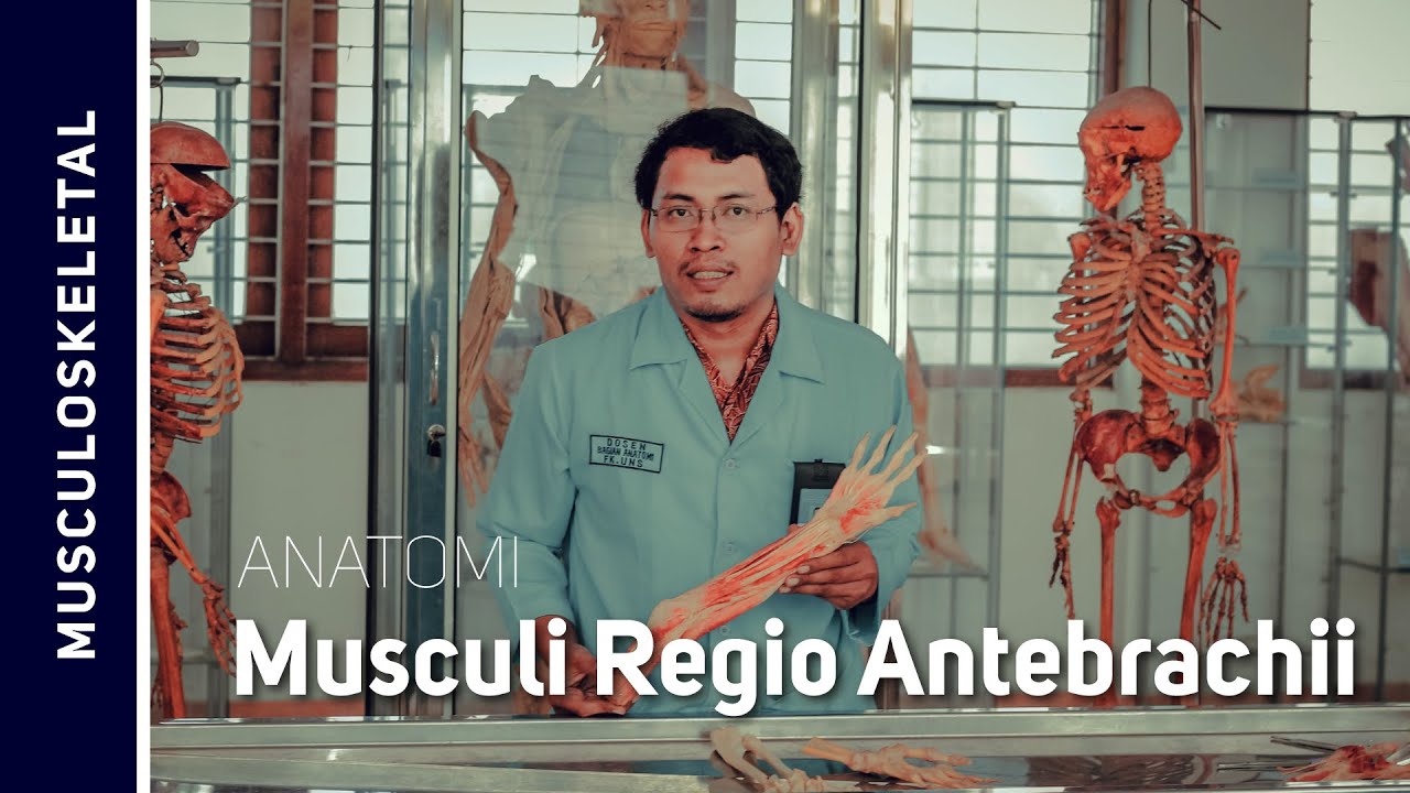


5.0 / 5 (0 votes)
