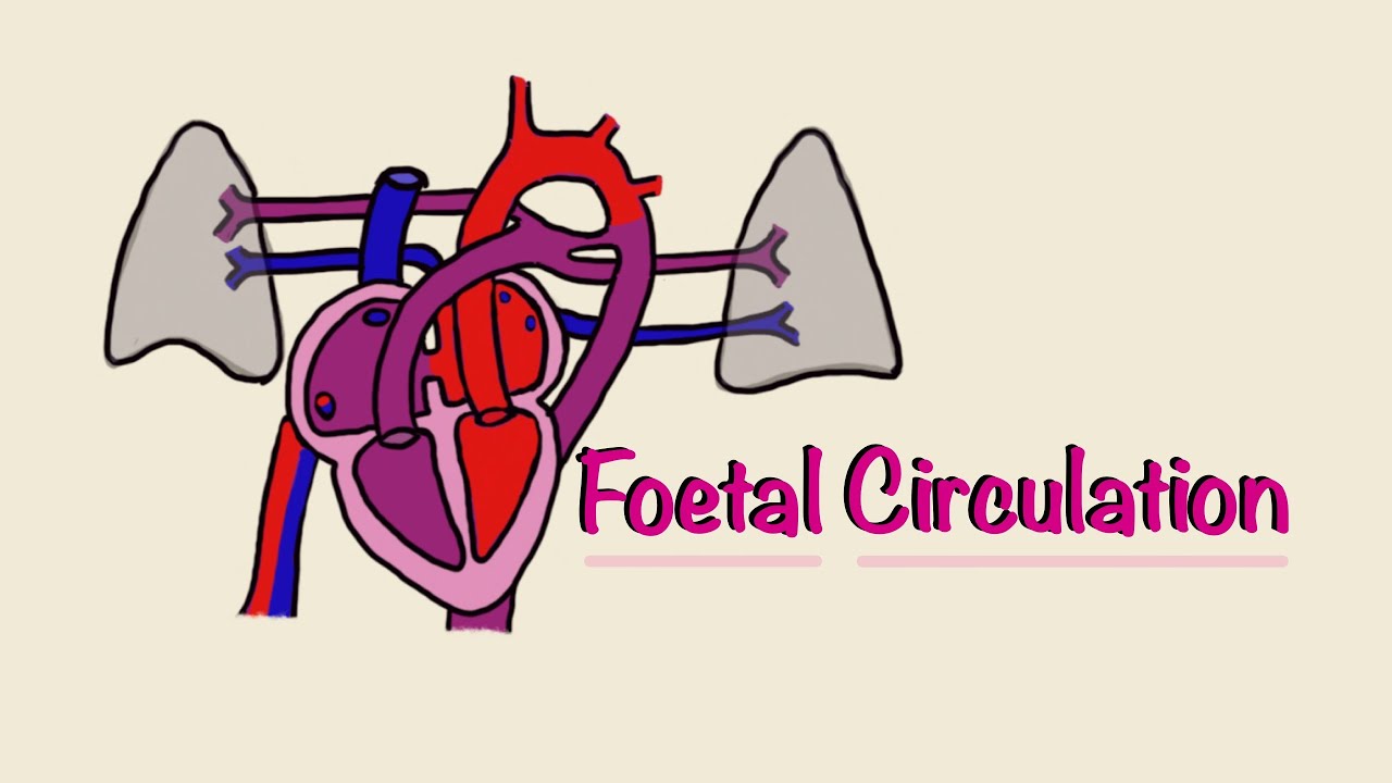
Foetal (Fetal) Circulation
Summary
TLDRThis video explains the complex process of fetal circulation, highlighting key differences from adult circulation. It covers the roles of the heart chambers, the foramen ovale, and the ductus arteriosus in allowing blood to bypass the lungs in a fetus. The placenta and umbilical cord are crucial for delivering oxygen and nutrients from the mother to the fetus, with fetal and maternal blood exchanging gases without mixing. The video concludes by describing how circulation changes after birth when the fetal structures close and adapt to adult circulation.
Takeaways
- 👶 Fetal circulation is different and more complicated than adult circulation, and relies on the placenta for oxygen instead of the lungs.
- ❤️ The fetal heart has four chambers, similar to an adult's heart, but blood bypasses the lungs through special openings.
- 🔄 Blood can flow from the right atrium to the left atrium through a hole called the foramen ovale, bypassing the lungs.
- 🩸 Another passage called the ductus arteriosus allows blood to bypass the lungs by moving from the pulmonary artery directly into the aorta.
- 🌬️ The fetal lungs have high pressure, which prevents blood from flowing into them, so the fetus depends on the placenta for oxygen.
- 👩🍼 Oxygen and nutrients are delivered to the fetus through the umbilical vein, which carries oxygenated blood from the placenta to the fetal liver.
- 🔗 The placenta is crucial as it allows maternal blood to transfer oxygen to fetal blood without mixing the two blood supplies.
- 🧬 Fetal hemoglobin has a higher affinity for oxygen than maternal hemoglobin, facilitating efficient oxygen transfer from mother to fetus.
- 🔄 Deoxygenated blood is returned to the placenta via two umbilical arteries, where it is reoxygenated by maternal blood.
- 👶 After birth, the fetal circulatory system transitions to adult circulation as the foramen ovale and ductus arteriosus close, and the umbilical vessels turn into ligaments.
Q & A
What is fetal circulation, and how does it differ from adult circulation?
-Fetal circulation is the system of blood flow in a fetus, which differs from adult circulation because the fetus does not use its lungs for oxygen. Instead, it relies on the placenta for oxygen, and it has special structures like the foramen ovale and ductus arteriosus that allow blood to bypass the lungs.
What are the key structures in the fetal heart that enable blood to bypass the lungs?
-The key structures in the fetal heart are the foramen ovale, an opening between the right and left atria, and the ductus arteriosus, which connects the pulmonary artery to the aorta. These allow blood to bypass the lungs since the fetus is not breathing air.
How does oxygenated blood from the placenta reach the fetus?
-Oxygenated blood from the placenta reaches the fetus through the umbilical vein. This vein carries the oxygen-rich blood to the fetal liver and then to the inferior vena cava, mixing with deoxygenated blood before entering the heart.
What role does the placenta play in fetal circulation?
-The placenta acts as the site of gas exchange between the maternal and fetal blood. It delivers oxygen and nutrients to the fetus via the umbilical cord and removes carbon dioxide and waste products.
How do the foramen ovale and ductus arteriosus facilitate the right-to-left shunt in fetal circulation?
-The foramen ovale allows blood to flow directly from the right atrium to the left atrium, bypassing the right ventricle and lungs. The ductus arteriosus connects the pulmonary artery to the aorta, allowing blood to bypass the lungs and flow into the systemic circulation.
Why is there no need for blood to go to the lungs in fetal circulation?
-There is no need for blood to go to the lungs in fetal circulation because the fetus does not breathe air. Oxygen is supplied through the placenta, so blood bypasses the lungs through the foramen ovale and ductus arteriosus.
How does the fetal body ensure it gets oxygen, and what makes fetal hemoglobin different from adult hemoglobin?
-Fetal hemoglobin has a higher affinity for oxygen than adult hemoglobin, allowing it to efficiently extract oxygen from maternal blood. This enables the fetus to get sufficient oxygen even from the lower-oxygen environment of the placenta.
What happens to the foramen ovale and ductus arteriosus after birth?
-After birth, the foramen ovale and ductus arteriosus close as the baby starts using its lungs for oxygen. The foramen ovale closes within hours to days, and the ductus arteriosus becomes a ligament, establishing the normal adult circulation pattern.
What is the function of the umbilical arteries in fetal circulation?
-The umbilical arteries carry deoxygenated blood and waste products from the fetus back to the placenta for reoxygenation and disposal via the maternal blood circulation.
How does the exchange of oxygen and carbon dioxide occur between maternal and fetal blood?
-Oxygen and carbon dioxide exchange between maternal and fetal blood in the placenta. Maternal blood delivers oxygen to fetal blood through diffusion, while fetal blood offloads carbon dioxide into the maternal circulation for elimination.
Outlines

Dieser Bereich ist nur für Premium-Benutzer verfügbar. Bitte führen Sie ein Upgrade durch, um auf diesen Abschnitt zuzugreifen.
Upgrade durchführenMindmap

Dieser Bereich ist nur für Premium-Benutzer verfügbar. Bitte führen Sie ein Upgrade durch, um auf diesen Abschnitt zuzugreifen.
Upgrade durchführenKeywords

Dieser Bereich ist nur für Premium-Benutzer verfügbar. Bitte führen Sie ein Upgrade durch, um auf diesen Abschnitt zuzugreifen.
Upgrade durchführenHighlights

Dieser Bereich ist nur für Premium-Benutzer verfügbar. Bitte führen Sie ein Upgrade durch, um auf diesen Abschnitt zuzugreifen.
Upgrade durchführenTranscripts

Dieser Bereich ist nur für Premium-Benutzer verfügbar. Bitte führen Sie ein Upgrade durch, um auf diesen Abschnitt zuzugreifen.
Upgrade durchführenWeitere ähnliche Videos ansehen
Foetal (Fetal) Circulation | Before and At Birth | Cardiac Physiology | Embryology

Sirkulasi Darah Fetus aka Janin

Sirkulasi Prenatal dan Postnatal

Fetal Circulation | Cardiovascular system | Step 1 Simplified
The Cardiovascular System (CVS) ❤️ 🩸 - A Simple Introduction - Biology, Anatomy, Physiology

Fetal Circulation by L. McCabe | OPENPediatrics
5.0 / 5 (0 votes)
