History of X-rays

Doctor Klioze
7 Dec 201324:20

Summary

TLDRThis episode explores the accidental discovery of X-rays by Wilhelm Conrad Roentgen in 1895, which revolutionized medicine, genetics, and manufacturing. The script delves into the history of X-rays, their initial use for entertainment, and the technological advancements that transformed them into an essential medical tool. It also discusses the physics behind X-ray production, their role in imaging, and Roentgen's decision not to patent his discovery, emphasizing his significant contribution to science and society.

Takeaways

  • 🔬 The discovery of X-rays revolutionized medicine and other fields like genetics and manufacturing.
  • 🏥 Halifax Medical Center houses the Halifax Radiology Museum, showcasing original radiology equipment from 1928.
  • 📸 X-rays are now a common part of society, used in security, manufacturing, and medical diagnostics.
  • 🧬 X-ray diffraction was key to understanding the DNA molecule's structure by scientists like Rosalind Franklin, Watson, and Crick.
  • 🌟 The first American medical X-ray was performed at Dartmouth College in 1896, marking a significant milestone.
  • 🎭 Early X-ray technology was used for entertainment, with 'bone portrait' studios offering X-ray images of body parts.
  • 🏬 The inventor of X-rays, Wilhelm Conrad Roentgen, worked at the University of Würzburg, where he made his groundbreaking discovery.
  • 🔋 The Crookes tube, an early experimental device, was used by Roentgen to produce X-rays and led to the first human radiograph.
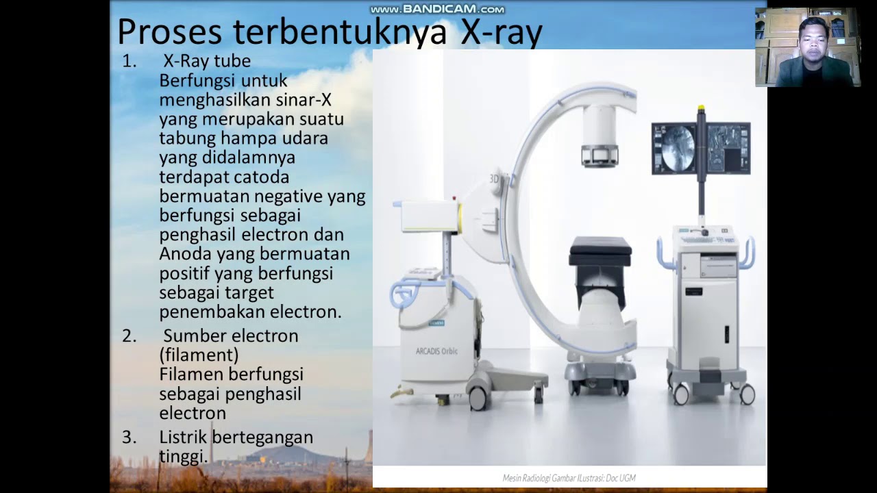
  • ⚙️ Modern X-ray tubes have evolved with a rotating anode and improved cathode design for more efficient X-ray production.
  • 🛠️ The voltage (kVp) and current (mA) settings on an X-ray machine are crucial for adjusting the image quality and penetrating power.
  • 🌌 Roentgen's discovery has had a profound impact on our understanding of the world, from genetics to material science and beyond.

Q & A

  • What significant discovery is discussed in the video script?

    -The video script discusses the discovery of X-rays, which revolutionized the field of medicine and transformed other fields such as genetics and manufacturing.

  • Where is the Halifax Medical Center located?

    -The Halifax Medical Center is located in Daytona Beach, Florida, approximately 2 miles from the world's most famous beach and a half a mile from the Daytona International Speedway.

  • Who made the Halifax Radiology Museum possible?

    -The Halifax Radiology Museum was made possible by Mr. Bud Hinkle, who was the Radiology manager from 1985 to 1995.

  • How did X-rays contribute to the understanding of DNA?

    -X-rays contributed to the understanding of DNA through the work of PhD scientist Rosalyn Franklin, who used X-ray diffraction to examine the DNA molecule, and later Watson and Crick used this information to define the double helix structure of DNA in 1953.

  • What was the term used to describe the initial public fascination with X-rays?

    -The initial public fascination with X-rays was termed 'X-ray Mania', where people were eager to get their hands on this new technology.

  • What was the first American Medical X-ray performed on?

    -The first American Medical X-ray was performed at Dartmouth College on February 3rd, 1896, and it was an image of a student's hand with a fractured distal phalanx.

  • Who was responsible for the discovery of X-rays?

    -Professor Wilhelm Conrad Roentgen, a physics professor at the University of Würzburg in Germany, is credited with the discovery of X-rays.

  • What was the Crooks tube and how was it used in the discovery of X-rays?

    -The Crooks tube was a device that produced cathode rays and was used by Roentgen in his experiments. It was a high-powered light bulb-like device evacuated of air with an anode and cathode connected to a high voltage DC power supply. Roentgen used it to discover X-rays when he noticed a faint glow from a piece of paper covered in phosphorescent material.

  • What was the purpose of the black box that Roentgen constructed during his experiments?

    -Roentgen constructed a black box to cover the Crooks tube to block any light that might interfere with his experiment to detect other types of rays.

  • How did the early X-ray technology lead to misconceptions about its capabilities?

    -Early X-ray technology led to misconceptions because people were afraid that it could be used for inappropriate purposes, such as seeing through clothing, due to its ability to see through walls and human bodies.

  • What was the first radiograph of a human taken by Roentgen?

    -The first radiograph of a human, purportedly of Roentgen's wife's hand, was taken by Roentgen after six weeks of characterizing the new X-ray. This image showed her hand with a wedding ring and is considered the first radiograph of a human hand.

Outlines

00:00

📡 The Discovery of X-rays

This paragraph discusses the accidental discovery of X-rays which revolutionized medicine, genetics, and manufacturing. The narrator stands in front of Halifax Medical Center, highlighting the Halifax Radiology Museum established by Bud Hinkle. The script describes the museum's collection of original radiology equipment from 1928 and the significant role X-rays play in modern society, from airport security to medical imaging. The historical context of X-ray's discovery in 1896 is provided, including the phenomenon of 'X-ray Mania' and early uses for entertainment. The first American medical X-ray is noted to have been performed at Dartmouth College in 1896, marking a milestone in medical imaging.

05:01

👨‍🔬 The Birth of X-ray Technology

The paragraph narrates the story of Wilhelm Conrad Röntgen, the physics professor who discovered X-rays at the University of Würzburg. The narrator recounts a visit to Würzburg to see the site of the discovery. Röntgen was experimenting with a Crookes tube, a high-vacuum tube through which a current was passed to produce cathode rays. His curiosity led him to block the tube's light to search for other types of rays. He noticed a faint glow from barium platinocyanide paper, which led to the discovery of X-rays. Röntgen conducted experiments to understand these new rays, observing how different materials affected their passage. His discovery was initially shared with the public in late 1895, and he is credited with taking the first human radiograph of his wife's hand.

10:03

👟 Early X-ray Applications and Risks

This section delves into the early applications of X-rays, such as shoe fitting fluoroscopes, which were popular in the 1940s but discontinued due to radiation exposure risks. The paragraph explains how Röntgen's Crookes tube produced X-rays and the phenomenon of bremsstrahlung, where high-speed electrons hit a surface and release X-ray energy. The inefficiency of the Crookes tube is highlighted, with most energy being lost as heat. The paragraph also discusses how the voltage between the tube's poles affects the energy of the produced X-rays, which is crucial for adjusting the power of modern X-ray tubes.

15:03

🔍 Advancing X-ray Technology

The paragraph outlines the evolution of X-ray technology from the basic cathode ray tube to more advanced designs. It describes how the anode was moved to directly face the cathode to focus X-rays in one direction, reducing the time needed to produce a radiograph. The transition from a single disc cathode to a heated element increased electron emission, improving X-ray production. Technologists learned to adjust voltage and current for optimal image quality. The paragraph concludes with the introduction of a rotating anode to distribute heat and prolong the life of the tube, essential for modern imaging techniques like CT scans.

20:05

🌌 X-rays in Medicine and Beyond

The final paragraph discusses how X-rays work, their place in the electromagnetic spectrum, and their ability to penetrate different tissues to varying degrees. It explains how X-rays create radiographs by casting shadows and how knowledge of normal radiographic anatomy aids in diagnosing diseases. The paragraph also reflects on Röntgen's decision not to patent X-rays, viewing his discovery as a gift to humanity for the benefit of medicine and industry. The script concludes by celebrating the impact of X-rays on our understanding of genetics, material science, and the universe.

Mindmap

Keywords

💡X-ray

X-rays are a form of electromagnetic radiation with wavelengths shorter than visible light but longer than gamma rays. They have the ability to penetrate many materials, which makes them useful for medical imaging. In the video, X-rays are discussed as a revolutionary discovery in medicine, allowing for the first time the visualization of the inside of the human body without invasive procedures. The script mentions the historical use of X-rays for medical diagnostics like chest and skull radiographs.

💡Wilhelm Conrad Röntgen

Wilhelm Conrad Röntgen was a German physicist who is credited with the discovery of X-rays. His work in the late 19th century led to a significant breakthrough in the field of diagnostic medicine. The script describes Röntgen's accidental discovery of X-rays while experimenting with cathode rays in a Crookes tube, which he was investigating for the possibility of other types of radiation.

💡Crookes tube

A Crookes tube is an early experimental electrical discharge tube, which contains a cathode and an anode in a mostly evacuated glass bulb. It was used by Röntgen during his experiments that led to the discovery of X-rays. The script describes how Röntgen used a Crookes tube to generate X-rays and how he noticed a glowing effect on a nearby screen, which initiated his investigation.

💡Radiology

Radiology is a medical specialty that uses imaging technology to diagnose and treat diseases. It plays a crucial role in modern healthcare, providing visual representations of the body's interior. The script discusses the establishment of radiology departments in hospitals, such as the one at Halifax Medical Center, and how radiology has evolved from its early days to become an integral part of medical diagnostics.

💡DNA molecule

The DNA molecule is the double helix structure that carries the genetic instructions for the development, functioning, growth, and reproduction of all known organisms and many viruses. In the script, it is mentioned that X-ray diffraction was used to study the structure of the DNA molecule, leading to the discovery of its double helix form by Watson and Crick, which was a monumental achievement in genetics.

💡Fluoroscope

A fluoroscope is a device that was used to obtain real-time moving images of the internal structures of the body利用X射线. The script references a handheld fluoroscope designed by Thomas Edison, which allowed people to see the bones in their hands. This illustrates the early fascination with X-rays and their use for both medical and entertainment purposes.

💡Radiation

Radiation refers to the emission of energy as electromagnetic waves or as moving subatomic particles. In the context of the video, it specifically refers to the ionizing radiation produced by X-rays, which can be harmful in high doses. The script discusses the early use of X-rays for entertainment, such as shoe fitting fluoroscopes, without understanding the risks of radiation exposure.

💡Mania

In the script, 'mania' is used to describe the frenzy and excitement that surrounded the discovery and application of X-rays. People were eager to use this new technology for various purposes, including entertainment, without fully understanding its potential dangers. This term captures the cultural impact of X-rays when they were first introduced.

💡Bremsstrahlung

Bremsstrahlung, German for 'braking radiation,' is the process by which X-rays are produced when high-speed electrons are rapidly decelerated. The script explains that this occurs when electrons hit the glass of a Crookes tube, releasing energy in the form of X-rays. This concept is fundamental to understanding how X-rays are generated.

💡Anode

In an X-ray tube, the anode is the positively charged electrode towards which electrons are accelerated. The script describes the evolution of the anode's design, from an off-centered position to a rotating anode, which improved the efficiency and focus of X-ray production. This is crucial for modern imaging techniques that require precise and powerful X-ray beams.

💡Cathode

The cathode is the negatively charged electrode in an X-ray tube, which emits electrons when heated. The script mentions the transition from a simple disc cathode to a heated element, which increases the number of electrons available for X-ray production. This advancement improved the efficiency of X-ray tubes and is essential for modern radiology.

Highlights

The discovery of X-rays revolutionized the field of medicine and other diverse fields such as genetics and manufacturing.

Halifax Medical Center in Daytona Beach, Florida, houses the Halifax Radiology Museum showcasing the history of radiology.

The original radiology equipment from 1928 is on display at the Halifax Radiology Museum.

X-rays are now an integral part of modern society, used in security, manufacturing, and medicine.

Rosalyn Franklin used X-ray diffraction to examine the DNA molecule, contributing to the discovery of its structure.

The first American medical X-ray was performed at Dartmouth College in 1896.

Early X-rays were used for entertainment, with 'bone portrait' studios offering X-ray images of hands and skulls.

Wilhelm Conrad Röntgen, a physics professor, is credited with the accidental discovery of X-rays.

Röntgen's discovery took place in a small laboratory at the University of Würzburg in Germany.

The Crookes tube, a high-powered light bulb-like device, was central to Röntgen's X-ray experiments.

Röntgen noticed a faint glow from barium platinocyanide paper, leading to the discovery of X-rays.

Röntgen spent six weeks characterizing the new form of radiation, which he named 'X' for unknown.

The first human radiograph was allegedly taken by Röntgen of his wife's hand.

The physics community immediately recognized the potential of X-rays to revolutionize medicine.

X-ray technology was used in shoe stores to fit shoes, a practice that was later discontinued due to radiation concerns.

The production of X-rays involves high-speed electrons hitting a glass tube, a process called bremsstrahlung.

Modern X-ray tubes have evolved with a focus on efficiency and safety, including the use of a rotating anode.

Röntgen's decision not to patent X-rays has had an immeasurable impact on medicine and industry.

X-rays have provided a deeper understanding of genetics, material science, and the universe.

Transcripts

play00:01

on today's episode we're going to

play00:02

discuss a discovery that basically

play00:04

revolutionized the field of medicine

play00:06

overnight over the following decades

play00:08

this same Discovery also transformed

play00:11

Fields as diverse as genetics and

play00:13

Manufacturing and the most amazing part

play00:15

of all this is that the discovery was

play00:17

completely accidental join us for this

play00:19

next half hour as we discuss the history

play00:22

of the X-ray

play00:49

I'm standing in front of the fountain

play00:50

building entrance to Halifax Medical

play00:52

Center in Daytona Beach Florida this

play00:55

historic building opened its doors for

play00:57

operation in 1928 and has been witnessed

play01:00

to the Great Depression multiple Wars

play01:02

and a strategically located

play01:04

approximately 2 miles from the world's

play01:06

most famous Beach and a half a mile from

play01:08

the world famous Daytona International

play01:10

Speedway but we're not here to soak up

play01:12

the Rays or watch the races what we're

play01:15

interested in is right through those

play01:16

doors so come on take a walk with

play01:19

me as you walk in the front entrance of

play01:22

the fountain building of Halifax Medical

play01:23

Center a few feet in on the left is this

play01:26

place right here this is the Halifax

play01:28

Radiology Museum

play01:30

made possible by Mr Bud Hinkle who was

play01:32

the Radiology manager from 1985 to 1995

play01:37

and the great thing about bud is he was

play01:38

a bit of a pack rrap never threw

play01:40

anything away which is great for us you

play01:42

got a little bit of a Americana behind

play01:44

us as I said previously Halifax opened

play01:47

its doors in 1928 complete with a

play01:49

radiology department some of the

play01:51

equipment behind me right now is the

play01:54

original equipment from that original

play01:57

radiology department and it's

play01:58

interesting to look at the Radiology

play02:01

equipment from that eror and compare it

play02:03

to what we have

play02:06

today x-rays are an intrinsic part of

play02:08

our modern society so much so that we

play02:10

take them for granted we've all been to

play02:12

the airport and had our luggage scan

play02:14

during the security process in

play02:16

manufacturing x-rays are absolutely

play02:18

imperative to examine for defects in

play02:21

Structural Materials in Airplane Wings

play02:23

in years past a PhD scientists Rosalyn

play02:26

Franklin use x-ray defraction

play02:29

information to examine the DNA molecule

play02:32

Watson and Crick used the same

play02:34

information to define the double helix

play02:36

structure of the DNA molecule in

play02:42

1953 and of course there's medical

play02:44

x-rays we've all been to the hospital

play02:46

and had a chest or a skull radiograph no

play02:49

big

play02:53

deal but let me take you back over a 100

play02:56

years ago the years 1896 and the newly

play03:00

discovered x-ray has now become a

play03:02

absolute phenomenon x-ray Mania has

play03:05

gripped the world people couldn't wait

play03:07

to get their hands on this new

play03:09

technology you got to remember at the

play03:11

end of the 19th century electricity was

play03:14

first coming into the households you got

play03:16

machines that are doing work for us and

play03:18

all of a sudden there's this new Ray

play03:21

this x-ray that can see through walls it

play03:23

can see through the human body and

play03:25

everybody wanted a piece of it and a lot

play03:27

of the early X-rays were for

play03:29

entertainment purposes as a matter of

play03:31

fact you could go up into the studios in

play03:33

New York that were called bone portrait

play03:36

studios and get a radiograph of your

play03:38

hand or your head or possibly even your

play03:40

chest get it framed and hang it up on

play03:42

the wall there was a lot of paranoia of

play03:45

course people were afraid that goggles

play03:47

at the Opera would have x-ray potential

play03:49

and you could look through a woman's

play03:50

skirt in the Opera so a lot of

play03:53

misconceptions about what the x-ray

play03:55

could actually

play03:56

do however there are a few level-headed

play03:59

individual ual who recognize the

play04:01

advantages of this new discovery and the

play04:03

first American Medical x-ray was

play04:06

performed at Dartmouth College on

play04:07

February 3rd

play04:09

1896 if you look at the image that's on

play04:12

the screen right now there's a picture

play04:14

memorializing that whole event there's a

play04:16

student sitting in the chair and there's

play04:18

two gentlemen on either side of the

play04:20

table the one on the right is a

play04:22

physicist who's actually timing the

play04:24

X-ray exposure the gentleman to the left

play04:27

was the patient's physician and this was

play04:30

the first radiograph very very primitive

play04:32

by today's standards but you can clearly

play04:34

see the patient had a fracture of his

play04:37

distal

play04:38

owna now this is a classic you're not

play04:40

going to find these anymore this was

play04:42

called a handheld fluoroscope actually

play04:45

designed by Thomas Edison and when

play04:47

X-rays were first developed they were a

play04:49

bit of an enigma people were just

play04:51

enthralled x-ray Mania everyone wanted

play04:53

to see a picture of their own hand and

play04:55

you could go mail order buy this little

play04:57

fluoroscope and they would send it to

play04:59

your home and if you could find someone

play05:01

to set up a Crooks tube for you and

play05:03

produce x-rays you could take a look at

play05:05

your own hand and basically what you

play05:06

would do is walk up to one of those

play05:08

tubes put your hand out raise the

play05:10

fluoroscope up to your eyes and actually

play05:12

see the bones of your own hand or you

play05:14

could look at your brother's face or

play05:15

your mom's foot it didn't matter it was

play05:18

great entertainment the problem of

play05:20

course is not only exposing your hand to

play05:22

a lot of excessive radiation your eyes

play05:24

were getting a pretty good dose also

play05:26

you're not going to find these anymore

play05:32

the man responsible for the discovery of

play05:34

x-rays was Professor wilhem Conrad renin

play05:37

a physics professor at the University of

play05:39

Wartburg in Wartburg Germany I took a

play05:42

trip to Wartburg myself a couple of

play05:44

years ago to check out where X-rays were

play05:46

actually discovered and looking at the

play05:49

picture that you're looking at right now

play05:50

I was able to recognize the building

play05:53

where Professor renen had his office on

play05:55

the first floor and we get out of the

play05:57

car and we go up to one of the students

play05:59

there my wife who speaks fluent German

play06:01

asks him where Professor rin's museum is

play06:04

and he had no idea and everyone we

play06:06

talked to had no idea where the physics

play06:09

department was or more specifically the

play06:11

renen museum was in the building we were

play06:13

standing in front of which was

play06:15

absolutely amazing to me here was a

play06:17

discovery that completely revolutionized

play06:19

the world medicine manufacturing as we

play06:22

discussed before and he couldn't tell us

play06:24

where the museum was but we eventually

play06:27

found it very unassuming small little

play06:30

room on the first floor of the Wartburg

play06:32

physics uh building in Wartburg Germany

play06:35

inside of that office inside that

play06:38

laboratory renken was working with a

play06:40

small device called a Crooks tube the

play06:43

crooks tube was a device that was

play06:45

essentially like a high powerered light

play06:46

bulb it was completely evacuated of any

play06:49

air and on one side was the anode and on

play06:52

the other side was the cathode the

play06:54

cathode was connected to the negative

play06:57

pole of a DC voltage high voltage you're

play07:00

talking hundreds of thousands of volts

play07:02

of course the anode was then connected

play07:04

to the positive side of the high voltage

play07:07

when this device was appropriately

play07:09

connected the tube itself would glow

play07:11

with this bluish green light and these

play07:13

were referred to as cathode rays there

play07:16

were theories at the time that these

play07:18

devices would actually produce other

play07:20

types of Ray yet discovered and renken

play07:23

was interested in possibly looking at

play07:26

some of these other types of rays to

play07:28

that end he set up his experiment in his

play07:30

first floor laboratory in the Wartburg

play07:32

University Physics Department on

play07:34

November 8th of

play07:35

1895 in this animation you see how we

play07:38

set that up all the way to the right he

play07:41

had a coil which would Supply the high

play07:43

voltage to the crooks tube which was

play07:45

sitting right in the middle of the table

play07:48

this was connected again the cathode to

play07:50

the negative pole the anode to the

play07:53

positive

play07:54

pole once the tube was appropriately

play07:56

connected again it glowed with that

play07:58

bluish green light now renen was trying

play08:01

to find other Rays possibly admitted

play08:04

from the tube so he didn't want the

play08:06

bluish green light interfering with his

play08:08

experiment to that end he constructed a

play08:11

black box that would fit perfectly over

play08:13

the tube and block any of the light that

play08:15

possibly was emanated when the tube was

play08:17

turned on he went over to the laboratory

play08:20

light switch and he flipped it off went

play08:23

back over to the power supply and

play08:25

flipped that on and while he was looking

play08:27

around the box to see if there was any

play08:30

light that was emanating from the tube

play08:32

itself his eye caught a faint glow from

play08:35

a piece of paper that was sitting about

play08:37

8 ft away on another table he walked

play08:40

over to that piece of paper and looked

play08:42

down at the barium platinos cyanide of

play08:44

phosphorescent material and saw that it

play08:47

was now glowing with this Eerie greenish

play08:50

light and he wanted to see if the

play08:52

discharge from the tube was actually

play08:54

responsible for the glow so we went back

play08:57

over to the tube turned the power supply

play08:59

off the light from the barium platina

play09:01

cyanide paper went away flipped it back

play09:04

on again the light came back so he was

play09:06

absolutely convinced that whatever was

play09:09

going on inside the tube was causing the

play09:11

paper 8 ft away to Glow being a great

play09:14

scientist he decided not to stop there

play09:16

he picked up that piece of barium

play09:18

platina cyanide paper and he put it

play09:20

right next to the tube and he started

play09:22

sticking objects between the tube itself

play09:25

covered in the black box and the piece

play09:27

of barium platinos cyanide paper paper

play09:29

and he noticed stuff like paper really

play09:31

did nothing to block those Rays whereas

play09:34

if he took a key a metallic key and put

play09:36

it between the tube in the paper it

play09:38

would completely block those Rays

play09:40

eventually while holding an object

play09:42

between the tube and the piece of paper

play09:44

he saw the eery outline of the bones of

play09:46

his hands he wasn't really sure if

play09:49

that's what he was actually seeing he

play09:50

thought it might have been some trick of

play09:52

the eye or or some kind of physics uh

play09:55

phenomenon that he had yet to really

play09:57

delineate and work out to that end he

play10:00

spent the next six weeks characterizing

play10:02

this new Ray since he had never seen

play10:04

anything like it before he decided to

play10:06

call it X for the algebraic symbol of

play10:09

the unknown over the next 6 weeks renen

play10:12

purportedly took the first human

play10:14

radiograph of a hand which is shown here

play10:17

this is purportedly Mrs renen he brought

play10:19

her into the laboratory and he said

play10:21

honey sit down let me take a picture of

play10:22

your hand and this is supposed to be

play10:24

that radiograph showing her wedding ring

play10:26

there's no absolute proof that that was

play10:28

Miss renen or that it was the first

play10:30

radiograph but that's how history has

play10:32

dubbed it and we consider this the first

play10:34

radiograph of a human hand after his six

play10:38

weeks of Exquisite characterization of

play10:40

this new Ray he gave his first Public

play10:43

Presentation just before Christmas of

play10:45

1895 to the Wartburg physical medic

play10:48

society and during that presentation he

play10:52

brought up the renowned anatomist

play10:54

Rudolph Albert Von CER to the front of

play10:56

the room and he sat him down under the

play10:58

crooks

play10:59

and he took this now famous radiograph

play11:01

of Professor Ker's hand at the end of

play11:05

that presentation the crowd was

play11:07

absolutely amazed they had never seen

play11:09

anything like it Professor Von Kiker

play11:11

renowned anatomist thought that this was

play11:13

going to revolutionize mankind the field

play11:15

of medicine and immediately declared

play11:18

that the new Ray should be labeled Rin

play11:20

in honor of the

play11:22

discoverer what we're looking at now is

play11:24

a classic piece of equipment and if you

play11:27

had a father or mother or possibly

play11:29

grandfather or grandmother who lived in

play11:31

the

play11:32

1940s uh they probably recognized this

play11:34

particular piece of equipment you would

play11:36

find it in just about any shoe store

play11:38

that you went to and this was great

play11:40

because what you would do as a kid you'd

play11:42

come up and you'd step up on this

play11:44

platform and you would stick your feet

play11:46

right through the little hole that you

play11:48

have right in the front here and if you

play11:50

look at the top of the machine there's

play11:52

basically three viewports one two three

play11:54

one for you one for your mom and

play11:56

probably one for the salesman he's

play11:58

trying to sell you a new pair of shoes

play12:00

and as you're standing here they turn on

play12:01

a radiation Source underneath of your

play12:04

feet and it would show you the bones of

play12:06

your feet relative to the shoe and the

play12:08

salesman could say hey they fit

play12:10

perfectly look how those bones fit right

play12:12

inside that shoe great great stuff the

play12:14

kids loved it they'd stop on their way

play12:16

home from school and stop at the local

play12:18

shoe store just to see the bones of

play12:20

their feet of course we finally realized

play12:23

that this overexposure to radiation was

play12:25

probably not a good thing and this

play12:26

particular device was discontinued by

play12:28

the 19

play12:29

60s so how was rin's Crooks tube able to

play12:33

produce x-rays you got to remember an

play12:35

electron is a negatively charged

play12:37

particles so the electrons would fly

play12:40

from the negative sided cathode across

play12:43

the tube towards the positively charged

play12:47

anode okay Opposites Attract so a

play12:49

negatively charged electron is going to

play12:51

want to go towards the positively

play12:53

charged anode these electrons would pick

play12:56

up so much force because this was a a

play12:58

high high voltage uh power supply so the

play13:02

potential was tremendous and they would

play13:04

fly across so fast that often they would

play13:06

bypass the anode and actually hit the

play13:09

backside of the glass with this

play13:10

particular configuration once those

play13:13

electrons hit that glass they gave up

play13:15

all that energy just like a bullet being

play13:17

fired from a gun you shoot the bullet

play13:19

from the gun it comes out the end if

play13:21

it's a lead bullet it hits something

play13:23

hard and all the energy in that bullet

play13:26

basically is dissipated at once it heats

play13:28

the lead up and the lead becomes flat

play13:31

just like the bullet the electron gives

play13:33

up its energy most of the energy that

play13:35

the electron gives up is in the form of

play13:39

heat approximately 1% or less of the

play13:44

electrons when they give up their energy

play13:46

they give it up all at once instead of

play13:47

producing heat they actually produce an

play13:50

x-ray and like I said very very

play13:53

inefficient device most of the energy is

play13:55

dissipated in the form of heat 99% but

play13:58

every now and then one of these

play14:00

electrons will hit the right way and

play14:02

produce an x-ray this process is called

play14:04

brem strolling and brem strolling is

play14:07

German for breaking radiation now the

play14:10

energy of the electron depends

play14:13

tremendously on the voltage potential

play14:15

between the two poles so the higher the

play14:18

voltage the more energetic the x-rays

play14:21

coming out of the tube and that's the

play14:23

way we adjust the power of the X-ray

play14:25

tube even today you increase the voltage

play14:28

the x-rays coming out are more powerful

play14:30

so they'll be able to penetrate more but

play14:32

the crooks tube itself is a very

play14:35

inefficient way to produce x-rays if you

play14:38

look at it again the anode was actually

play14:40

positioned off center at the base of the

play14:43

tube so when the x-rays were produced

play14:45

they basically bounced off in all

play14:47

directions the two produced x-rays all

play14:49

over the

play14:51

place if you look at this picture here

play14:53

this actually shows a couple of

play14:55

scientists or average guys in the

play14:57

physics department using a crook tube

play14:59

and both looking at their hand one

play15:01

gentleman is looking at his hand on a

play15:03

standard radiographic plate the other

play15:05

one has got a handheld fluoroscope and

play15:08

he's holding it up and looking at his

play15:10

hand in front of the crooks tube and the

play15:11

reason they're able to do that is

play15:13

because the crooks tube basically bathe

play15:15

the whole room in x-ray light okay it

play15:18

was unfocused it didn't go in any

play15:20

particular direction the problem with

play15:23

that is if you're trying to make an

play15:24

x-ray you don't an x-ray image you don't

play15:27

have a lot of the x-rays available to

play15:29

take a picture if you look at this

play15:31

picture an x-ray of a foot in a shoe

play15:33

this picture was fabulous it showed up

play15:35

in a popular Journal right around the

play15:37

turn of the century people always like

play15:39

to see these new radiographic images but

play15:41

it took almost 20 minutes to make this

play15:43

single image so the person that posed

play15:45

for this radiograph had to sit still for

play15:48

20 minutes to make this simple

play15:54

photograph the first major development

play15:56

for the Improvement in the X-ray tube

play15:59

from the basic cathode ray tube or

play16:01

Crooks tube was the movement of the

play16:04

anode from an off-centered position on

play16:06

the bulbas side of the tube to directly

play16:09

across from the cathode itself that way

play16:11

all the electrons that are being

play16:13

admitted from the cathode go directly

play16:16

into the anode the anode was then

play16:19

beveled and that bevel basically focused

play16:22

the x-rays in One Direction so instead

play16:24

of having x-rays produced all over the

play16:26

room now you had X-rays that were

play16:28

essentially coming out in One Direction

play16:31

and because they were coming out in One

play16:33

Direction you could significantly reduce

play16:35

the amount of time that it needed to

play16:37

produce a single radiograph uh the next

play16:40

major advancement was on the cathode

play16:42

side of the tube and on the cathode side

play16:45

we went from a single basic disc to a

play16:48

heated element just like the heated

play16:50

element inside of a light and when you

play16:52

turn on the light bulb you'll notice

play16:54

that the filament glows with this

play16:56

reddish glow eventually producing a

play16:58

tremendous amount of light and lighting

play16:59

up the room but the glowing element

play17:02

actually boils electrons off of the

play17:05

metal and they kind of rotate or Cloud

play17:08

around the filament itself this is

play17:10

called thermionic emission the hotter

play17:13

you make that particular element the

play17:15

more of these electrons are boiled off

play17:18

so once you heat that element off you

play17:20

will see the electrons boiling around

play17:23

and when you connect the tube to the

play17:25

high voltage power source you will have

play17:27

more electrons available for x-ray

play17:31

production in summary even on a modern

play17:34

x-ray tube the adjustable settings are

play17:36

the voltage or kvp and the current or

play17:40

Mas increasing the voltage increases the

play17:43

average strength or power of each

play17:45

individual x-ray coming out of the

play17:54

tube increasing the current makes the

play17:56

cathode filament hotter B boils more

play17:59

electrons off and allows the tube to

play18:01

produce more x-rays over a set time

play18:04

period the Radiology technologist is

play18:06

trained to adjust these settings and

play18:09

maximize image quality for each patient

play18:12

depending on the body part being imaged

play18:14

and the overall size or thickness of the

play18:16

patient

play18:17

themselves the final design change

play18:19

involved the anode if you look at a Old

play18:22

Tube like the one you see behind me here

play18:25

with the angled anode and the element

play18:27

for the uh the element for the cathode

play18:30

over time this constant bombardment of

play18:33

these high energy electrons would cause

play18:35

the anode to basically pit and if that

play18:39

got bad enough then the x-rays would

play18:41

start spreading out they wouldn't be

play18:42

focused in One Direction and the anode

play18:45

would be considered blown the whole tube

play18:47

would be considered bad the final design

play18:49

involved a rotating anode so instead of

play18:52

depositing all of that heat energy right

play18:55

into one point into that angled anode

play18:57

you basically have a disc that was

play19:00

angled and spun around to deposit that

play19:03

heat and uh and basically uh spread out

play19:06

the heat energy over a wider area and

play19:09

have the tube last longer and longer we

play19:12

couldn't do our modern Imaging without

play19:14

some kind of rotating anode because the

play19:17

energies that's produced in the CT scan

play19:18

is so tremendous it would completely

play19:20

melt any known metals that we have so

play19:23

absolutely imperative and when you go

play19:24

and get an X-ray done the first thing

play19:26

you'll hear is a little motor start up

play19:29

and that little rotating motor is the

play19:31

anode that's spinning around in

play19:33

preparation for the production of x-rays

play19:35

now it's interesting I've describe to

play19:37

you what an anode is and what a cathode

play19:40

is and what an x-ray tube actually is

play19:42

itself if you look at a cathode ray tube

play19:46

or the tube that was inside your old TV

play19:49

set it's basically an electron gun and

play19:52

the electrons just like in the X-ray

play19:54

tube are fired from the back of the gun

play19:57

and then they're shot onto a fossil

play19:59

fluorescent screen just like rin's

play20:01

screen that he was using in his

play20:03

experiment and this is what produces the

play20:05

picture on the TV screen and you can

play20:07

remember your mom or your dad telling

play20:09

you back away from that TV there's

play20:11

radiation coming out of that thing

play20:13

they're absolutely right it's basically

play20:15

an x-ray tube and the X-ray radiation

play20:18

that's produced is minimal but there is

play20:20

a finite amount of x-rays that are

play20:22

produced from the front of a standard TV

play20:25

tube with the new LCD screens or or the

play20:29

new technologies that we have to make

play20:30

TVs that's not an issue anymore but the

play20:33

older TVs if you still have a tube if

play20:35

you sit close enough you'll actually get

play20:37

some x-ray

play20:42

exposure we've looked at the process of

play20:44

making an x-ray from high-speed

play20:45

electrons the next step is to make an

play20:48

actual radiographic image x-rays are

play20:51

part of the electromagnetic spectrum

play20:53

just like the light that allows you to

play20:55

see this image on the screen right now

play20:57

in fact what we humans refer to as

play21:00

colors actually make up a very small

play21:02

portion of that Spectrum moving from one

play21:05

end to the other we start off with low

play21:07

energy waves like AM radio the visible

play21:10

portion of the spectrum is actually a

play21:13

small slice almost right in the center

play21:15

at the far right end there are the high

play21:18

energy X-rays and gamma rays while these

play21:20

Rays can cast Shadows just like visible

play21:23

light their higher energies make them

play21:25

capable of passing through some objects

play21:27

as well are bodies are made up of many

play21:29

tissues that can block these Rays to a

play21:31

varying degree depending on their

play21:33

thickness and density as seen on this

play21:35

image the air filled lungs block very

play21:38

little X-rays and allow most of the

play21:39

energy to pass through and expose the

play21:41

sheet of film The Heart on the other

play21:43

hand is a dense muscle and blocks more

play21:46

x-rays than the air filled lungs but

play21:48

less x-rays than the hard calcium-rich

play21:50

bones of the

play21:52

chest putting these all

play21:57

together an x-ray exposure allows us to

play22:00

produce a composite anatomic image of

play22:02

X-ray Shadows that we refer to as a

play22:05

radiograph with a firm knowledge of

play22:07

normal radiographic Anatomy we can use

play22:10

x-ray images to actually diagnose

play22:12

disease processes like this pneumonia in

play22:14

the right

play22:17

lung Ron died in 1923 despite urging

play22:21

from friends and family he never pursued

play22:24

patent protection on x-rays he felt that

play22:26

any scientific discovery needed a stay

play22:28

with the scientific Community to be

play22:30

thoroughly investigated and developed by

play22:32

Future Generations his generosity to

play22:35

humankind is absolutely immeasurable in

play22:37

terms of lives saved both in medicine

play22:40

and Industry from a scientific

play22:42

standpoint x-rays have provided us a

play22:44

clear understanding of the world around

play22:46

us from genetics Material Science and

play22:48

even the universe itself pretty amazing

play22:51

credits for a chance Discovery in a

play22:53

small German laboratory in November of

play22:55

1895 thanks for watching

play23:58

e

Rate This

5.0 / 5 (0 votes)

Ähnliche Tags
X-ray HistoryMedical DiscoveryAccidental InventionScientific RevolutionWilhelm Conrad RöntgenRadiologyInnovationHealthcarePhysicsTechnology
Benötigen Sie eine Zusammenfassung auf Englisch?