What is skin? (Epidermis) | Integumentary system physiology | NCLEX-RN | Khan Academy
Summary
TLDRThis video delves into the fascinating layers of human skin, starting with the epidermis, which is composed of five distinct layers: the stratum basale for cell generation, stratum spinosum with desmosomes, stratum granulosum for keratin-handling proteins, the clear stratum lucidum, and the outermost stratum corneum, made up of dead keratinocytes. The script also touches on the role of melanocytes in determining skin color and the unique shedding process of reptiles, providing an engaging exploration of skin's complex structure.
Takeaways
- 💀 The outermost layer of the skin, the stratum corneum, is composed of dead cells.
- 🌈 The skin has three main layers: the epidermis, the dermis, and the subcutaneous tissue (hypodermis).
- 🌱 The epidermis is divided into five layers, known as strata, each with unique characteristics.
- 📚 The deepest layer of the epidermis, the stratum basale, is where new skin cells (keratinocytes) are generated.
- 🌞 Melanocytes in the stratum basale produce melanin, which determines an individual's skin color.
- 🔗 The stratum spinosum is named for the desmosomes that connect cells, giving a 'spiny' appearance when cells shrink.
- 🛡 Langerhans cells in the stratum spinosum are part of the immune system, helping to protect against pathogens.
- 🍂 The stratum granulosum contains keratohyalin granules that produce proteins to handle keratin.
- 💧 The stratum granulosum also releases lamellar bodies, which form a lipid layer to protect the skin from pathogens.
- 👻 The stratum lucidum contains dead keratinocytes that have lost their nuclei and organelles, appearing clear under a microscope.
- 🐍 Reptiles shed the entire stratum corneum in one piece, unlike humans where cells slough off individually.
Q & A
What is the outermost layer of the skin called?
-The outermost layer of the skin is called the stratum corneum.
How many layers does the epidermis have, and what are they called?
-The epidermis has five layers, known as strata. They are the stratum basale, stratum spinosum, stratum granulosum, stratum lucidum, and stratum corneum.
What type of cells are produced in the stratum basale and what is their function?
-The stratum basale produces keratinocytes, which are involved in the production of the protein cytokeratin, contributing to the skin's tough outer layer.
What is the role of melanocytes in the stratum basale?
-Melanocytes in the stratum basale produce melanin, a pigment that determines the skin color of an individual.
Why is the stratum spinosum also known as the spiny layer?
-The stratum spinosum is called the spiny layer because of the presence of desmosomes that connect the keratinocytes, giving them a spiky appearance when cells lose water.
What are Langerhans cells and where are they located within the skin?
-Langerhans cells are immune cells that reside in the stratum spinosum, where they help protect the body by identifying and consuming foreign bodies such as bacteria and fungi.
What are keratohyalin granules and what do they produce?
-Keratohyalin granules, found in the stratum granulosum, produce keratin-handling proteins that assist in the management of cytokeratin within the cell.
What is the function of the lipid layer formed by lamellar bodies in the stratum granulosum?
-The lipid layer formed by lamellar bodies in the stratum granulosum is impermeable and provides a water-tight barrier to prevent pathogens from penetrating deeper into the skin.
Why are the cells in the stratum lucidum considered dead?
-The cells in the stratum lucidum are considered dead because they have lost their nuclei and other organelles, which typically give cells their color and functionality.
How does the stratum corneum differ from the other layers of the epidermis?
-The stratum corneum differs from other layers as it is composed entirely of dead keratinocytes that continuously slough off, making way for new cells to rise to the surface.
What is unique about how reptiles shed their stratum corneum compared to human skin?
-Reptiles shed their entire stratum corneum in one piece, unlike human skin where the dead keratinocytes of the stratum corneum slough off individually and continuously.
Outlines

Dieser Bereich ist nur für Premium-Benutzer verfügbar. Bitte führen Sie ein Upgrade durch, um auf diesen Abschnitt zuzugreifen.
Upgrade durchführenMindmap

Dieser Bereich ist nur für Premium-Benutzer verfügbar. Bitte führen Sie ein Upgrade durch, um auf diesen Abschnitt zuzugreifen.
Upgrade durchführenKeywords

Dieser Bereich ist nur für Premium-Benutzer verfügbar. Bitte führen Sie ein Upgrade durch, um auf diesen Abschnitt zuzugreifen.
Upgrade durchführenHighlights

Dieser Bereich ist nur für Premium-Benutzer verfügbar. Bitte führen Sie ein Upgrade durch, um auf diesen Abschnitt zuzugreifen.
Upgrade durchführenTranscripts

Dieser Bereich ist nur für Premium-Benutzer verfügbar. Bitte führen Sie ein Upgrade durch, um auf diesen Abschnitt zuzugreifen.
Upgrade durchführenWeitere ähnliche Videos ansehen

Gingival Epithelium | Layers | Microscopic features | Differences | Periodontology | Animated
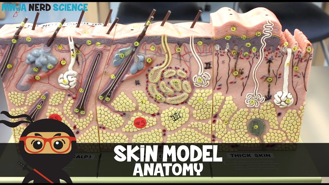
Integumentary System | Skin Model Anatomy

Integumentary System

Signs of over-moisturizing the skin // Can you MOISTURIZE TOO MUCH? @DrDrayzday

REWIND the *HANDS* of time (how to reverse hand aging)

Epiderme: Sistema Tegumentar 2/4 | Anatomia etc
5.0 / 5 (0 votes)
