Peripheral chemoreceptors | Respiratory system physiology | NCLEX-RN | Khan Academy
Summary
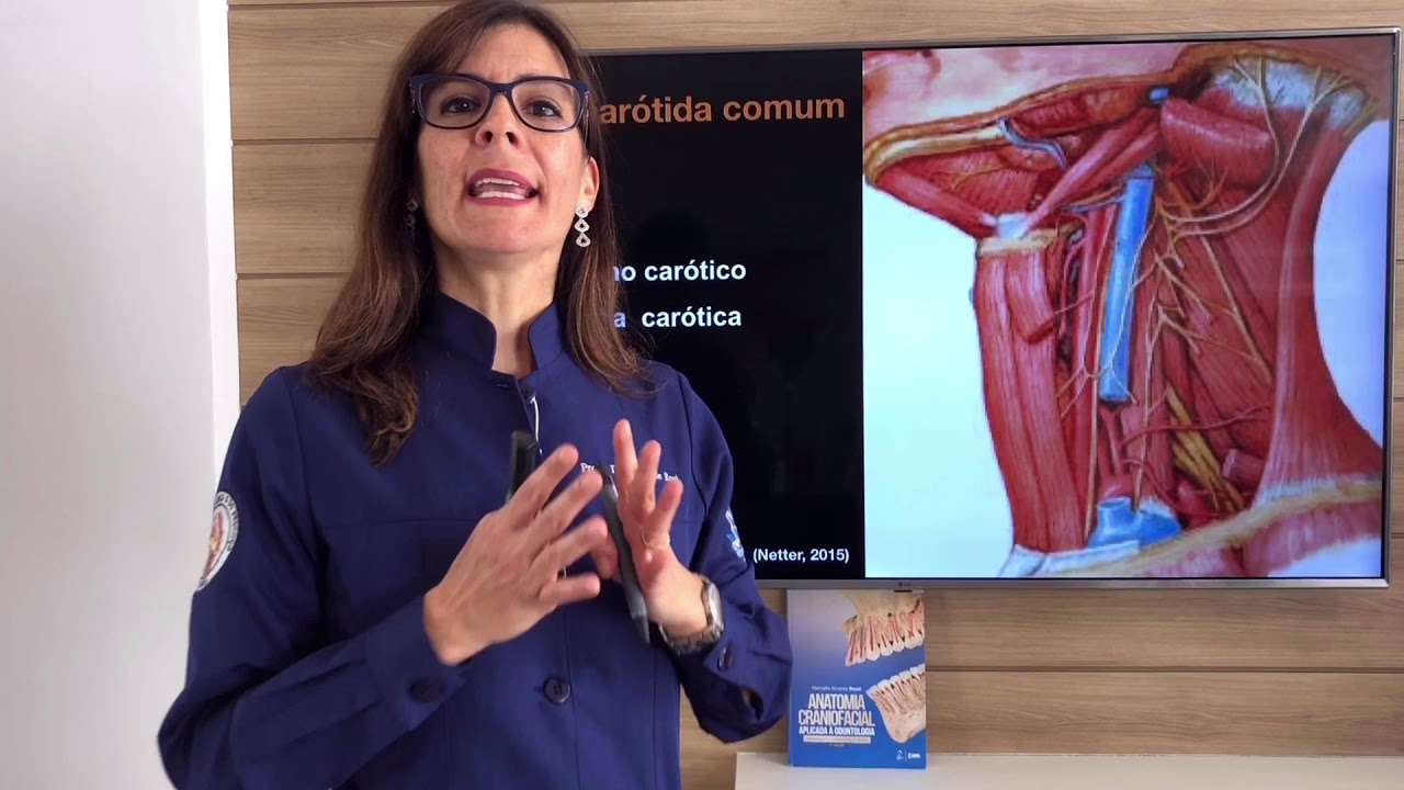
TLDRThe script explains the anatomy of the human heart, focusing on the aorta and its branches, including the right and left common carotid arteries. It details the division of these arteries into internal and external branches and highlights the carotid sinus and aortic arch as locations for baroreceptors, which regulate blood pressure. The script then delves into chemoreceptors, specifically the carotid and aortic bodies, which detect oxygen, carbon dioxide levels, and pH in the blood. These chemoreceptors, comprised of glomus cells, communicate with neurons via neurotransmitters to signal changes in blood chemistry to the brain, with the glossopharyngeal and vagus nerves transmitting this information.
Takeaways
- 💓 The human heart is sketched with emphasis on the aorta and its key branches, particularly those leading to the head, neck, and arms.
- 🔍 The right common carotid artery is highlighted, which splits into the internal and external carotid arteries.
- 🔎 A similar process is described for the left side, with the left common carotid artery also splitting into internal and external branches.
- 🧐 The carotid sinus, an open area in the carotid artery, is identified as the location for baroreceptors, which detect blood pressure and stretch.
- 🌡️ Baroreceptors are crucial for blood pressure regulation by sending information about vessel stretch or pressure back to the brain.
- 🧬 The focus shifts to chemoreceptors, which are responsible for detecting oxygen levels, carbon dioxide levels, and blood pH.
- 📍 Chemoreceptors are located in the aortic body and carotid body, which are slightly different from the baroreceptors' location.
- 🌀 The carotid body is described as receiving some of the highest blood flow rates in the body, emphasizing its importance.
- 🩸 The script explains the role of glomus cells in the carotid and aortic bodies, which detect changes in oxygen and carbon dioxide levels and initiate a response.
- 🚨 A low oxygen level in the blood triggers depolarization in glomus cells, leading to the release of neurotransmitters and action potentials to signal the brain.
- 🔋 The glomus cells' response to high carbon dioxide levels involves the buildup of CO2 in the tissue, leading to increased neurotransmitter release and action potential signaling.
Q & A
What is the primary vessel discussed in the script?
-The primary vessel discussed in the script is the aorta, specifically the aortic arch.
What are the key branches of the aortic arch that are mentioned?
-The key branches of the aortic arch mentioned are those that go up to the head and neck and those that go out to the arms.
What is the function of the right common carotid artery?
-The right common carotid artery eventually bulges and splits into the internal and external branches, which serve different parts of the head and neck.
What are the carotid sinuses and where are they located?
-The carotid sinuses are open areas or spaces located in the internal side of the carotid arteries where baroreceptors are found.
What is the role of baroreceptors in the body?
-Baroreceptors are nerves that detect stretch or pressure in the vessels and send information back to the brain to help regulate blood pressure.
What are chemoreceptors and what information do they provide?
-Chemoreceptors are specialized cells that provide information about oxygen levels, carbon dioxide levels, and pH of the blood.
Where are chemoreceptors located in relation to the aortic arch and carotid arteries?
-Chemoreceptors are located in the aortic body and carotid body, which are slightly different from the locations of baroreceptors.
What is the significance of the carotid body's blood flow rate?
-The carotid body has one of the highest blood flow rates in the human body, approximately 2 liters per minute for 100 grams of tissue.
What are glomus cells and how do they relate to oxygen detection?
-Glomus cells are specialized cells in the carotid and aortic bodies that detect low oxygen levels by depolarizing when oxygen molecules diffuse into them.
How do glomus cells respond to high carbon dioxide levels?
-When carbon dioxide levels are high, it becomes difficult for CO2 to diffuse out of the glomus cells, leading to a buildup of CO2 and a subsequent increase in neurotransmitter release and action potentials.
What is the connection between glomus cells and the nervous system?
-Glomus cells communicate with neurons through the release of neurotransmitters in response to changes in oxygen and carbon dioxide levels, sending signals to the brain via the vagus nerve and glossopharyngeal nerve.
Outlines
🫀 Anatomy of the Heart and Arteries
The paragraph begins with a sketch of the human heart, focusing on the aorta and its branches. The aortic arch is highlighted, with emphasis on the right common carotid artery and its division into the internal and external branches. The left common carotid artery is similarly described. The paragraph then introduces the concept of carotid sinuses and their role in housing baroreceptors, which are nerves that detect pressure changes in the blood vessels to regulate blood pressure. The focus shifts to chemoreceptors, which are located in the aortic body and carotid body and are responsible for detecting oxygen levels, carbon dioxide levels, and pH in the blood. The paragraph concludes with a visual aid, zooming in on the carotid and aortic bodies to illustrate their structure and function.
🔬 Functioning of Chemoreceptors and Glomus Cells
This paragraph delves into the functioning of chemoreceptors, specifically the glomus cells found in the carotid and aortic bodies. It explains how these cells detect low oxygen levels by the diffusion of oxygen molecules into the cell. The paragraph describes the process of depolarization that occurs when oxygen levels are low, leading to the release of neurotransmitters and the generation of action potentials. The role of carbon dioxide in this process is also discussed, explaining how high CO2 levels can lead to increased neurotransmitter release due to difficulty in diffusing out of the cell. The paragraph further explores the chemical reaction of CO2 binding with water to form H2CO3, which can lead to a high proton concentration and low pH, both of which can trigger more action potentials. The paragraph concludes with a note on the evolutionary relationship between glomus cells and nerve cells, sharing a common ancestor in the neuroectoderm.
🌐 Nervous System Integration of Chemoreceptors
The final paragraph discusses the integration of chemoreceptor information into the nervous system. It describes how the neurons from the carotid and aortic bodies converge to form the glossopharyngeal nerve (cranial nerve number 9) and the vagus nerve (cranial nerve number 10), respectively. These nerves carry information from the peripheral chemoreceptors to the brain. The paragraph emphasizes that these nerves are not part of the brain but are essential for transmitting chemical information detected by the chemoreceptors to the central nervous system. The summary underscores the importance of these peripheral chemoreceptors in the body's ability to respond to changes in blood oxygen, carbon dioxide, and pH levels.
Mindmap
Keywords
💡Aorta
💡Aortic Arch
💡Carotid Arteries
💡Baroreceptors
💡Chemoreceptors
💡Carotid Sinus
💡Carotid Body
💡Aortic Body
💡Glomus Cells
💡Neurotransmitters
💡Vagus Nerve
💡Glossopharyngeal Nerve
Highlights
Introduction to sketching the human heart and labeling key vessels.
Identification of the aorta as the largest vessel and its arch.
Description of the aortic arch's branches to the head, neck, and arms.
Focus on the right common carotid artery and its internal and external branches.
Explanation of the carotid artery's bulging and splitting into internal and external branches.
Naming convention for the internal and external branches of the left common carotid artery.
Discussion on the carotid sinus as an open area for baroreceptors.
Function of baroreceptors in detecting pressure and stretch in vessels to regulate blood pressure.
Introduction to chemoreceptors and their role in sensing oxygen, carbon dioxide, and pH levels.
Location of chemoreceptors in the aortic body and carotid body, distinct from baroreceptors.
Visual representation of the carotid body and aortic body with their respective vessels and tissues.
Explanation of the carotid body's high blood flow rate and its significance.
Description of the glomus cells in the carotid and aortic bodies and their function.
Mechanism by which glomus cells detect low oxygen levels and depolarize.
Process of neurotransmitter release from glomus cells in response to low oxygen or high carbon dioxide levels.
Connection between glomus cells and neurons through the release of neurotransmitters.
Impact of high carbon dioxide levels on the glomus cell and its response.
Chemical reaction of carbon dioxide with water forming H2CO3 and its breakdown into bicarbonate and a proton.
Evolutionary connection between glomus cells and nerve cells through the neuroectoderm.
Role of the vagus nerve and glossopharyngeal nerve in transmitting information from peripheral chemoreceptors to the brain.
Transcripts
I'm going to quickly sketch out the human heart.
We're also going to label some vessels coming off of it.
So the big vessel, of course, is the aorta.
This is the giant aortic arch.
And the aortic arch has a couple of key branches
that go, for example, up to the head and neck.
It has other branches as well that go out to the arms.
But these branches that are going up
are the ones I'm going to focus on.
So out here on this right side, we
have the right common carotid artery.
And it's called the common because, eventually,
what's going to happen is it's going to bulge here,
and then it's going to split.
And it's going to split into the internal branch-- this is going
inside-- and the external branch over here.
So this would be called, for example,
the right external carotid artery.
And the same thing is happening on the other side,
and we name it kind of the same way.
We say, OK, there's an internal branch and an external branch.
This would be the internal, and this
might be the external branch of the left common carotid artery.
So I think you're getting the idea now.
These are named exactly the same way.
And these are the ones we're going to focus on.
Now, previously, we had talked about how,
in these particular locations, in the internal side and then
this bulgy side, we have what are called the carotid sinus.
Or sinuses, I suppose.
But the carotid sinus is right there.
And the sinus refers to any open area or open space.
And there's also an area over here in the aortic arch.
And these two areas, they are the home for our baroreceptors.
Our baroreceptors are basically little nerves
that are going to detect pressure.
So they're going to detect stretch, or pressure,
that is in the vessels.
And they're going to give information back to the brain.
And that's going to help regulate our blood pressure.
Now, in this video, we're actually
going to focus on chemoreceptors.
Chemoreceptors are also important in giving us
information, but they're going to give us
information about things like oxygen levels, carbon dioxide
levels, pH of the blood, things like that.
So these chemoreceptors-- and this
gets confusing-- they're located in a similar region, but not
exactly the same region.
I'm actually going to shade in where our chemoreceptors might
be, and then also you might get some over here.
So these three areas are where chemoreceptors are.
And they're very, of course, closely
related to where the baroreceptors are,
but they're actually in slightly different locations.
And we call them the aortic body and the carotid body.
And the reason we use the term "body" is
that it's a body of tissue.
So that's why that word gets used.
And this is actually-- you can see now
a slightly different location, and certainly a different job.
So let me blow up some of these regions
and show you, close up, what this might look like.
So let me draw for you the carotid body on this side,
and on the other side, we'll do the aortic body.
And I'm basically just zooming in on it,
so you can see up close what this might look like,
so you can visualize it.
So for the carotid body, you might have the external artery,
the internal artery.
And coming off of the external artery,
you might have little branches, little branches serving
this tissue that's in the middle.
And these branches, of course, are going to branch some more.
And you're going to get all the way down
to the capillary level.
And once you have little capillaries in here,
there's going to be a bunch of little cells.
And these cells are, of course, going
to get the nutrition from the capillary.
And taken together, all these cells--
if you zoom out of this picture, this
would be a little body of cells or body of tissue.
And that's why we call this the carotid body.
And really, the same thing is going on on the aorta side.
So on the aorta side, you've got little branches
coming off of the aorta, of course.
And these branches are going to branch again,
and again, and again, and again.
And eventually, you're going to get
lots and lots of little capillaries.
And these capillaries are going to serve
all these little blue cells that I'm drawing here,
and these are the chemoreceptors that we're talking about.
So these blue cells together make up a body of tissue,
and that's where we get the term "aortic
body" and "carotid body."
Now, on the carotid side, one interesting fact
is that this body of tissue gets a lot of blood flow,
in fact, some of the highest blood
flow in the entire human body.
It's about 2 liters per minute for 100 grams.
And just to put that in perspective
for the carotid body, imagine that you
have a little 2-liter bottle of soda.
I was thinking of something that would be about 2 liters,
and soda came to mind.
And you can imagine pouring this soda out
over something that's about 100 grams-- maybe a tomato.
That's about a 100-gram tomato.
And if you could do this in one minute,
if you could pour out this bottle in one minute,
imagine how wet this tomato's going
to get, how much profusion, in a sense,
this tomato is going to get.
That is how much profusion your carotid body gets.
So it really puts it in perspective
how much blood flow's going into that area.
So let's now zoom in a little bit further.
Let's say I have a capillary.
And inside my capillary, I've got a little red blood cell
here, floating around.
And my red blood cell, of course,
has some hemoglobin in it, which is a protein.
And this protein has got some oxygen bound to it.
I'm going to draw little blue oxygen molecules.
And of course, there's some oxygen
out here in the plasma itself as well.
And if we're in our carotid body or aortic body,
you might have these special little blue cells
that I've been drawing, our peripheral chemoreceptor cells.
And specifically they have a name.
These things are called glomus cells.
I had initially misstated it as a globus cell.
But actually it's an M-- glomus.
And these oxygen molecules-- these
are oxygen molecules over here-- are
going to diffuse down into the tissue
and get into our glomus cell.
It's going to look something like that.
And if you have a lot of oxygen in the blood, of course,
a lot of molecules are going to diffuse in.
But if you don't have too much in here, then not too much
is going to make its way into the cell.
And that's actually the key point.
Because what our cell is going to be able to do is
start to detect low oxygen levels.
Low oxygen levels in the glomus cell
tells this cell that, actually, there
are probably low levels in the blood.
And when the levels are low, this cell
is going to depolarize.
Its membrane is going to depolarize.
And what it has on the other side
are little vesicles that are full of neurotransmitter.
And so when these vesicles detect that, hey, there's
a depolarization going on, these vesicles
are going to dump their neurotransmitter out.
And what you have waiting for them
is this nice little neuron.
So there's a nice little neuron waiting patiently for a signal,
and that signal is going to come in the form
of a neurotransmitter.
So this is how the communication works.
There's going to be a depolarization,
the vessels release their neurotransmitter,
and that is going to send an action potential down
to our neuron.
And if the oxygen levels fall really low,
let's say they get dangerously low,
where the cell is very unhappy, then
you're going to get much more neurotransmitter getting dumped
out, and you're going to get many more action potentials.
So this is how the glomus cell helps to detect oxygen.
And in fact, it also detects carbon dioxide.
Because, remember, this cell is going
to be making carbon dioxide.
Let's say this is a little molecule of CO2,
and that CO2 is going to diffuse out and into the blood.
Well, let's say that the blood has a lot of carbon dioxide
already.
Let's say that it's loaded with carbon dioxide, lots and lots
of it.
In this situation, it's going to be very difficult for carbon
dioxide to make its way from the glomus cell
all the way out into the plasma.
And as a result, carbon dioxide starts building up.
The tissue starts gathering more and more CO2,
because it can't go anywhere.
And this glomus cell is going to say, hey, wait a second.
Our CO2 levels are starting to rise.
There are high CO2 levels.
And again, that's going to make the cell unhappy,
and it's going to send out more neurotransmitter,
and it's going to, of course, send out
more action potentials.
So two different reasons why you might get action
potentials coming out of this glomus cell.
And now I want to remind you that there's
this little formula.
There's this formula where carbon dioxide binds
with water, and it forms H2CO3.
And that's going to break down into bicarbonate and a proton.
So this is our formula.
So if CO2 levels are rising, like the example
I just offered, then the proton level must be high as well.
So a high proton concentration-- I'm
going to put it in brackets, to indicate concentration.
Or another way of saying that would be a low pH.
So these are the things that are going
to make our glomus cell send off more action potentials.
So if you're like me, you're thinking, well, wait a second.
This is really interesting.
Our cell is depolarizing.
It can depolarize.
It also has this neurotransmitter
that I mentioned.
Our glomus cell, then, right here in blue,
is basically sounding a little bit
like it has properties of a nerve cell.
This is a nerve cell.
And the reason for that is that, if you actually take a look,
these two cells have a common ancestor cell.
And so in development, when the fetus is developing,
there is a type of tissue called the neuroectoderm.
And both of these cells, this nerve cell
and this glomus cell, both are derived
from this neuroectoderm.
So it makes sense that they would
have a lot of common features.
So we know the glomus cell is not a neuron,
but it's going to be talking to neurons.
In fact, you're going to have many neurons working together
in this area.
And they're going to join up, both in the aortic body
and the carotid body.
And these neurons, going back to the original picture,
are going to meet up into a big nerve.
And this nerve is going to be called the vagus nerve.
The vagus nerve is going to be the one for our aortic body,
sometimes also called cranial nerve number 10.
And up here with the carotid body, we have a nerve as well.
This is another nerve.
This one, we call the glossopharyngeal nerve.
So these two nerves, the vagus nerve
and the glossopharyngeal nerve-- this one,
this glossopharyngeal nerve, by the way,
is cranial nerve number 9-- these two nerves are not
part of the brain.
They're headed to the brain, right?
So these two nerves are fundamentally
taking information from chemoreceptors
that are outside of the brain.
They're not located in the brain, right?
They're peripheral, and they're taking information
about chemicals and taking that information to the brain.
That's why we call them-- these blue areas, the carotid body
and the aortic body-- we call them peripheral chemoreceptors.
Weitere ähnliche Videos ansehen
Carbon Dioxide | HOMEOSTASIS | Easy to understand
Internal Carotid Artery - Anatomy (Circle of Willis)
Coronary arteries Anatomy / Blood supply of Heart / Arterial supply of heart : Animation
Sistema Cardiovascular - Toda Matéria
IMAT Biology Lesson 6.4 | Anatomy and Physiology | Circulatory System I
CVS 3 The Heart
5.0 / 5 (0 votes)