MITOSIS, CYTOKINESIS, AND THE CELL CYCLE
Summary
TLDRThis script delves into the cell cycle, detailing the process of cell duplication through division. It explains the phases G1, S, G2, and M, emphasizing the importance of accurate DNA replication and regulatory systems. The script clarifies the distinction between chromosomes and chromatids, highlighting the role of the mitotic spindle and centrosomes in mitosis. It concludes with cytokinesis, the cytoplasmic division, completing the cell cycle.
Takeaways
- 🌱 The cell cycle is the process of duplicating a cell, resulting in two genetically identical daughter cells from a parent cell.
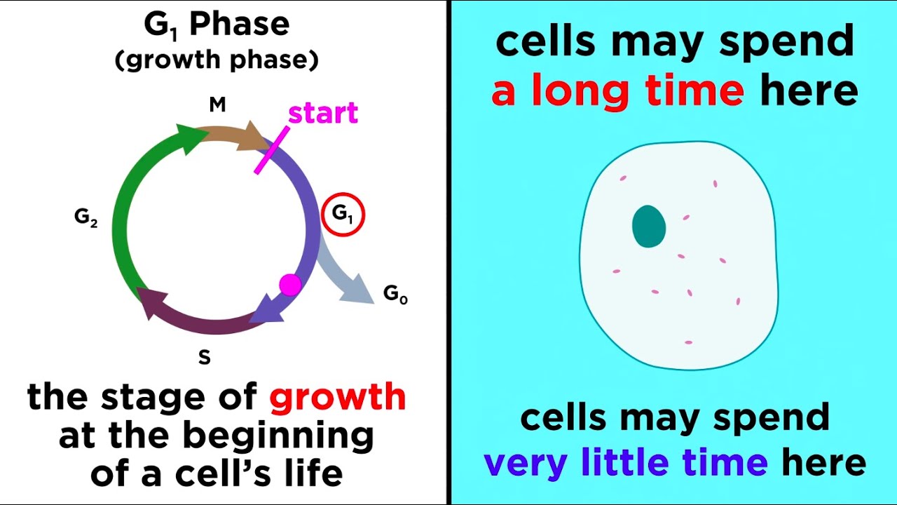
- 🔄 The cell cycle consists of four phases: G1, S, G2, and M, with G1 and G2 being growth phases known as interphase.
- 📈 The purpose of G1 and G2 phases is to allow cell growth, doubling of organelles, and assessment of environmental conditions for division.
- 🛡️ Eukaryotic cells have a cell-cycle control system of regulatory proteins to ensure correct and appropriate cell division.
- 🧬 The S phase is crucial for DNA replication and chromosomal duplication, requiring high fidelity to avoid errors.
- 🔬 Chromosomes can be a single DNA strand or two sister chromatids linked by a centromere, with the latter forming after DNA replication.
- 🤝 Sister chromatids are identical copies of the original chromosome, which separate during cell division to form two daughter chromosomes.
- 🧬 In diploid cells, each chromosome has two copies, one from each parent.
- 🕊️ The M phase includes mitosis and cytokinesis, where mitosis divides the genetic material and cytokinesis divides the cytoplasm.
- 🧭 The mitotic spindle, composed of microtubules, is essential for separating chromosomes and is anchored by centrosomes.
- 🔗 Kinetochores are protein structures that attach to sister chromatids and facilitate their separation by the mitotic spindle.
- 📊 Mitosis has five stages: prophase, prometaphase, metaphase, anaphase, and telophase, each with specific actions for chromosome separation and nuclear reformation.
- 💧 Cytokinesis involves the formation of a contractile ring that pinches the cell into two, facilitated by actin and myosin filaments.
Q & A
What is the only way to create a new cell?
-The only way to create a new cell is to duplicate a pre-existing one through a process called cell division.
What are the two new cells produced after cell division called?
-The two new cells produced after cell division are called daughter cells, which are genetically identical to the original cell, known as the parent cell.
What is the cell cycle?
-The cell cycle is the series of steps leading to and involving the duplication of a cell, including cell growth, chromosome replication, chromosome segregation, and cell division.
What are the four sequential phases of the cell cycle?
-The four sequential phases of the cell cycle are G1, S, G2, and M, with the first three phases collectively known as interphase.
Why do cells have G1 and G2 phases in the cell cycle?
-G1 and G2 phases, the gap phases, are present in the cell cycle to allow cells more time to grow, double their organelles and biomolecules, and assess whether the environment is favorable for division and if all preparations for cell division have been completed.
What is the role of the cell-cycle control system in eukaryotic cells?
-The cell-cycle control system in eukaryotic cells is a complex system of regulatory proteins that ensures cell division occurs correctly and only when appropriate.
What happens during the S phase of the cell cycle?
-The S phase is when DNA replication and chromosomal duplication take place, which is critical for the accurate copying of every nucleotide.
What is the difference between a chromosome and chromatids?
-A chromosome can be a continuous strand of DNA or, after DNA replication, two sister chromatids linked together by a centromere. Sister chromatids include the original chromosome and its identical replica.
What is the significance of the M phase in the cell cycle?
-The M phase includes mitosis and cytokinesis, which are the processes by which sister chromatids are split up equally between two new daughter nuclei and the cytoplasm is divided, respectively.
What are the functions of the mitotic spindle and centrosomes during cell division?
-The mitotic spindle is a bipolar array of microtubules that separates chromosomes into two new daughter cells, while centrosomes, consisting of centrioles and the pericentriolar matrix, nucleate microtubules and help position the spindle in the cell.
What are the five stages of mitosis?
-The five stages of mitosis are prophase, prometaphase, metaphase, anaphase, and telophase, each involving specific events in the division of the cell's nucleus.
What is cytokinesis and how does it occur?
-Cytokinesis is the division of the cytoplasm that occurs through the formation of a contractile ring of actin and myosin filaments, which pinch the cell into two new cells.
What is the role of kinetochores in mitosis?
-Kinetochores are large protein structures that allow the plus ends of kinetochore microtubules to attach correctly to sister chromatids, ensuring proper chromosome segregation during mitosis.
Outlines
🌱 Cell Cycle and Division Process
The paragraph explains the fundamental process of cell division, emphasizing the necessity of duplicating a pre-existing cell to create new ones. It details the cell cycle, which includes growth, chromosome replication, segregation, and division, occurring across four phases: G1, S, G2, and M, with the first three being interphase. The paragraph highlights the importance of the cell-cycle control system in eukaryotic cells to ensure accurate division. It also distinguishes between chromosomes and chromatids, explaining the process of DNA replication leading to sister chromatids that form a chromosome. The M phase involves mitosis and cytokinesis, with mitosis ensuring equal distribution of genetic material and cytokinesis dividing the cytoplasm. The paragraph further describes the mitotic spindle, centrosomes, and kinetochores, which play crucial roles in chromosome separation and cell division.
🔬 Mitosis and Cytokinesis Stages
This paragraph delves into the specifics of mitosis, the process dividing the genetic material between two nuclei. It outlines the five stages of mitosis: prophase, prometaphase, metaphase, anaphase, and telophase. During prophase, chromosomes condense and the mitotic spindle forms. Prometaphase begins with the breakdown of the nuclear envelope, allowing chromosomes to attach to spindle microtubules. In metaphase, chromosomes align at the spindle equator, while anaphase sees the separation of sister chromatids towards the spindle poles. Telophase concludes mitosis with the arrival and decondensation of daughter chromosomes at the spindle poles and the formation of two nuclei. Following mitosis, cytokinesis occurs, dividing the cytoplasm through the action of a contractile ring composed of actin and myosin filaments. The paragraph outlines the four steps of cytokinesis: initiation, contraction, membrane insertion, and completion, detailing the role of intracellular vesicles in providing additional plasma membrane during cell division.
Mindmap
Keywords
💡Cell Cycle
💡Parent Cell
💡Daughter Cells
💡Interphase
💡Chromosome Replication
💡Chromosomes
💡Chromatids
💡Mitosis
💡Cytokinesis
💡Mitotic Spindle
💡Kinetochore
💡Centromeres
Highlights
The cell cycle is the process of cell duplication, resulting in genetically identical daughter cells from a parent cell.
The cell cycle consists of four phases: G1, S, G2, and M, with G1 and G2 being growth phases known as interphase.
Cells require time in G1 and G2 phases for growth and to assess environmental conditions for division.
Eukaryotic cells have a cell-cycle control system of regulatory proteins to ensure accurate and appropriate division.
The S phase is critical for accurate DNA replication and chromosomal duplication.
Chromosomes can be a single DNA strand or two sister chromatids linked by a centromere after replication.
Diploid cells contain two copies of each chromosome, which are crucial for genetic diversity.
M phase includes mitosis and cytokinesis, the processes that lead to the formation of two genetically identical daughter cells.
The mitotic spindle is a bipolar array of microtubules essential for separating chromosomes during cell division.
Centromeres and kinetochores play a key role in attaching microtubules to chromosomes and ensuring proper segregation.
Mitosis is divided into five stages: prophase, prometaphase, metaphase, anaphase, and telophase.
During prophase, chromosomes condense and the mitotic spindle assembles between centrosomes.
Prometaphase involves the breakdown of the nuclear envelope, allowing chromosomes to attach to spindle microtubules.
Metaphase aligns chromosomes at the spindle equator with microtubules attached to opposite poles.
Anaphase is characterized by the separation of sister chromatids and their movement towards spindle poles.
Telophase marks the arrival of daughter chromosomes at spindle poles and the beginning of nuclear envelope formation.
Cytokinesis follows mitosis, involving the division of the cytoplasm through a contractile ring mechanism.
The contractile ring is composed of actin and myosin filaments, facilitating the physical separation of the cell.
Cytokinesis includes steps of initiation, contraction, membrane insertion, and completion, leading to two new cells.
Transcripts
The only way to create a new cell is to duplicate a pre-existing one.
The original cell is called the parent cell, and the two new cells, which are genetically
identical, are called daughter cells.
The series of steps leading to and involving the duplication of a cell is called the cell cycle.
The cell cycle involves cell growth, chromosome replication, chromosome segregation, and cell division
These processes take place over the course of the four sequential phases of the cell
cycle: G1, S, G2, and M. The first three phases are together called interphase.
Most cells need more time to grow and double their organelles and biomolecules than
they need to duplicate and divide up their chromosomes, the cell cycle has the extra
gap phases, called G1 and G2.
These gap phases, in addition to compensating for the time needed for growth, also give
the cell time to assess whether the environment is favourable for division and whether all
the preparations for cell division have been appropriately completed.
Eukaryotic cells have a complex system of regulatory proteins, called the cell-cycle
control system, to make sure that cell division occurs correctly and only when appropriate.
Taking place between G1 and G2, S phase is when DNA replication and chromosomal duplication
takes place.
It is critical that this replication is accurate, and that every nucleotide is copied exactly
once.
Here it’s important to discuss two closely-related, and commonly confused terms – chromosomes
and chromatids.
A chromosome can be one of two things: a continuous strand of DNA or two sister chromatids linked
together with a centromere.
These sister chromatids include the original chromosome and its identical replica.
So to recap: you have a chromosome, then after DNA replication you have a chromosome made
up of sister chromatids.
When cell division takes place, the sister chromatids will be separated, and you now
have two daughter chromosomes.
Also, keep in mind that in diploid cells, there would be two copies of
each chromosome present.
Finally, we come to M phase, which includes mitosis and cytokinesis.
Mitosis is the process by which sister chromatids are split up equally between the two new daughter
nuclei, so that each new cell can have an identical and complete version of the genome.
Cytokinesis is the division of the cytoplasm.
Before we describe what happens during mitosis, it is important to define the mitotic spindle
and centrosomes.
The mitotic spindle is what eukaryotes use to separate chromosomes into two new daughter
cells.
It is a bipolar array of microtubules that pulls sister chromatids apart.
These microtubules have minus ends focused at the “spindle poles” and plus ends that
radiate outward.
Microtubules are composed of tubulin subunits, which can get added or removed from the plus
end of the microtubule, making it grow or shrink.
There are three types of microtubules in the mitotic spindle.
Interpolar microtubules are those whose plus ends interact with the plus ends of interpolar
microtubules originating from the opposite spindle pole.
Astral microtubules radiate outward from the poles to contact the cell cortex.
Their job is to position the spindle in the cell.
Finally, there are the kinetochore microtubules, whose positive ends are attached to sister
chromatid pairs at kinetochores.
Kinetochores are large protein structures located at the centromere of each sister chromatid.
The kinetochores are built on the heterochromatin that forms at the centromeric region of the
chromosome.
The kinetochores allow the plus ends of kinetochore microtubules to be embedded in special attachment
sites in the kinetochore.
An animal cell kinetochore can have 10-40 of these attachment sites.
The attachment sites contain a protein collar that secures the microtubule but still allows
the addition or removal of tubulin subunits to its end.
Kinetochores allow kinetochore microtubules to attach correctly to sister chromatids.
How?
Well, if there is an imbalance in microtubule attachment, such as if two microtubules from
opposite spindle poles attach to the same sister chromatid, the attachment is unstable.
However, the correct attachment gets locked in place.
In animal cells, at each spindle pole, there is a single centromere, which consists of
two centrioles oriented perpendicular to one another, surrounded by the pericentriolar
matrix.
The pericentriolar matrix nucleates a radial array of microtubules, whose negative ends
are associated with the centromere.
Like the genome, the centromere must be duplicated exactly one time prior to mitosis.
An incorrect number of centromeres would result in errors in spindle assembly and chromosome
segregation.
This duplication occurs at the start of S phase.
Finally, we come to mitosis, the division of identical genomic information between two
new nuclei!
Mitosis has 5 stages: prophase, prometaphase, metaphase, anaphase, and telophase.
During prophase, chromosomes condense within the nucleus, with each chromosome consisting
of two linked sister chromatids.
Outside the nucleus, the mitotic spindle assembles between two centrosomes.
Next we have prometaphase, which begins when the nuclear envelope breaks down.
This means that chromosomes can now attach to spindle microtubules.
This occurs via kinetochores and results in the movement of the chromosomes.
Metaphase takes place when the chromosomes are aligned along the equator of the spindle,
with microtubules attaching sister chromatids to opposite poles of the spindle.
Anaphase involves the synchronized separation of sister chromatids to form two daughter
chromosomes, which are pulled towards the spindle poles.
During this process, not only do the kinetochore microtubules shorten, but the spindle poles
also move apart.
Telophase is the last stage of mitosis.
At this point, the two sets of daughter chromosomes arrive at the spindle poles and decondense.
The formation of the two nuclei, which occurs when a nuclear envelope assembles around the
two genomes, means that mitosis is over.
Mitosis is followed by cytokinesis, which is the division of the cytoplasm.
This occurs thanks to the formation of a contractile ring of actin and myosin filaments, which
form a rapidly deepening furrow and pinch the cell into two new cells.
The contractile ring actually begins to assemble during anaphase, just beneath the plasma membrane.
As the ring contracts, there is fusion of intracellular vesicles to add more plasma
membrane to compensate for the extra surface area.
So cytokinesis has 4 steps: initiation, contraction, membrane insertion, and completion.
5.0 / 5 (0 votes)