A-Level Biology - Measuring cells : Calibrate Eyepiece graticule, Magnification, Resolution
Summary
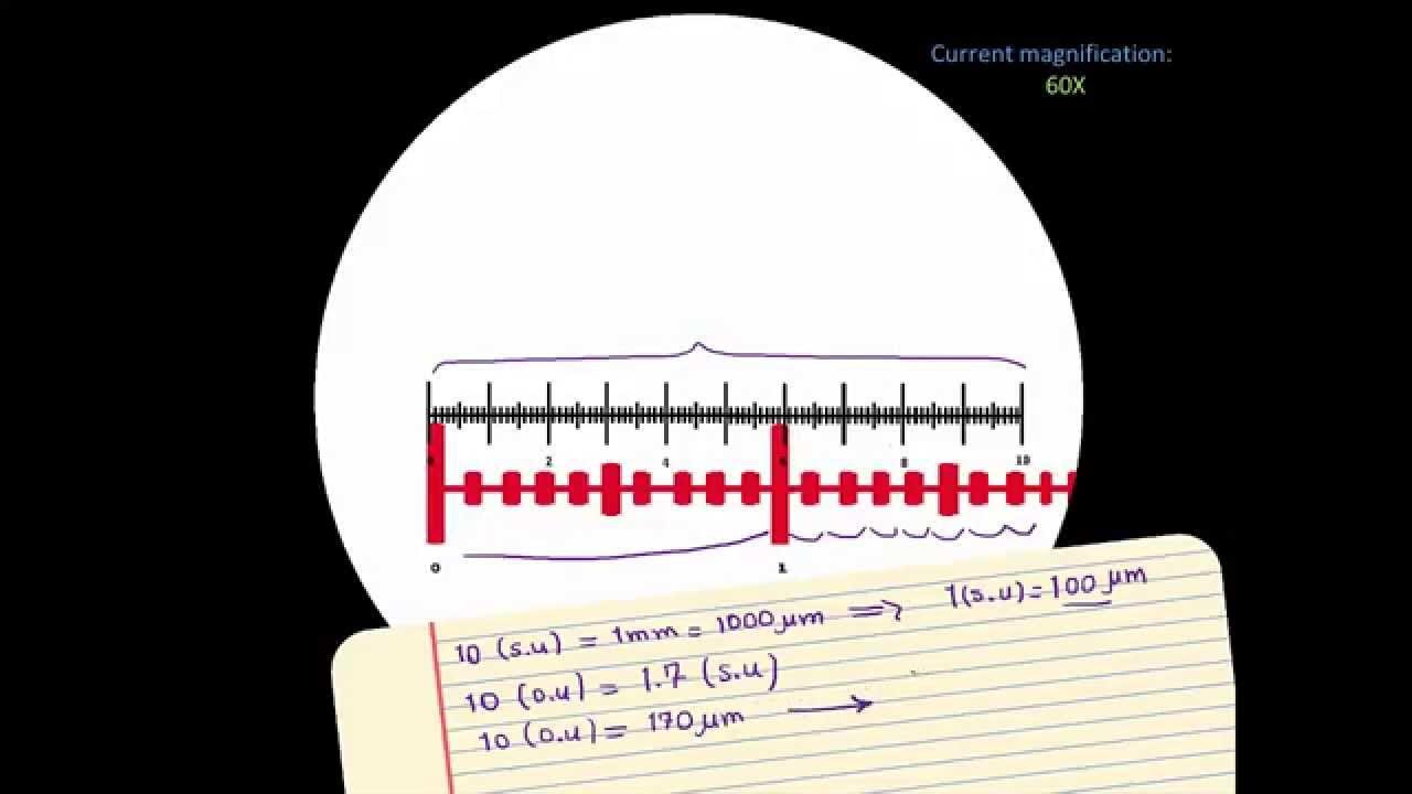
TLDRIn this educational video, David explains the importance of calibrating the eyepiece graticule using a stage micrometer for accurate cell measurements. He illustrates how to measure objects under a microscope, emphasizing that calibration is necessary when changing magnifications. The video also covers the concept of magnification, resolution, and how they relate to the ability to distinguish small structures. David highlights the differences between light and electron microscopes, explaining how their resolutions are determined by the wavelengths of light and electrons. This insightful guide equips viewers with essential knowledge for effective microscopy.
Takeaways
- 🔍 Calibration of the eyepiece graticule is essential for accurate measurements in microscopy.
- 📏 A stage micrometer serves as a standard to calibrate the eyepiece graticule, ensuring measurements are accurate across different specimens.
- 🔄 It's necessary to recalibrate the eyepiece graticule whenever you switch objective lenses on the microscope.
- 🔬 The eyepiece graticule typically contains 100 divisions, which correspond to actual measurements after calibration.
- 📐 Each division on the stage micrometer is often 0.1 mm, which aids in determining the size of objects viewed through the microscope.
- 🧪 Magnification compares the size of an image to its actual size, calculated using the formula: magnification = image size / actual size.
- ⚖️ To find actual size from an image, rearrange the magnification formula to: actual size = image size / magnification.
- 🔬 Resolution determines the ability of a microscope to distinguish between two closely situated objects; a smaller resolution value indicates better clarity.
- 🌈 Light microscopes have a maximum resolution of 200 nanometers due to the longer wavelength of visible light.
- ⚡ Electron microscopes have a significantly higher resolution of 0.5 nanometers, allowing for the visualization of much smaller structures.
Q & A
What is the purpose of calibrating the eyepiece graticule?
-Calibrating the eyepiece graticule ensures accurate measurements when using a microscope, as the scale on the graticule must match the actual scale of the specimen observed.
How does the analogy of the camera lens and the ruler help explain the need for calibration?
-The analogy illustrates that placing a ruler at the lens doesn't provide accurate measurements of objects on the stage due to distance distortion, similar to how an eyepiece graticule needs to be calibrated to measure objects correctly.
What is a stage micrometer, and why is it used in calibration?
-A stage micrometer is a slide with a precisely marked scale, which is used to calibrate the eyepiece graticule by aligning its divisions with the graticule to establish a known measurement reference.
How many divisions does a typical eyepiece graticule have?
-A typical eyepiece graticule has 100 divisions.
What measurement does each division on the eyepiece graticule represent after calibration?
-After calibration, each division on the eyepiece graticule represents 2.5 micrometers.
What is the formula for calculating magnification?
-The formula for calculating magnification is magnification = image size / actual size.
Why is it important to keep units consistent when performing measurements?
-It's important to keep units consistent to avoid confusion and ensure accurate calculations; for example, using millimeters for rulers and micrometers for cell measurements.
What is resolution in the context of microscopy?
-Resolution is the ability of a microscope to distinguish two separate objects as distinct from one another; higher resolution allows for clearer images of smaller objects.
How does the wavelength of radiation affect resolution in microscopes?
-The smaller the wavelength of radiation used to view a specimen, the better the resolution; shorter wavelengths, like those of electrons, allow for distinguishing smaller details compared to longer wavelengths of visible light.
What is the maximum resolution of light microscopes compared to electron microscopes?
-The maximum resolution of light microscopes is approximately 200 nanometers, while electron microscopes can achieve a maximum resolution of 0.5 nanometers, allowing for the observation of much smaller structures.
Outlines

هذا القسم متوفر فقط للمشتركين. يرجى الترقية للوصول إلى هذه الميزة.
قم بالترقية الآنMindmap

هذا القسم متوفر فقط للمشتركين. يرجى الترقية للوصول إلى هذه الميزة.
قم بالترقية الآنKeywords

هذا القسم متوفر فقط للمشتركين. يرجى الترقية للوصول إلى هذه الميزة.
قم بالترقية الآنHighlights

هذا القسم متوفر فقط للمشتركين. يرجى الترقية للوصول إلى هذه الميزة.
قم بالترقية الآنTranscripts

هذا القسم متوفر فقط للمشتركين. يرجى الترقية للوصول إلى هذه الميزة.
قم بالترقية الآنتصفح المزيد من مقاطع الفيديو ذات الصلة

AS Biology - How to calibrate a microscope

Eyepiece graticule and stage micrometer A level Biology - BioTeach

How to study cells - Microscopes, magnification and calibrating the eyepiece graticule

Microscope Calibration: a short tutorial [New version]

HOW TO READ YOUR MICROMETER SCREW AND CALIBRATION ❗️

Mengukur Keausan Blok silinder sepeda Motor
5.0 / 5 (0 votes)
