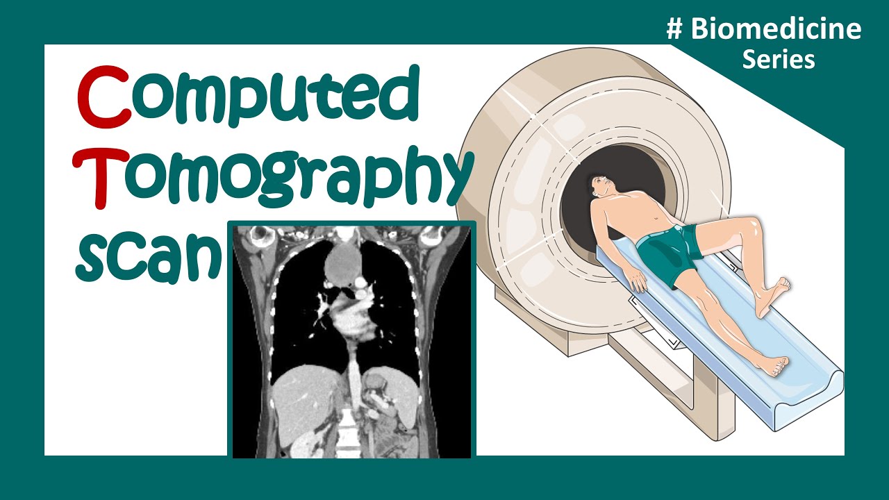
CT (Computed Tomography) Scans - A Level Physics
Summary
TLDRThis video discusses CT (computed tomography) scans, explaining how they use X-rays to produce a 3D image of the body. Unlike regular X-rays, which give a flat, 2D image, CT scans provide detailed views by rotating an X-ray source around the patient, capturing multiple angles. The body is divided into tiny volumes called voxels, each assigned a value based on how much it attenuates X-rays. The computer processes these values to create a 3D representation of internal structures, making CT scans powerful diagnostic tools, though they expose patients to higher radiation than standard X-rays.
Takeaways
- 💡 CT (Computed Tomography) scans use X-rays to create three-dimensional images of the inside of the body.
- 📸 Traditional X-ray images are two-dimensional and do not provide information on the relative positions of different organs.
- 🔄 A CT scan works by rotating an X-ray tube and detector around the patient, capturing images from multiple angles.
- 🧠 The patient remains still on a bed while the CT scanner collects data from different perspectives.
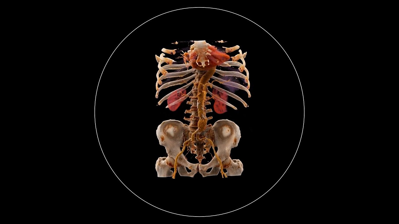
- 🔬 The data collected is processed by a computer to create a 3D model of the body's internal structures.
- 🧩 CT scans divide the body into small units called 'voxels,' each representing a tiny volume of tissue.
- 🔢 Each voxel is assigned a number indicating how much it attenuates (reduces) the X-ray intensity passing through it.
- 📊 The computer applies an algorithm to reconstruct the 3D image by analyzing how X-rays are absorbed at different angles.
- 🖥️ The calculations involve summing and subtracting values for different voxel arrays to accurately map the internal structures.
- ⚠️ CT scans expose the patient to more X-ray radiation compared to standard two-dimensional X-rays due to multiple scans from various angles.
Q & A
What is the key difference between a regular X-ray and a CT scan?
-A regular X-ray provides a two-dimensional image, whereas a CT scan produces a three-dimensional image by taking multiple X-rays from different angles.
How does a CT scan create a three-dimensional image?
-A CT scan takes X-rays at different angles around the patient using a rotating X-ray tube. The data is then processed by a computer to construct a 3D image of the body by dividing it into small volumes called voxels.
What are voxels, and how do they contribute to the CT scan process?
-Voxels are small volumes within the body that are analyzed during a CT scan. Each voxel has a number that represents the extent to which X-rays are attenuated as they pass through it, helping construct a 3D image of the body.
Why do bones have a high voxel number compared to soft tissue?
-Bones attenuate X-rays significantly, meaning they block or absorb more X-rays, leading to a higher voxel number. Soft tissue, like skin, has a lower voxel number because it allows more X-rays to pass through with less attenuation.
How many angles are used to scan a patient during a CT scan in this example?
-In the example, X-rays are taken at four different angles, each contributing to a clearer and more accurate 3D reconstruction of the body.
What is the purpose of the algorithm applied by the computer in a CT scan?
-The algorithm combines the X-ray data from different angles, adding and subtracting values to calculate the attenuation for each voxel, ultimately reconstructing a 3D image of the body.
How does the process of adding and subtracting X-ray attenuation values work?
-The computer combines the total attenuation values from X-rays passing through different angles, and then it subtracts the total initial attenuation to refine the voxel values, providing a more accurate image.
Why is it necessary to divide the final values by three in this CT scan example?
-The final values are divided by three because the data includes three sets of additional scans from different angles. This step adjusts the voxel numbers to reflect the combined effects of all the scans.
What is one significant downside of a CT scan compared to a regular X-ray?
-One downside is that CT scans expose patients to more X-rays compared to regular X-rays since multiple scans are taken from different angles to build a 3D image.
Why is the amount of X-ray exposure higher in a CT scan than in a standard X-ray?
-The exposure is higher because the patient undergoes multiple X-ray scans from different angles, whereas a standard X-ray only involves a single scan.
Outlines

هذا القسم متوفر فقط للمشتركين. يرجى الترقية للوصول إلى هذه الميزة.
قم بالترقية الآنMindmap

هذا القسم متوفر فقط للمشتركين. يرجى الترقية للوصول إلى هذه الميزة.
قم بالترقية الآنKeywords

هذا القسم متوفر فقط للمشتركين. يرجى الترقية للوصول إلى هذه الميزة.
قم بالترقية الآنHighlights

هذا القسم متوفر فقط للمشتركين. يرجى الترقية للوصول إلى هذه الميزة.
قم بالترقية الآنTranscripts

هذا القسم متوفر فقط للمشتركين. يرجى الترقية للوصول إلى هذه الميزة.
قم بالترقية الآنتصفح المزيد من مقاطع الفيديو ذات الصلة
CT scan | computerized tomography (CT) scan |What is a CT scan used for? | Clinical application

Biomedical instrumentation- CT scan (Computed Tomography)

How X-rays see through your skin - Ge Wang

UQx Bioimg101x 3.2.4 CT Reconstruction & Back Projection
What is Computed Tomography (CT) and how does it work?
Computed Tomography | CT Scanners | Biomedical Engineers TV |
5.0 / 5 (0 votes)
