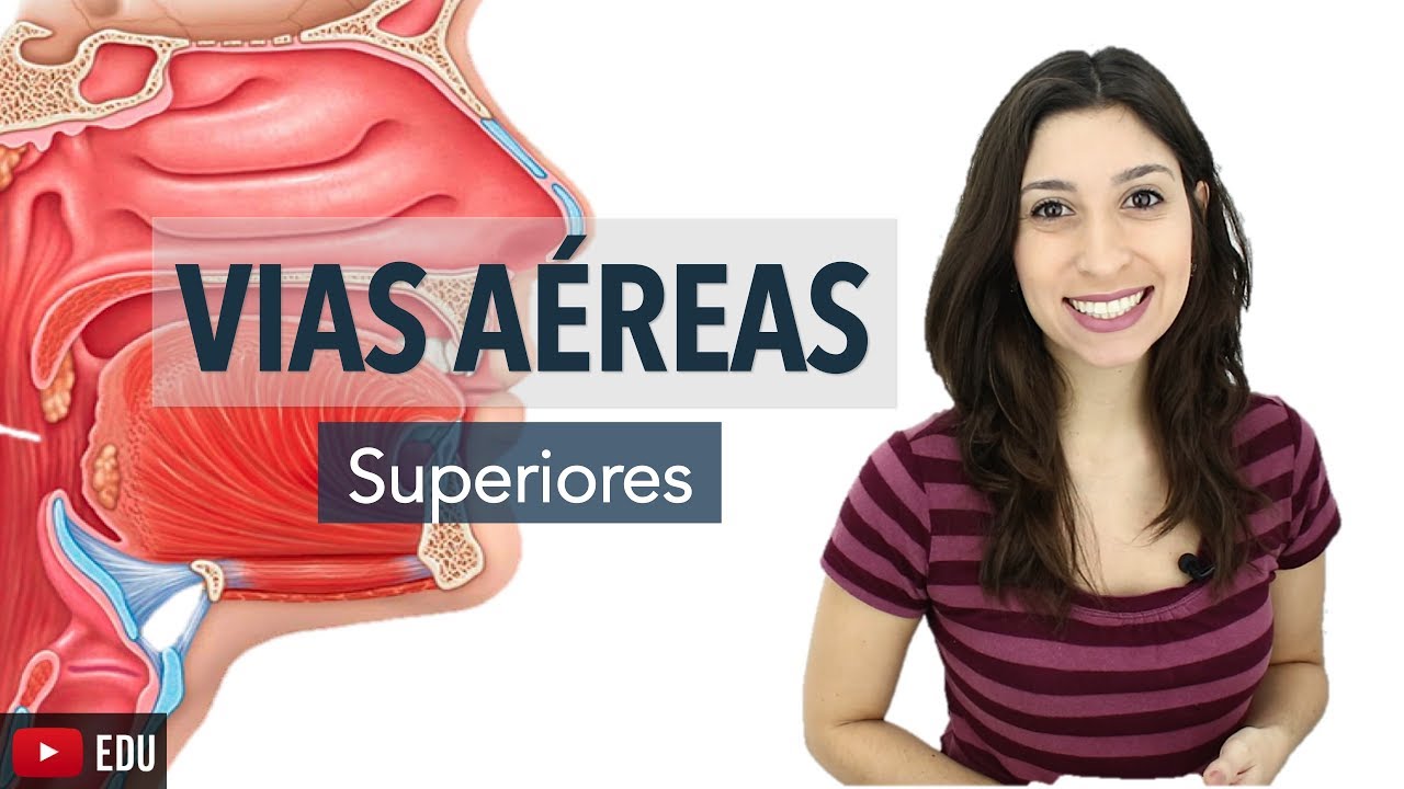
Nasal Anatomy (Cartilage, Nasal Cavity, Sinuses, Meatuses, Nasal Mucosa)
Summary
TLDRThis video script delves into the anatomy of the respiratory system, focusing on the nose. It explains the external and internal structures of the nose, including the nasal bones, cartilages, and the nasal cavity. The script describes the nasal septum, nasal vestibule, and the proper nasal cavity divided into olfactory and respiratory parts. It also covers the sinuses' functions, such as warming and humidifying air, and the importance of the nasal mucosa in filtering, smelling, and defense mechanisms. Conditions like sinusitis and allergies are briefly touched upon.
Takeaways
- 👃 The respiratory system includes the nose, pharynx, larynx, trachea, bronchi, and lungs.
- 👂 The external nose consists of the root (radix nasi), dorsum, apex, and ala nasi.
- 🦴 The nose is composed of bone, cartilage, and fat, including the nasal bones and various cartilages.
- 🔄 The nasal septum is made up of a cartilaginous part and a bony part, providing flexibility.
- 🌿 The nasal cavity is divided into the nasal vestibule and the nasal cavity proper.
- 👃🏼 The nasal cavity proper is further divided into the olfactory part for smell and the respiratory part for breathing.
- 🧠 The olfactory part senses smell due to the first cranial nerve and contains olfactory cells with receptors.
- 🌀 The respiratory part features conchae or turbinates and meatuses, which facilitate sinus drainage.
- 💧 Sinuses help reduce the skull's mass, resonate sound, warm and humidify air, and produce mucus.
- 🚦 The maxillary sinus drains into the middle meatus, while the posterior ethmoidal air cells drain into the superior meatus.
- 🔬 The nasal mucosa has a respiratory epithelium with cilia and goblet cells, and olfactory epithelium with olfactory cells and supporting cells.
Q & A
What are the primary components of the respiratory system?
-The primary components of the respiratory system include the Nose, Pharynx, Larynx, Trachea, Bronchi, and Lungs.
What is the function of the nasal anatomy in the respiratory system?
-The nasal anatomy is responsible for filtering, warming, and humidifying the air that enters the body, as well as playing a role in the sense of smell.
What are the external structures of the nose?
-The external structures of the nose include the Root of the nose (radix nasi), Dorsum of the nose, Apex, and the wings of the nose (ala nasi).
What are the different parts of the nose's cartilage?
-The nose's cartilage consists of the Nasal bones, Lateral nasal cartilages, Major alar cartilage, Minor alar cartilage, Accessory nasal cartilages, and the Septal Nasal Cartilage.
What is the role of the nasal septum?
-The nasal septum divides the nasal cavity into two parts and consists of a cartilaginous part and a bony part, providing support and flexibility to the nose.
How is the nasal cavity divided?
-The nasal cavity is divided into the Nasal Vestibule and the nasal cavity proper, separated by the nasal Limen.
What are the two main parts of the nasal cavity proper?
-The nasal cavity proper is divided into the Olfactory Part, responsible for smell, and the Respiratory Part, responsible for breathing.
What are the functions of the Conchae or Turbinates in the nasal cavity?
-The Conchae or Turbinates increase the surface area of the nasal cavity, helping to warm and humidify the air, and they also help filter out particles from the air.
What is the significance of the sinuses in the nasal anatomy?
-The sinuses help reduce the weight of the skull, contribute to voice resonance, warm and humidify the air, and produce mucus to keep the nasal passages moist.
How do the sinuses connect to the nasal cavity?
-The sinuses connect to the nasal cavity through various meatuses; for example, the Sphenoid sinus opens into the Spheno-Etmoidal Recess, and the Maxillary sinus drains into the Middle Meatus.
What is the function of the nasal mucosa?
-The nasal mucosa lines the nasal cavity and is responsible for producing mucus, trapping irritants, and playing a role in the sense of smell.
What could potentially block the sinuses?
-Conditions such as the common cold, allergies, nasal polyps, or a deviated septum can cause inflammation and swelling, leading to blocked sinuses.
Outlines
👃 Anatomy of the Nose and Nasal Cavity
This paragraph introduces the respiratory system, emphasizing the anatomy of the nose and nasal cavity. The external nose is composed of the root (radix nasi), dorsum, apex, and alar wings (ala nasi). Beneath the skin are the nasal bones, lateral nasal cartilages, major and minor alar cartilages, accessory nasal cartilages, and the septal nasal cartilage. The nasal cavity is divided into the nasal vestibule and the proper nasal cavity, separated by the nasal limen. The proper nasal cavity is further divided into the olfactory part, responsible for smell via the olfactory nerve, and the respiratory part, characterized by conchae or turbinates and meatuses. The sinuses, including the sphenoid, frontal, and ethmoidal sinuses, open into these meatuses, and their functions include reducing the skull's weight, resonating sound, warming and humidifying air, and producing mucus.
🌀 Sinuses and Nasal Mucosa
This section delves into the sinuses' anatomy and function, highlighting how they communicate with the nasal cavity. The sphenoid sinus drains into the spheno-ethmoidal recess, the frontal sinus into the middle meatus, and the ethmoidal sinuses, which are unique for their numerous air compartments called ethmoid air cells, drain into both the superior and middle meatuses. The maxillary sinus, visible from an anterior view, drains into the middle meatus. Sinuses serve to lighten the skull, resonate sound, warm and humidify air, and produce mucus. The paragraph also discusses the lacrimal duct, which carries tears into the nasal cavity, and the structure of the nasal mucosa, distinguishing between the respiratory part with its cilia and mucus-producing cells and the olfactory part with olfactory cells and glands. The importance of sustentacular cells in olfaction is noted, along with the potential for nasal conditions like allergies, nasal polyps, and a deviated septum to cause sinus blockages and inflammation.
🤧 Common Causes of Nasal and Sinus Issues
The final paragraph discusses common conditions that affect the nasal and sinus mucosa, leading to inflammation and mucus blockage. These include the common cold, allergies caused by allergens like pollen, nasal polyps, and a deviated septum. The importance of understanding the anatomy of the meatuses and sinus openings is emphasized for managing such conditions. The video concludes with an invitation for viewers to engage with the content and a预告 of the next video, which will cover the anatomy of the larynx.
Mindmap
Keywords
💡Respiratory System
💡Nasal Cavity
💡Nasal Septum
💡Olfactory Part
💡Respiratory Part
💡Conchae or Turbinates
💡Meatus
💡Spheno-Ethmoidal Recess
💡Sinuses
💡Nasal Mucosa
💡Nasolacrimal Duct
Highlights
The respiratory system consists of the Nose, Pharynx, Larynx, Trachea, Bronchi, and Lungs.
The detailed anatomy of the nose and nasal cavity is covered, starting with the external structures.
The external nose includes the Root, Dorsum, Apex, and Alae Nasi.
The nose is composed of bone, cartilage, and fat, with specific names for each part.
The nasal bones, lateral nasal cartilages, major and minor alar cartilages are key components of the nose's structure.
The Septal Nasal Cartilage divides the nose and is supported by accessory cartilages.
The nasal septum is made up of a cartilaginous part and a bony part, providing flexibility.
The nasal cavity is situated in front of the pharynx and above the hard palate.
The nasal cavity is divided into the Nasal Vestibule and the nasal cavity proper.
The nasal Limen separates the Nasal Vestibule from the nasal cavity proper.
The nasal cavity proper is divided into the Olfactory Part for smell and the Respiratory Part for breathing.
The olfactory part senses smell thanks to the 1st cranial nerve.
The respiratory part features Conchae or Turbinates and meatuses for sinus openings.
The sinuses help reduce the skull's mass, resonate sound, warm, and humidify air, and produce mucus.
The lacrimal duct drains tears from the eyes into the nasal cavity.
The nasal mucosa is lined with respiratory epithelium containing cilia and goblet cells.
The Olfactory Epithelium consists of bipolar neurons with receptors for smell.
COVID-19 may damage sustentacular cells, affecting the olfactory nerves.
Inflammation and swelling of the nasal and sinus mucosa can block sinuses, leading to sinusitis.
Transcripts
Hey what’s up, meditay here.
Let’s talk about the anatomy of the respiratory system.
In this segment, we will be talking about the Nasal anatomy.
Alright, so the respiratory system consist of all the organs involved in breathing.
These are the Nose, Pharynx, Larynx, Trachea, Bronchi and the Lungs.
Our goal is to cover the anatomy of all the structures you see here, step by step, and
we’ll start by covering the detailed anatomy of the nose and nasal cavity.
So in this video, we’ll start by covering the external structures of the nose, including
the cartilage.
Then we’ll split open the head and try to cover the anatomy of the nasal cavity, where
we’ll be talking about the parts of the nasal cavity, the sinuses, and then talk about
the layers of the nasal cavity’s wall.
So let’s now start with the external nose.
So here you see a side view of the nose.
There are anatomical names to each of segment of the external nose
And here each are separated by color.
So here we have the Root of the nose, the radix nasi.
Then we have the Dorsum of the nose, and an apex.
Laterally we have the wings of the nose, or ala nasi, which go around the nostrils.
Now, if we’d remove the skin and muscle layers, we’ll get to the cartilage and bone.
The nose is actually build up of bone, cartilage and fat. So the bony part, is called the Nasal bones, the right and left nasal bones
I’ll show you another scheme after this.
Under the bones, you’ll find the Lateral nasal cartilages, then there’s the major
alar cartilage, and the minor alar cartilage.
And in between the major alar cartilage and the lateral nasal cartilage, you’ll find
some accessory nasal cartilages supporting the nose.
And straight in the middle, you’ll find the Septal Nasal Cartilage.
Now let’s do that again, but in a better model.
Let’s zoom in to keep you focused on this.
We have two nasal bones.
One, Two.
Then we have the Lateral nasal cartilages, then there’s the major alar cartilage, and
the minor alar cartilage.
And in between the major alar cartilage and the lateral cartilage, you’ll find some
accessory nasal cartilages supporting the nose.
And straight in the middle, you’ll find the Septal Nasal Cartilage, which divides
the nose.
The rest of the nose, is called Alar fibrofatty tissue, which aids by giving the nasal wings
more flexibility.
Now.
Let’s remove all of this, and look at it from a sideview to talk a little bit more
about the nasal septum.
As you see here.
So the nasal septum is here.
It consists of a Cartilaginous part, and a bony part.
And they reach all the way to the back of the nasal cavity, until it reached the beginning
of the pharynx.
The cartilaginous part is a little bit more flexible than the bony part, which is essentially
good, giving the nose little bit of flexibility without breaking.
Now, let’s take the skull, split it in half, and look at it from a side view.
We’ll see this.
Let’s now continue with the anatomy of the nasal cavity.
Here is the nasal cavity, situated in front of the pharynx, and above the hard palate.
Under here you’ll find the oral cavity.
So the nasal cavity is divided into two parts.
There’s the Nasal Vestibule, as the nostrils, and there’s the nasal cavity proper.
What separates these two?
Well, there’s a ridge on the mucosa of the nasal cavity, called the nasal Limen, or Limen
Nasi, which is generally used as a landmark to separate these two parts.
Now again, The area just inside the nostril, the nasal vestibule, is supported by the cartilage
of the nose and lined with tissue that contains hair.
So the hair you see in your nostrils are only found in the nasal vestibule.
The rest of the nasal cavity, which is the nasal cavity proper doesn’t have any nasal
hair.
It’s lined by mucosa.
So let’s talk about the nasal cavity proper a little bit.
The nasal cavity proper is also divided into two parts, since their function differ.
We divide it into the Olfactory Part for smell, and a Respiratory Part, for breathing.
Let’s cover these two, starting with the olfactory Part.
The main reason why the olfactory part is able to sense smell, is thanks to your 1st
cranial nerve that reaches out towards it.
It has a Olfactory Tract, and a Olfactory Bulb.
And from the olfactory bulb, there’s gonna be numerous nerves coming down, piercing through
the ethmoidal bone, which are fibers from the olfactory nerve.
And if we take a small section, and zoom in a little bit.
You’ll be able to see how the nerves reach down towards the nasal mucosa.
There’s gonna be epithelial cells supporting the neurons as they reach down with their
olfactory receptors aswell.
Pretty cool process, right?
Now the respiratory part differs a little bit, so let’s go ahead and talk about that.
The first thing you notice when you look at the respiratory part, are these bumps on the
nasal mucosa.
And these are called Conchae, or Turbinates.
We have a superior Conchae, Middle Conchae and an inferior Conchae.
And in between them, we have meatuses, which are spaces that have openings for the sinuses.
There’s the Superior Meatus, between the Superior and middle chonchae, there’s the
middle meatus, and there’s the inferior meatus, underneath the inferior conchae.
And above the superior conchae, right at the junction between the sphenoid bone and the
etmoidal bone, there’s a recess called the Spheno-Etmoidal Recess.
And here’s an anterior view of the nasal cavity, just to give you a different view
of these structures Here are the Superior Conchae, Middle Conchae
and inferior Conchae.
In between them there’s the Superior Meatus, Middle Meatus, and Infeiror Meatus, and at
the top here, you’ll find the Spheno-Etmoidal Recess.
Now.
Why is this relevant?
Well, here’s one of the sinuses we have, the sphenoid Sinus.
It has a tube that opens straight into the sphenoetmoidal recess.
In front here, we have the frontal sinus, which open into the middle meatus, as you
see here.
Then in between the Sphenoid and the Frontal bone, there are the ethmoidal sinus.
The ethmoidal bone is a little different in that, this sinus has numerous walls within
the sinus, making air compartments, called ethmoid air cells.
So the etmoidal air cells are numerous thin-walled cavities .
They’re generally divided into the anterior etmoidal air cells, middle etmoidal air cells
and posterior etmoidal air cells.
The posterior etmoidal air cells, will open up into the Superior Meatus.
While the Anterior and Middle etmoidal air cells, open up into the Middle meatus.
So again.
Posterior etmoidal air cells to superior meatus, and anterior and middle to middle meatus.
Now, if we look at an anterior view of all the sinuses, you’ll see another sinus, which
weren’t visible from a lateral view.
And this one is the Maxillary sinus.
Which drains into the middle meatus, as you see here.
Now why do we have these sinuses?
Well, your skull is pretty heavy, and sinc these sinuses are relative cavities, they
are thought to decrease the relative mass of the skull, as well as help resonating the
sound as you speak.
And since they are lined with mucus with a lot of blood, they’ll also help warming
up the air and humidifying it as the air goes further down your respiratory system.
Lastly, and one of very important functions of the sinuses, is to produce mucus, to keep
the nose from drying out.
Now, there’s one more structure that open up into the nasal cavity, and that’s the
lacrimal duct, or the nasolacrimal duct.
Which lead the tears from the eyes down into the inferior meatus, as you see here.
Now let’s talk about the nasal mucosa, or the wall of the nasal cavity.
Let’s take one section from up here, and one section from down here.
This is the Olfactory part, and this is the Respiratory part.
Now I’m not gonna go detailed into the histology of them, but you need to understand some basic
principles in order to get the full anatomy of the nasal cavity.
So both parts have a tunica mucosa.
But the respiratory part have a so called respiratory epithelium, which consist of pseudostratified
columnar epithelium, which have numerous amount of cilia on them, to catch any type of irritants
in the air, and trap them so that you can sneeze them out.
In between the epithelium, you’ll find goblet cells, that produce lubricating mucus on the
surface.
And in the Tela submucosa, you’ll find some mucus glands, mixed type pof mucus glands
that produce mucus.
The Olfactory Part also has Tunica Mucosa, but here it’s lined by Olfactory Epithelium,
which consist of Olfactory Cells, which are bipolar neurons with receptors for smell.
Between these neurons, there are supporting cells called sustentacular cells which are
tall columnar epithelium that provide support.
You’ve probably heard by now that the novel corona virus may lead to los of smell.
And that is because the covid 19 may damage these supporting cells, the sustentacular
cells.
This just shows how important the sustentacular cells are to our olfactory nerves.
Other cells you’ll find in the olfactory mucosa are basal cells, which are essentially
stem cells to replace the olfactory and sustentacular cells if they get destroyed.
And on the top here, there are Olfactory cilia, that the olfactory cells give off containing
receptors.
In the Tela Submucosa, there are Olfactory Glands, or also called Bowman’s glands,
which also produce mucus.
So, as you see each part of the nasal cavity has their own specific function in respiration,
smelling and even defence against any unwanted particles, since the nasal cavity has so rich
blood flow.
Although sometimes, certain conditions may lead to an excessive inflammation and swelling
of the nasal and sinus mucosa, leading to an increased secretion of mucus which may block
the sinuses as you see here.
These conditions may be caused by the common cold, or certain allergies, where in this
case certain allergens like pollen, may cause a hypersensitivity response and cause swelling
of the mucosa. Other causes could be Nasal Polyps which may block the outflow of mucus
from one of the sinuses, leading to inflammation and sinusitis
Or even other less common conditions, like a deviated septum, meaning the septum of the
nose is a little deviated leading to an increase chance of blocking for the sinuses.
There are of course many reasons for this, but this give you a little bit of understanding
in regards to why it’s so important to know the meatuses and where the sinuses open up to
So
And so that was everything I had regarding the nasal anatomy.
If you found this video helpful please put a like, share, comment
whatever you find convinient to you
The next video will be about the anatomy of the Larynx.
تصفح المزيد من مقاطع الفيديو ذات الصلة
Clinical Anatomy - Nasal Cavity and Sinuses
Nerves of nasal cavity (preview) - Human Anatomy | Kenhub
Quiz on Respiratory System || Interactive MCQ on Respiratory System
THE RESPIRATORY SYSTEM | Introduction to structures and fucntions
Upper Respiratory Tract Anatomy | Anatomy & Physiology for Nurses
Anatomi Fisiologi Sistem Respirasi
5.0 / 5 (0 votes)