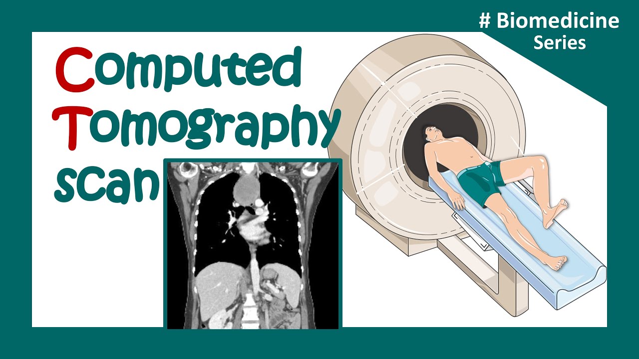
How Does a CT Scan Work?
Summary
TLDRA CT scan is an advanced x-ray procedure that creates detailed cross-sectional images of bones, soft tissues, and organs. Unlike conventional x-rays, which use a fixed x-ray beam, a CT scanner utilizes a motorized x-ray source that rotates around the patient, capturing images from multiple angles. These images are collected by digital detectors and processed by a computer to form either 2D slices or 3D images, helping physicians detect abnormalities and plan treatments with greater precision.
Takeaways
- 🔍 A CT scan uses x-rays and computer processing to create detailed cross-sectional images.
- 🏥 CT imaging provides more detail than traditional x-rays, showing both bones and soft tissues.
- 🔬 Conventional x-rays use a fixed tube, while CT scanners use a rotating motorized x-ray source.
- 🌀 The CT scanner's x-ray source moves around the patient, emitting narrow x-ray beams.
- 📡 Digital x-ray detectors are positioned opposite the source to capture the x-rays as they pass through the patient.
- 💻 The captured x-rays are sent to a computer for processing and image reconstruction.
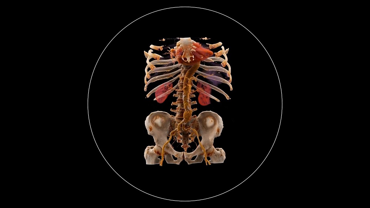
- 📑 CT scans can display images in 2D or stack them to create a 3D view for a comprehensive analysis.
- 🏠 CT imaging is essential for revealing abnormal structures within the body.
- 🩺 Physicians use CT scan results to plan and monitor treatments effectively.
- 🔎 The technology allows for a detailed examination of internal structures without invasive procedures.
Q & A
What is a CT scan?
-A CT scan is an x-ray procedure that creates cross-sectional images with the help of computer processing.
How are CT images different from conventional x-ray images?
-CT images are more detailed than conventional x-ray images and can reveal not only bones but also soft tissue and organs.
What is the difference between the x-ray source in a conventional x-ray and a CT scanner?
-A conventional x-ray uses a fixed tube that sends x-rays in only one direction, while a CT scanner uses a motorized x-ray source that shoots narrow beams of x-rays as it rotates around the patient.
What role do digital x-ray detectors play in a CT scan?
-Special digital x-ray detectors are located directly opposite the x-ray source and pick up the x-rays as they pass through the patient.
How are the x-rays detected and processed in a CT scan?
-The x-rays detected by the digital x-ray detectors are transmitted to a computer, where they are processed to create image slices.
Can CT scan images be displayed in two-dimensional or three-dimensional form?
-Image slices can either be displayed individually in two-dimensional form or stacked together to generate a three-dimensional image.
What is the purpose of generating a three-dimensional image from CT scan slices?
-A three-dimensional image can reveal abnormal structures, which helps physicians plan and monitor treatments.
How does the rotation of the x-ray source in a CT scanner contribute to image detail?
-The rotation of the motorized x-ray source allows for a comprehensive scan of the patient from multiple angles, contributing to the detailed nature of CT images.
What is the advantage of CT scans over conventional x-rays in terms of diagnostic capabilities?
-CT scans provide more detailed images that can reveal soft tissues and organs, which are not visible in conventional x-rays, enhancing diagnostic capabilities.
How does the computer processing in a CT scan contribute to the final image quality?
-Computer processing in a CT scan helps in reconstructing the x-ray data into detailed cross-sectional images, improving the image quality and diagnostic accuracy.
What are some potential applications of CT scans in medical diagnostics?
-CT scans are used in medical diagnostics to detect and monitor a variety of conditions, including tumors, internal injuries, and diseases affecting bones and soft tissues.
Outlines

هذا القسم متوفر فقط للمشتركين. يرجى الترقية للوصول إلى هذه الميزة.
قم بالترقية الآنMindmap

هذا القسم متوفر فقط للمشتركين. يرجى الترقية للوصول إلى هذه الميزة.
قم بالترقية الآنKeywords

هذا القسم متوفر فقط للمشتركين. يرجى الترقية للوصول إلى هذه الميزة.
قم بالترقية الآنHighlights

هذا القسم متوفر فقط للمشتركين. يرجى الترقية للوصول إلى هذه الميزة.
قم بالترقية الآنTranscripts

هذا القسم متوفر فقط للمشتركين. يرجى الترقية للوصول إلى هذه الميزة.
قم بالترقية الآنتصفح المزيد من مقاطع الفيديو ذات الصلة
CT scan | computerized tomography (CT) scan |What is a CT scan used for? | Clinical application

Diagnostic Imaging Explained (X-Ray / CT Scan / Ultrasound / MRI)
What is Computed Tomography (CT) and how does it work?

Biomedical instrumentation- CT scan (Computed Tomography)

How X-rays see through your skin - Ge Wang

Comprendre le Contraste Radiologique : Fondements Physiques et Applications Diagnostiques
5.0 / 5 (0 votes)
