Vision: Anatomy and Physiology, Animation
Summary
TLDRThis script explores the human visual system, detailing how eyes detect light through a narrow spectrum and convert it into neural signals. It highlights the roles of the cornea, lens, iris, and retina, explaining how rods and cones enable night and color vision, respectively. The process of light absorption by visual pigments and the subsequent neural signaling to the brain is described. The script also addresses color blindness, the brain's role in filling visual gaps, and the pathway of visual information from the retina to the brain, emphasizing the trade-off between sensitivity and resolution in vision.
Takeaways
- 👀 Vision is the ability to perceive objects through light they emit or reflect, with human eyes detecting a specific range of wavelengths (400-750 nm) in the electromagnetic spectrum.
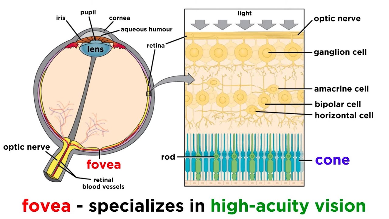
- 👁️ The eye functions similarly to a camera, with optical components like the cornea, lens, and iris focusing light onto the retina, and neural components like the optic nerve transmitting visual information to the brain.
- 🌀 The iris adjusts the pupil's size to control light intake, acting as an aperture and protecting the eye from excessive light.
- 🌈 Photoreceptor cells in the retina, known as rods and cones, are responsible for night and day vision, respectively, with cones also detecting color through three types sensitive to red, green, and blue light.
- 👁️🗨️ The fovea, at the retina's center, provides the sharpest central vision, while the optic disk, lacking photoreceptors, corresponds to the blind spot in our visual field.
- 🧠 The brain compensates for the blind spot by filling in the missing visual information from surrounding areas, creating a seamless visual experience.
- 🌑 Rod cells, sensitive to low light, have a high degree of convergence, making them highly sensitive but providing low-resolution images.
- 🌞 Cone cells, active in bright light, have a lower degree of convergence, allowing for high-resolution vision but requiring more light to function.
- 🔬 Visual pigments in photoreceptor cells, like rhodopsin in rods and iodopsins in cones, are responsible for detecting light and initiating the visual signal transmission process.
- 🔄 The process of light absorption by visual pigments triggers a series of biochemical reactions that convert the light signal into electrical signals, which are then sent to the brain via the optic nerve.
- 🧬 Other cell types in the retina contribute to detecting light intensity changes and provide information about contrast and object edges, enhancing our visual perception.
Q & A
What is the range of wavelengths that the human eye can detect?
-Human eyes can detect visible light, which is a narrow range of electromagnetic radiation, roughly from 400 to 750 nm in wavelengths.
How do the optical components of the eye function?
-The main optical components of the eye, including the cornea, lens, and iris, work like a camera to capture and focus images. The cornea and lens refract light, while the iris controls the amount of light entering the eye by adjusting the size of the pupil.
What is the role of the retina in the process of vision?
-The retina is a light-sensitive tissue lining the inner surface of the eye. It absorbs light through photoreceptor cells, and the optical information is then converted into action potentials and sent via the optic nerve to the visual cortex of the brain.
Why is the fovea significant for central vision?
-The fovea is the central part of the retina where the sharpest central vision is achievable. It has a high concentration of cones, which are responsible for high-resolution color vision.
What causes the blind spot in the visual field?
-The blind spot corresponds to the optic disk, where the optic nerve leaves the eye and has no photoreceptor cells. If an object falls on this spot, it would generate no visual information.
How does the brain handle the blind spot in vision?
-The brain fills in the blind spot with visual information from around the object, so instead of leaving a black hole in the vision, the blind spot is imperceptible to the viewer.
What are the two types of photoreceptor cells in the retina and their functions?
-The two types of photoreceptor cells are rods and cones. Rods are responsible for night vision and can detect dim light but provide low-resolution images and cannot differentiate colors. Cones function in bright daylight, detect colors, and provide high-resolution details.
What causes color blindness and how is color vision perceived?
-Color blindness occurs when a person lacks a certain kind of cones. Color vision is perceived based on the proportions of signals coming from the three types of cones that absorb best: red, green, and blue.
What are visual pigments and their components?
-Visual pigments are light-receptor molecules in photoreceptor cells, consisting of a protein called opsin and a vitamin A-derivative called retinal. Rhodopsin is found in rods, and iodopsins are found in cones. Different opsins absorb different wavelengths, allowing for the detection of different colors.
How does the process of light absorption affect the neurotransmitter glutamate?
-In the dark, the presence of cGMP allows a constant influx of sodium, and the cells release glutamate. When light is absorbed, the retinal changes form and dissociates from opsin, which deactivates the enzyme, stops the dark current, and reduces glutamate secretion, signaling to bipolar cells that light has been detected.
What is the difference between the signal processing of rods and cones in terms of sensitivity and resolution?
-Rods have a high degree of convergence, making them highly sensitive but providing low resolution. Cones have a lower degree of convergence, particularly in the fovea, leading to high-resolution images but lower sensitivity because each cone must be stimulated with a strong enough signal to generate action potentials in the ganglion cell.
Outlines
👀 Vision and the Human Eye
Vision is the ability to perceive objects through light they emit or reflect. Human eyes are sensitive to a specific range of light wavelengths, approximately 400 to 750 nm. The eye's optical components, including the cornea, lens, and iris, function similarly to a camera to capture and focus light. The iris adjusts the pupil's size to control light entry. The neural components involve the retina, which contains photoreceptor cells sensitive to light, and the optic nerve, which transmits visual information to the brain. The fovea, located centrally in the retina, is responsible for sharp central vision, while the optic disk, lacking photoreceptors, corresponds to the blind spot in our vision. The brain fills in the blind spot with surrounding visual information. Photoreceptor cells, rods, and cones, are responsible for night and day vision, respectively. Rods are sensitive to low light but provide less detail and no color differentiation, while cones, of which there are three types sensitive to red, green, and blue light, offer color perception and high-resolution vision. Color blindness arises from the absence of one or more types of cones. Visual pigments in the retina, such as rhodopsin in rods and iodopsins in cones, initiate the process of light detection. These pigments consist of opsin and retinal, with different opsins allowing for the detection of various light wavelengths. In darkness, a 'dark current' due to cGMP allows sodium influx, but light absorption changes the retinal's conformation, halting this current and neurotransmitter release, signaling light detection to the brain.
🌐 Neural Processing of Visual Information
Beyond the initial photoreception, other cell types in the retina, such as bipolar cells, contribute to the detection of light intensity changes and provide information about contrast and object edges. Some ganglion cells, which are second-order neurons, absorb light directly but serve non-imaging functions like controlling pupil size and regulating the sleep-wake cycle. The axons of ganglion cells form the optic nerve, which converges at the optic chiasm where fibers partially cross to the opposite side of the brain. Most fibers proceed to the thalamus and then to the primary visual cortex, while others are involved in reflexes like the pupillary light reflex. It's noted that the left visual field is processed by the right side of the brain, which also controls the left side of the body's motor responses.
Mindmap
Keywords
💡Vision
💡Electromagnetic radiation
💡Cornea
💡Lens
💡Iris
💡Retina
💡Photoreceptor cells
💡Visual pigments
💡Fovea
💡Optic nerve
💡Visual cortex
Highlights
Vision is the perception of objects based on light they emit or reflect.
Human eyes detect a narrow range of visible light, from 400 to 750 nm in wavelengths.
The eye functions like a camera with optical and neural components for image capture and processing.
The cornea, lens, and iris are the main optical components, focusing light on the retina.
The iris controls light entry by adjusting the pupil size.
Photoreceptor cells in the retina absorb light and convert it into nerve impulses.
The fovea is the retina's central part for sharp central vision.
The optic disk, lacking photoreceptor cells, corresponds to the blind spot in the visual field.
The brain fills in the blind spot with visual information from surrounding areas.
Rods and cones are the major photoreceptor cells, responsible for night and day vision respectively.
There are three types of cones, sensitive to red, green, and blue light, contributing to color perception.
Color blindness occurs due to the absence of one or more types of cones.
Visual pigments in rods and cones, like rhodopsin and iodopsins, are responsible for light detection.
The dark current in photoreceptor cells is due to the presence of cGMP, allowing sodium influx.
Light absorption changes retinal from cis to trans-form, affecting neurotransmitter release.
Ganglion cells receive signals from rods and cones and send them to the brain as action potentials.
High convergence of rod signals in ganglion cells increases sensitivity but reduces resolution.
Cones have low convergence, providing high-resolution images but requiring strong light stimulation.
Other cell types detect changes in light intensity, contributing to contrast and edge detection.
Some ganglion cells absorb light for reflex control and sleep-wake cycle regulation, not image formation.
The optic nerves from both eyes converge at the optic chiasm, with fibers crossing to the opposite side of the brain.
The primary visual cortex processes visual information from the thalamus, while some fibers control reflexes.
Transcripts
Vision is the perception of objects based on the light that they emit or reflect.
Human eyes can only detect visible light - a narrow range of electromagnetic radiation,
roughly from 400 to 750 nm in wavelengths.
The eye consists of optical components, which work like a camera, capturing and focusing images;
and neural components that convert these images into nerve impulses and send them to the brain.
The main optical components are the cornea, the lens, and the iris. The cornea and the
lens refract light and focus the image on the retina. The iris acts as an aperture,
it controls the amount of light that enters the eye by adjusting the size of the pupil.
The neural components are the retina - a light-sensitive tissue lining the inner
surface of the eye, and the optic nerve. Light is absorbed by photoreceptor cells in the retina.
The optical information is then passed through several cell layers, where it is converted into
action potentials and sent, via the optic nerve, to the visual cortex of the brain.
The fovea is the central part of the retina where the sharpest central vision is achievable.
The optic disk, where the optic nerve leaves the eye, has no photoreceptor cells.
It corresponds to the blind spot in the visual field. If an object falls on that spot,
it would generate no visual information. However, instead of leaving a black hole in the vision,
the brain fills it in with visual information from around the object.
The major photoreceptor cells of the retina are rods and cones. Rod cells are responsible
for night vision. They can detect dim light, but provide low-resolution images and cannot
differentiate colors. Cones function in bright day light. They detect colors, and provide high
resolution details. There are 3 kinds of cones named after the color that they absorb best:
red, green and blue. A color is perceived based on proportions of signals coming from these cones.
Color blindness occurs when a person lacks a certain kind of cones.
The ability of photoreceptor cells to detect light
is due to their light-receptor molecules, called visual pigments.
It’s rhodopsin in rods, and iodopsins in cones. These molecules consist of 2 components:
a protein called opsin, and a vitamin A-derivative called retinal. The retinal component is
identical for all visual pigments, but the opsin is different for rods and each type of cones.
Different opsins absorb different wavelengths, allowing detection of different colors.
In the dark, there is a so-called dark current in photoreceptor cells.
This is due to the presence of cGMP, which permits a constant influx of sodium. The cells are
depolarized, they release the neurotransmitter glutamate at the synapse with bipolar cells.
The retinal exists in 2 conformations: cis and trans. In the dark,
the cis-form is bound to opsin, keeping it inactive. As the retinal absorbs light,
it changes to trans-form and dissociates from the opsin, which now becomes an active enzyme. The
enzyme degrades cGMP, sodium channel closes, dark current stops and so does glutamate secretion.
The drop in glutamate tells the bipolar cells that light has been absorbed.
The information is then transmitted to ganglion cells - the only cells
within the retina that generate action potentials and send them to the brain.
On average, each ganglion cell receives signals from over a hundred of rods. This
degree of convergence is at the basis of the high sensitivity of rod cells. A dim light produces
only a weak signal in a rod, but together, hundreds of these signals converge and become
one strong signal acting on a single ganglion cell. However, as the signal comes from a large
area of the retina, the image resolution is poor. The cones have a much lower degree of convergence.
The fovea in particular has only cones and no rods, and each cone conveys signal to one
ganglion cell. Because one ganglion cell receives input from a very small area of the retina, this
setup produces high resolution images. But high resolution comes with low sensitivity, because
each cone must be stimulated with a signal strong enough to generate action potentials
in the ganglion cell. This also explains why there is no color vision in dim light.
There exist other cell types that form connections between photoreceptor cells,
or bipolar cells. They detect changes in light intensity within an image and provide additional
information about contrast and edges of objects. Some of the ganglion cells also absorb light
directly, but not for the purpose of forming images. They transmit information about light
intensity to the brainstem, as part of the reflex that controls pupil size; and to the hypothalamus,
as input for the sleep-wake cycle. The bipolar cells are first-order neurons,
and ganglion cells are second-order neurons. The axons of ganglion cells form the optic nerve.
The 2 optic nerves from the 2 eyes converge at the optic chiasm.
Here, the medial half of nerve fibers from each eye cross to the other side of the brain.
Most of the fibers then continue to the thalamus and synapse with third-order neurons, whose axons
project to the primary visual cortex. Some fibers take a different route: they terminate in the
midbrain and are responsible for pupillary light reflex and accommodation reflex, among others.
Note that objects in the left visual field are perceived by the right side of the brain,
which also controls motor responses of the body’s left side – the same side as the objects.
تصفح المزيد من مقاطع الفيديو ذات الصلة
10-Minute Neuroscience: Visual Pathways
Vision: Crash Course Anatomy & Physiology #18
What Is Color? | Physics in Motion
Sensation and Perception: Crash Course Psychology #5
Fisiologi Melihat (Refraksi & Visual Pathway) #1 BASIC OPTHALMOLOGY
A Level Biology Revision (Year 13) "Rod Cells and Cone Cells as Light Receptors"
5.0 / 5 (0 votes)