Anatomy of the Pericardium
Summary
TLDRThis video explains the anatomy of the pericardium, covering its three main layers: fibrous pericardium, parietal serous pericardium, and visceral serous pericardium (epicardium). It discusses their development, functions, and clinical significance, such as the role of pericardial fluid and conditions like pericardial effusion. The video also highlights the differences between the parietal and visceral layers, including their innervation and pain sensitivity. The script provides essential insights for understanding heart protection and related medical conditions, making it valuable for both learners and healthcare professionals.
Takeaways
- 😀 The pericardium is a double-walled sac surrounding the heart, consisting of fibrous and serous layers.
- 😀 The fibrous pericardium is the outermost tough layer that provides structural support and remains unaffected during heart development.
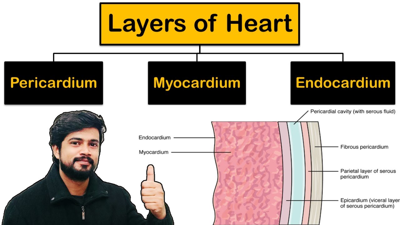
- 😀 The serous pericardium has two layers: parietal (lines the fibrous pericardium) and visceral (covers the heart surface, also called epicardium).
- 😀 The pericardial cavity lies between the parietal and visceral layers and contains approximately 50 mL of pericardial fluid.
- 😀 Excess pericardial fluid results in a condition called pericardial effusion, similar to pleural effusion in the lungs.
- 😀 During heart development, the growing heart compresses the serous pericardium, bringing its layers close together.
- 😀 The parietal pericardium is adherent to the fibrous pericardium, develops from somatic mesoderm, and is innervated by the phrenic nerve, making it pain-sensitive.
- 😀 The visceral pericardium (epicardium) is adherent to the myocardium, develops from splanchnic mesoderm, and is innervated by autonomic nerves, making it pain-insensitive.
- 😀 The differences between parietal and visceral pericardium include adherence, developmental origin, innervation type, and pain sensitivity.
- 😀 Clinical relevance includes understanding that chest pain from pericarditis arises from the pain-sensitive parietal layer, while the visceral layer does not produce pain.
Q & A
What are the main layers of the pericardium?
-The pericardium has three main layers: the fibrous pericardium (outer layer), the parietal layer of the serous pericardium (middle layer lining the fibrous pericardium), and the visceral layer of the serous pericardium (inner layer covering the heart surface, also called epicardium).
What is the pericardial cavity and what does it contain?
-The pericardial cavity is the space between the parietal and visceral layers of the serous pericardium. It contains approximately 50 mL of pericardial fluid, which reduces friction during heart movements.
What is the function of the fibrous pericardium?
-The fibrous pericardium provides structural support, protects the heart, and prevents overexpansion during increased blood volume. It is pain-sensitive and innervated by the phrenic nerve.
How do the parietal and visceral layers of the serous pericardium differ in their adherence?
-The parietal layer adheres to the fibrous pericardium, while the visceral layer (epicardium) is closely attached to the myocardium, the muscle layer of the heart.
Which layer of the pericardium is pain-sensitive and which is pain-insensitive?
-The fibrous pericardium and the parietal layer of the serous pericardium are pain-sensitive, while the visceral layer (epicardium) is pain-insensitive.
What is another name for the visceral layer of the pericardium?
-The visceral layer of the pericardium is also called the epicardium.
From which embryonic tissue do the parietal and visceral layers of the serous pericardium develop?
-The parietal pericardium develops from the somatic mesoderm, while the visceral pericardium (epicardium) develops from the splanchnic mesoderm.
What nerves innervate the parietal and visceral layers of the serous pericardium?
-The parietal pericardium is innervated by somatic fibers from the phrenic nerve, making it pain-sensitive. The visceral pericardium is innervated by autonomic fibers and is pain-insensitive.
What is pericardial effusion and how does it occur?
-Pericardial effusion is the accumulation of excess fluid in the pericardial cavity beyond the normal ~50 mL. It can occur due to inflammation, infection, trauma, or other medical conditions affecting the pericardium.
How does the developing heart influence the formation of the serous pericardium?
-During heart development, the heart pushes the serous pericardium, creating two layers (parietal and visceral) with a cavity in between. The fibrous pericardium remains undisturbed.
Why is it important to distinguish between parietal and visceral layers clinically?
-Understanding the differences is crucial because pain originating from the parietal layer can indicate pericardial inflammation or other pathology, whereas the visceral layer does not produce pain. It also helps in procedures like pericardiocentesis.
Outlines

此内容仅限付费用户访问。 请升级后访问。
立即升级Mindmap

此内容仅限付费用户访问。 请升级后访问。
立即升级Keywords

此内容仅限付费用户访问。 请升级后访问。
立即升级Highlights

此内容仅限付费用户访问。 请升级后访问。
立即升级Transcripts

此内容仅限付费用户访问。 请升级后访问。
立即升级浏览更多相关视频
Layers of Heart - Pericardium, Myocardium, Endocardium in Hindi | Cardiovascular System

Body Cavities and Membranes (Dorsal, Ventral)- Anatomy and Physiology

Anatomi Jantung

Cardiovascular System (Part 1) - Heart

IMAT Biology Lesson 6.5 | Anatomy and Physiology | Circulatory System II

Cardiac Tamponade - pericardial effusion, causes, pathophysiology, investigations and treatment
5.0 / 5 (0 votes)
