How to Interpret a Chest X-Ray (Lesson 2 - A Systematic Method and Anatomy)
Summary
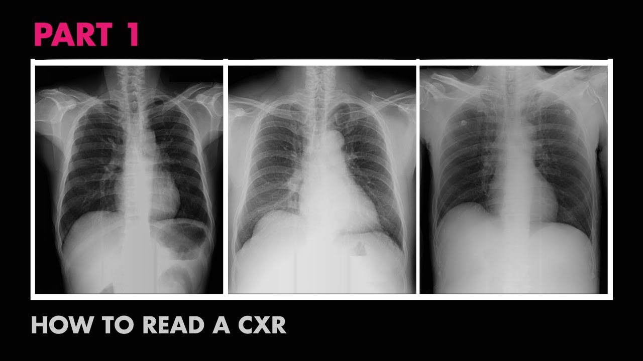
TLDRThis educational video introduces a systematic approach to interpreting chest x-rays, emphasizing the ABCDEF method for assessing technical quality, airways, bones, cardiac silhouette, diaphragm, and lung fields. It highlights the importance of recognizing normal anatomy to identify abnormalities and discusses the significance of each anatomical structure visible on x-rays, including airways, bones, cardiac silhouette, diaphragm, and lung lobes.
Takeaways
- 🔍 The systematic approach to interpreting chest x-rays is crucial for clinicians, especially those with less experience, to ensure important findings are not missed.
- 📚 The ABCDEF system is a common method for interpreting chest x-rays, making it easier to remember the sequence: Airways, Bones, Cardiac silhouette and mediastinum, Diaphragm, Pleura, and Lung fields.
- 📏 Assessing the technical quality of the x-ray film is the first step before starting the ABCDEF system.
- 🌐 'A' in ABCDEF stands for Airways, focusing on the trachea and the right and left main bronchi, which are typically visible on a normal x-ray.
- 🦴 'B' represents Bones, including ribs, clavicles, sternum, and vertebral bodies, which are essential to examine on a PA and lateral x-ray set.
- ❤️ 'C' is for the Cardiac silhouette and mediastinum, where various anatomical structures compose the silhouette, including the aortic pulmonary window.
- 🏋️♂️ 'D' stands for Diaphragm, which is normally higher on the right than the left due to the liver's position, and is crucial for diagnosing conditions like pneumothorax.
- 🌬️ 'E' is for Pleura, a double membrane surrounding the lungs, which is normally invisible but essential for diagnosing certain lung conditions.
- 🌌 'F' is for Fields, referring to the lung fields where the focus is on the fissures between the lobes and the lobes themselves.
- 🔎 The lungs are examined near the end of the ABCDEF system because they are the most likely to be abnormal and often the area of greatest interest.
- 📈 Understanding the normal anatomy and appearance of each anatomical structure on the x-ray is fundamental before identifying any pathology.
Q & A
What is the main purpose of a systematic approach to interpreting chest x-rays?
-The main purpose of a systematic approach to interpreting chest x-rays is to ensure that all aspects of the x-ray are examined in a logical and memorable sequence, reducing the chance that important findings will be missed, especially by clinicians with less experience.
What does the acronym ABCDEF stand for in the context of interpreting chest x-rays?
-In the context of interpreting chest x-rays, ABCDEF is a mnemonic that stands for Airways, Bones and soft tissue, Cardiac silhouette and mediastinum, Diaphragm, Pleura, and Lung fields.
Why is it important to assess the technical quality of a chest x-ray before anything else?
-Assessing the technical quality of a chest x-ray before anything else is important because it ensures the image is clear and properly aligned, which is crucial for accurate interpretation of the x-ray.
What are the three anatomic airway structures typically visible on a normal chest x-ray?
-The three anatomic airway structures typically visible on a normal chest x-ray are the trachea, the right main bronchus, and the left main bronchus.
Why are aspirated foreign bodies more likely to end up in the right lung than the left?
-Aspirated foreign bodies are more likely to end up in the right lung than the left because the left main bronchus tends to take off from the trachea at a slightly more horizontal angle compared to the right, which is more vertical.
What are the four types of bones easily visualized on a PA and lateral chest x-ray set?
-The four types of bones easily visualized on a PA and lateral chest x-ray set are the ribs, clavicles, sternum, and vertebral bodies.
What is the significance of the aortopulmonary window in chest x-ray interpretation?
-The aortopulmonary window is an important space between the aortic arch and the pulmonary artery where the recurrent laryngeal nerve and lymph nodes are located. It is significant for diagnosing certain conditions and is part of the cardiac silhouette evaluation.
Why is it normal for the right hemidiaphragm to be slightly higher than the left on a chest x-ray?
-It is normal for the right hemidiaphragm to be slightly higher than the left due to the liver being located directly beneath the right hemidiaphragm, which pushes it upward.
What are the three fissures in the lungs and which ones are commonly visible on normal x-rays?
-The three fissures in the lungs are the horizontal fissure, and the right and left oblique fissures. The horizontal fissure on the right side is the only one commonly visible in normal x-rays because a significant portion of its plane is parallel to the direction of the x-ray beams.
Why is it difficult to determine the lobe location of a visualized nodule or mass from only PA or AP views without a lateral view?
-It is difficult to determine the lobe location of a visualized nodule or mass from only PA or AP views without a lateral view because the oblique fissures, which help differentiate between lobes, run obliquely and are not easily visible in these views.
What is the significance of the gastric air bubble in assessing a chest x-ray?
-The gastric air bubble, usually located under the left hemidiaphragm, is significant as it represents air in the stomach and can help in assessing the position of the diaphragm and differentiating between gas in the stomach and intestines.
Outlines
📚 Systematic Approach to Chest X-ray Interpretation
This paragraph introduces the systematic approach to interpreting chest x-rays, emphasizing its importance for clinicians, especially those with less experience. It outlines the ABCDEF system for remembering the sequence: Airways, Bones and soft tissue, Cardiac silhouette and mediastinum, Diaphragm, Fusions, and Fields (lung fields). The paragraph also stresses the need to assess the technical quality of the x-ray film first. It explains each component of the ABCDEF system, providing insights into normal anatomy and potential abnormalities. For instance, it discusses the visibility of the trachea and bronchi, noting the angle at which the left main bronchus branches off, which is relevant for understanding aspiration of foreign bodies and endotracheal tube placement. The paragraph concludes by highlighting the importance of knowing the normal appearance of anatomical structures on x-rays to identify pathology.
🔍 Detailed Anatomy of Chest X-ray Structures
The second paragraph delves into the detailed anatomy visible on a chest x-ray, focusing on the cardiac silhouette and mediastinum, diaphragm, and lungs. It discusses the various structures that make up the cardiac silhouette, including the aortic arch and pulmonary artery, and introduces the aortopulmonary window, an important anatomical space. The paragraph also covers the diaphragm and pleura, explaining their appearance on PA and lateral views, and the significance of the costophrenic angles. It touches on the gastric air bubble, a common finding under the left hemidiaphragm, and its implications for identifying the stomach. Lastly, it describes the anatomy of the lungs, including the fissures and lobes, and the challenges of identifying lung nodules or masses on PA or AP views without a lateral view. The paragraph concludes with a teaser for the next video, which will discuss the assessment of chest film technical quality.
Mindmap
Keywords
💡Systematic Approach
💡Chest X-ray
💡Anatomy
💡Airways
💡Bones and Soft Tissue
💡Cardiac Silhouette
💡Diaphragm
💡Pleura
💡Lung Fields
💡Lines, Tubes, Devices
💡Technical Quality
Highlights
Introduction to the systematic approach to interpreting chest x-rays and its importance for clinicians.
The ABCDEF system as a common method for interpreting chest x-rays, emphasizing its memorability and effectiveness.
Assessment of film technical quality as the initial step in the systematic approach.
Importance of examining all elements of the chest x-ray in a logical and memorable sequence.
The significance of the systematic approach in reducing the chance of missing important findings.
Description of the Airways as the 'A' in ABCDEF, including the trachea and main bronchi.
Explanation of why aspirated foreign bodies are more likely to end up in the right lung.
Details on the four types of bones visible on a chest x-ray: ribs, clavicles, sternum, and vertebral bodies.
The cardiac silhouette and mediastinum as part of the 'C' in the ABCDEF system.
Identification of the aortopulmonary window and its anatomical significance.
The diaphragm and pleura as 'D' and 'E' in the ABCDEF system, including the assessment of the gastric air bubble.
Importance of recognizing that no structures in the body are truly flat, especially when evaluating the diaphragm.
Explanation of the costophrenic angles and their significance in chest x-ray interpretation.
The lungs as 'F' in the ABCDEF system, focusing on fissures and lobes.
Visibility of the horizontal fissure on the right side and its parallel alignment with the x-ray beams.
Challenge of identifying the lobe location of a nodule or mass without a lateral view.
Conclusion summarizing the systematic approach and normal chest x-ray anatomy.
Encouragement for viewers to like or share the video and预告 of the next video on assessing chest film technical quality.
Transcripts
[Music]
hello everyone this is the second video
in this series on interpreting chest
x-rays the topic is the systematic
approach and normal chest x-ray
Anatomy the learning objectives of this
video are to be familiar with the
systematic approach to interpreting
chest x-rays and to know the correlation
between anatomy and normal Shadows on
the
X-ray before just presenting a
systematic approach I first wanted to
mention a couple of important principles
about it a systematic approach is most
important for the clinician's least
experience with reading chess xrays
since it reduces the chance that
important findings will be
missed all aspects of Chess x-ray
interpretation should be
included the individual elements of the
approach should be examined in a
sequence that's either logical and or
easy to remember
and there is no one best system though
all should begin with an assessment of
the film's technical
quality so the system I teach trainees
is informally referred to as the
ABCDEF system it's not the only one but
it's certainly the most common at least
in the US it's also not perfect but it's
easy to remember each of those six
letters refers to a specific anatomic
structure even before the a we need to
assess the technical
quality then a stands for
Airways B for bones and soft tissue C
for the cardiac silhouette and
medyum D for diaphragm which also
includes assessment of the gastric air
bubble usually located under the left
Hemi
diaphragm e for fusions in other words
assessment of the plur which actually
includes findings Beyond just plural
fusions
and F for fields that is the lung
Fields lastly although it's not
explicitly part of the neonic is an
assessment of lines tubes devices and
prior surgeries such as sternotomies and
valve
Replacements aside from the fact that
it's easy to remember another nice thing
about this pneumonic is that the lungs
are examined near the end this is a good
idea because normally the lungs are the
area of greatest interest and the most
likely to be abnormal therefore once the
clinician finds an abnormality there
it's very easy for him or her to forget
examining the rest of the film I've seen
more than one rib fracture missed due to
distraction over acute lung
pathology you may have noticed that the
list of items here lines up really
nicely with the remaining videos in this
series which of course is not a
coincidence but before you can identify
pathology of each of these anatomic
structures you first need to know where
where they are on the X-ray and what
they normally look like so let's go
through the X-ray anatomy of a to f one
at a
time as is for the Airways there are
three anatomic Airway structures that
are typically visible on a normal x-ray
they are the trachea which is normally
in the midline and the right and left
main bronchus remember that the
patient's right will be on the left side
of the screen to help you visualize
these structures let me superimpose a
drawing of them
the left main bronchus tends to take off
from the trachea at a slightly more
horizontal angle as compared to the
right which is why aspirated foreign
bodies if small enough are more likely
to end up in the right lung than the
left and it's also why endot tral tubes
if Advance too far end up in the right
main broncus blocking off ventilation to
the left rather than vice
versa I'll take the drawing away again
so you can try to see the Airways on
your own
the next set of structures is the
bones there are four types of Bones
easily visualized on a PA and lateral
x-ray set first is this structure which
as you probably know or could guess is a
rib there are two radiographic
components of the rib on the PA film the
posterior rib which is easier to see and
is oriented horizontally and the
anterior rib which is harder to see and
oriented at a 45°
angle the next bone is the clavicle
located up here there's obviously a
right and a left one the sternum is
sometimes visible on the lateral film
but is often obscured by soft tissue in
the anterior chest
wall then are the vertebral bodies more
easily visible on the lateral film but
usually seen on the PA as well provided
that the technical quality is
adequate then there is the cardiac
silhouette and the medus dyum which I
think logically should be evaluated
together there are a lot of anatomic
structures which compose the silhouette
here I'll start by outlining it and then
label what each bump knob or side
corresponds to you will usually also be
able to see another vertical line
running just to the patient's left of
the vertebral bodies which corresponds
to the left side of the descending aorta
to make it easier to visualize here's a
drawing of the heart and great
vessels the one structure on the
patient's left called the aoro pulmonary
window may be unfamiliar to some viewers
this is an important space between the
aortic Arch and the pulmonary artery
where one will find the recurrent lenial
nerve and lymph
nodes and then I'll take the drawing
away and try to appreciate those various
structures
moving on the diaphragm and plora which
also make sense to evaluate together so
here are the right and left Hemi
diaphragms as seen on the PA view it is
normal for the right Hemi diaphragm to
be slightly higher than the left
presumably due to the liver directly
beneath it while looking at the
diaphragms it's important to remember
that there aren't any truly flat
structures in the body for example the
Hemi diaphragms are curved in
three-dimensional space as you might be
able to appre iate from comparing the PA
to the lateral the heavy diaphragms
that's seen on the PA really just
represent the superiormost aspect of the
diaphragms the plora which is a double
membrane that surrounds each lung is
outlined here but is normally invisible
due to its
thinness however knowledge that the
plora is there is essential for
diagnosing a pneumothorax plural plaques
from asist exposure and plural
thickening in addition there are spaces
on either side where the diaphragm meets
the thoracic wall these two spaces seen
on the PA are usually called the right
and left costophrenic angles though the
term costophrenic sulai would probably
be more semantically accurate in most
circumstances the space in the back on
the lateral film has several very
similar names the most common of which
is the posterior costophrenic
angle there's also usually a small
pocket of air visible under the left h
diaphragm this usually represents air in
the stomach in which case it's called
the gastric air bubble although
occasionally it can be unclear as to
whether the gas is in the stomach or in
the
intestines finally the last structures
on the chest x-ray are of course the
lungs the anatomic structures in the
lungs to worry about when interpreting
x-rays are the fissures between the loes
and the loes
themselves first is the horizontal
fissure located here there's only one
and it's on the right
side here is its location on the lateral
film it's the only one of the three
fissures that is commonly visible in
normal x-rays which is because a
significant portion of the plane of the
fissure is parallel to the direction of
the X-ray
beams however it's only visible on the
PA which is why the fissure there is
represented by a solid line and the
fissure on the lateral is represented by
a dotted
line then there are the right and left
oblique fissures
neither of which is usually visible in
either
view as you can probably guess if you
didn't already know it the fact that
there are two fissures on the patient's
right meaning that the right long is
divided into three loes while the one
fissure on the left divides the left
long into two loes here's the location
of the right upper
lobe and the right middle
lobe and finally the right lower
lobe notice that because the oblique
visure runs obliquely as seen in the
lateral view the lower lobe extends
almost all the way to the apex of the
lung this will be true on the left side
as well as a consequence if only the PA
or AP views are presented without a
lateral it's almost impossible to tell
what lobe a visualized nodule or mass is
located in unless it's seen in the
uppermost part above the lower lob's
most Superior extent
here's the left upper
lobe and finally the left lower
lobe that concludes this video on the
systematic approach and normal chest
x-ray Anatomy if you found it helpful
please remember to like or share it the
next video will discuss the assessment
of a chest film's technical quality
浏览更多相关视频
Anatomy of a Chest X-Ray - How to Read a Chest X-Ray (Part 1)
ABCs of Reading a Chest X-ray - How to Read a Chest X-Ray (Part 2) - MEDZCOOL
Chest X-ray: Introduction and Approach
Radiology of Thorax (Chest)
¿Cómo interpretar una radiografía de tórax?
How to Interpret a Chest X-Ray (Lesson 5 - Cardiac Silhouette and Mediastinum)
5.0 / 5 (0 votes)