PET/CT Basics
Summary
TLDRThis video explains the principles and clinical applications of Positron Emission Tomography (PET) imaging, highlighting its role in diagnosing and monitoring various conditions like cancer, neurological disorders, and cardiovascular issues. The script covers how PET works with radiotracers like FDG, the process of metabolic imaging, and the integration with CT scans for better anatomical localization. It also discusses the limitations of PET, including false positives and negatives, and the challenges posed by normal physiological uptake in certain tissues. Emerging PET tracers and their potential applications are also explored.
Takeaways
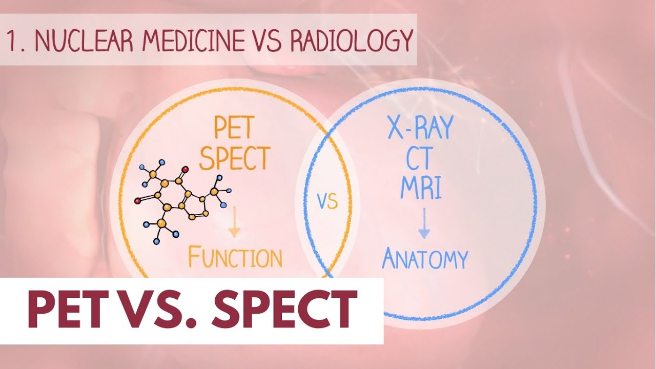
- 😀 Structural imaging (e.g., x-ray, CT) detects physical changes in body structures, while functional imaging (e.g., PET) identifies abnormal physiological or metabolic processes.
- 😀 PET imaging relies on radioactive tracers that emit positrons, which annihilate with electrons, producing gamma ray photons detected to create a 3D image map.
- 😀 The most commonly used PET tracer is FDG (fluorodeoxyglucose), a glucose analog that accumulates in metabolically active cells, highlighting areas of increased glucose metabolism.
- 😀 PET imaging is widely used in diagnosing, staging, and assessing treatment responses for cancers, but its effectiveness varies based on cancer type.
- 😀 PET imaging combined with CT (PET/CT) improves anatomical localization of abnormal tracer uptake and compensates for artifacts like scatter and absorption.
- 😀 FDG PET is highly sensitive for detecting cancers like lung, esophageal, and colorectal cancers, but less effective for low-grade tumors or certain cancer types like breast cancer.
- 😀 False positives in FDG PET can occur due to infections, inflammatory diseases, or even exercise-induced muscle activity, while false negatives may occur in tumors with low glucose metabolism.
- 😀 Normal tissues like the brain, liver, kidneys, and bowel naturally show high FDG uptake, which must be considered when interpreting results to avoid false interpretations.
- 😀 PET imaging with FDG is limited for diagnosing some cancers, such as renal, bladder, and prostate cancers, due to high background glucose utilization or excretion in these areas.
- 😀 New radio tracers are being developed for better sensitivity in diagnosing cancers like liver or kidney cancers, including carbon-11 acetate and fluorine-18 sodium fluoride.
- 😀 PET imaging, particularly FDG PET, plays a crucial role in assessing cancer recurrence, metastasis, and response to treatments, including immune modulation and targeted therapies.
Q & A
What is the difference between structural and functional medical imaging modalities?
-Structural imaging modalities, like X-ray or CT, are used to diagnose problems when a disorder changes the morphology of an organ or structure. Functional imaging modalities, like PET, are used to diagnose disorders that affect the physiological or metabolic processes within the body.
Why is PET imaging not routinely used for diagnosing renal malignancies and breast cancer?
-PET imaging is not routinely used for renal malignancies and breast cancer because, in these cases, PET imaging has a limited ability to identify certain types of cancers or it may be influenced by factors like high background glucose uptake in normal tissue, which can obscure the tumor.
How does PET imaging work at a fundamental level?
-PET imaging works by introducing radioactive tracers (radio nuclides) into the body. These tracers emit positrons, which annihilate electrons, producing gamma ray photons. Detectors capture these photons to reconstruct a 3D image of where the annihilation events occurred in the body.
What role does glucose metabolism play in PET imaging?
-Glucose metabolism is central to PET imaging as the tracer used, Fluorodeoxyglucose (FDG), mimics glucose. Cells take up FDG in a similar way to glucose, and when the tracer accumulates in hypermetabolic cells (such as cancer cells), it produces detectable signals, helping to identify areas of abnormal activity.
Why are patients asked to fast before a PET scan?
-Patients are asked to fast before a PET scan to reduce the competitive uptake of glucose by cells, which ensures that the FDG tracer is taken up efficiently by cells. Fasting also helps prevent the heart from using glucose as a fuel, which could obscure other signals.
What is the Standardized Uptake Value (SUV) in PET imaging, and how is it calculated?
-The Standardized Uptake Value (SUV) in PET imaging represents the ratio of radio tracer concentration in a given pixel relative to the average concentration in the body, normalized by the injected dose and the patient's body weight. It serves as a proxy for glucose utilization in the body.
What are the advantages of combining PET with CT in PET/CT scans?
-Combining PET with CT improves anatomical localization of abnormal radio tracer uptake, compensates for issues like photon undercounting in deep or high-attenuation tissues, and allows for more accurate interpretation of PET images.
Why can FDG PET imaging be difficult in diagnosing brain tumors?
-FDG PET imaging can be challenging for brain tumors because the brain naturally has high glucose metabolism, making it hard to distinguish between normal tissue and potential malignancies. It can also be difficult to differentiate between benign conditions and malignant tumors in the brain using FDG PET.
How is PET imaging used in oncology, specifically for cancer detection?
-In oncology, PET imaging is useful for detecting rapid cellular growth, a common trait of cancer. It is used in various stages of cancer care, including diagnosis, staging, and assessing treatment response. FDG PET is particularly effective for detecting tumors with high glucose metabolism, such as in lung, colorectal, and lymphoma cancers.
What are some limitations of FDG PET in cancer imaging?
-FDG PET has limitations, including false positives in conditions like infections, inflammation, and certain benign tumors. It can also have false negatives in cancers with low glucose metabolism or low tumor density, such as small or minimally invasive tumors.
Outlines

This section is available to paid users only. Please upgrade to access this part.
Upgrade NowMindmap

This section is available to paid users only. Please upgrade to access this part.
Upgrade NowKeywords

This section is available to paid users only. Please upgrade to access this part.
Upgrade NowHighlights

This section is available to paid users only. Please upgrade to access this part.
Upgrade NowTranscripts

This section is available to paid users only. Please upgrade to access this part.
Upgrade NowBrowse More Related Video


5.0 / 5 (0 votes)