RADIODICAS - Formação do feixe de Raios X
Summary
TLDRThis video provides a detailed exploration of the x-ray imaging process, from electron acceleration in the x-ray tube to the interaction of x-rays with the human body. It covers key concepts such as photon energy generation, the role of metal filters in optimizing the beam, and the importance of heat dissipation in the x-ray tube. The script also touches on the dynamics of braking radiation, the effect of focal spot size on image resolution, and the filtering process that ensures high-quality diagnostic images. This informative presentation delves into the physics of x-rays and their medical applications.
Takeaways
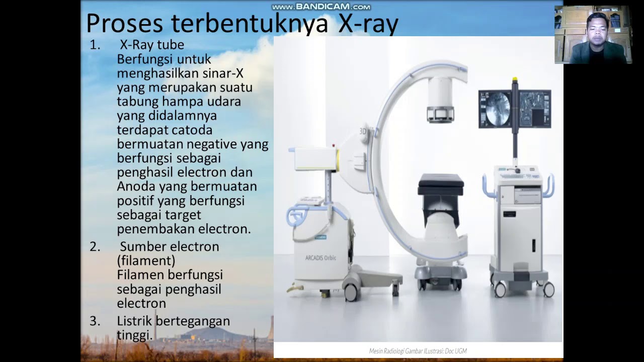
- 😀 X-ray beams are formed through the interaction of high-energy electrons with a metal target (typically tungsten) inside the X-ray tube.
- 😀 The energy of the electrons is accelerated and injected into the tube, where it interacts with the atoms of the target, producing X-ray photons.
- 😀 X-rays are produced when electrons eject inner-shell electrons from the target's atoms, causing energy release in the form of X-ray photons.
- 😀 When an electron does not have enough energy to eject an atom's electron, it may excite the atom, causing radiation emission in forms like ultraviolet, infrared, or visible light.
- 😀 The interaction between the electron and the atom can lead to heat generation, which is why the X-ray tube needs to dissipate heat efficiently.
- 😀 Braking radiation (or Bremsstrahlung) occurs when electrons are deflected by the positively charged nucleus of the atom, resulting in low-energy X-ray photons.
- 😀 Low-energy X-rays produced by braking radiation have limited diagnostic value, as they are absorbed by the skin and contribute to image degradation.
- 😀 X-ray systems are designed to filter out low-energy photons by using a metal filter, such as lead, aluminum, or molybdenum, to ensure only useful X-rays reach the patient.
- 😀 The efficiency of X-ray production is low, with about 99% of the energy being lost as heat and only about 1% contributing to the formation of useful X-ray photons.
- 😀 The ideal target material for X-ray production is one with a high atomic number, high melting point, and good conductivity to handle the heat generated during interactions.
- 😀 The focal spot size in the X-ray tube can be adjusted to optimize image quality and heat dissipation, with smaller spots providing higher definition but requiring careful heat management.
Q & A
What is the main purpose of the X-ray tube in the video?
-The main purpose of the X-ray tube is to accelerate electrons and direct them onto a target material (anode), where they interact to produce X-ray photons for diagnostic imaging.
What is the difference between characteristic radiation and bremsstrahlung?
-Characteristic radiation occurs when an electron ejects an inner electron from an atom, causing an energy gap that is filled by an electron from a higher energy level, emitting X-rays. Bremsstrahlung, on the other hand, is produced when electrons are deflected by the nuclei of atoms, losing kinetic energy in the process and emitting low-energy X-rays.
Why is tungsten preferred as the target material in the X-ray tube?
-Tungsten is preferred because of its high atomic number, high melting point, and excellent electrical conductivity, which makes it efficient in producing X-rays and dissipating the significant heat generated during the process.
What role does filtration play in X-ray imaging?
-Filtration is used to remove low-energy photons from the X-ray beam, which do not contribute to the diagnostic image and would only increase radiation exposure to the patient.
How much of the energy in an X-ray tube is converted into useful X-rays?
-Only about 1% of the energy in an X-ray tube is converted into useful X-rays, with the remaining 99% being lost as heat.
What is the relationship between focal spot size and image resolution?
-The size of the focal spot affects the image resolution. A smaller focal spot provides higher resolution and more definition, but it also generates more heat, making heat dissipation more critical.
Why is the energy distribution of the X-ray beam important?
-The energy distribution of the X-ray beam determines its ability to penetrate the patient and contribute to the diagnostic image. The beam includes both high-energy photons that are useful and low-energy photons that are filtered out.
What happens to the low-energy photons in the X-ray beam?
-Low-energy photons are filtered out of the X-ray beam, as they do not contribute to the diagnostic image and would only increase the radiation exposure to the patient.
How does the X-ray tube ensure that only useful photons reach the patient?
-The X-ray tube uses a filter (typically made of aluminum or molybdenum) to remove low-energy photons and a window to direct the filtered high-energy photons toward the patient.
What is the main challenge in the X-ray process, as discussed in the video?
-The main challenge in the X-ray process is managing the significant amount of heat generated during X-ray production. Efficient heat dissipation is essential to prevent damage to the X-ray tube and ensure safe operation.
Outlines

This section is available to paid users only. Please upgrade to access this part.
Upgrade NowMindmap

This section is available to paid users only. Please upgrade to access this part.
Upgrade NowKeywords

This section is available to paid users only. Please upgrade to access this part.
Upgrade NowHighlights

This section is available to paid users only. Please upgrade to access this part.
Upgrade NowTranscripts

This section is available to paid users only. Please upgrade to access this part.
Upgrade NowBrowse More Related Video


5.0 / 5 (0 votes)